SlideShare a Scribd company logo
1 of 156
Ultrasound of thyroid nodules
Samir Haffar MD
Internal medicine – Ultrasound
Ultrasound of thyroid nodules
① Epidemiology & etiologies of thyroid nodules
② Ultrasound features of thyroid nodules
③ Ultrasound of cervical lymph nodes
④ Ultrasound systems for thyroid nodule risk stratification
⑤ Pseudo-nodules of thyroid
⑥ Fine needle aspiration of thyroid nodules
① Epidemiology & etiology of thyroid nodules
Epidemiology of thyroid nodules
• Very common: Palpation 4 – 8%, US 10 – 40%, autopsy 50%
Increases with age
• Malignancy risk: Patients younger than 20 or older than 60 years
History of neck irradiation
History of familial thyroid cancer
• Cancer prevalence: 10-13% no matter how many nodules at US
No decrease of cancer risk in multiple nodules
Cancer found in dominant nodules in 2/3 of cases
Frates MC et al. Ultrasound Quarterly 2006;22(4):231–238.
Most thyroid nodules are benign hyperplastic nodules
Thyroid cancer not common compared to very frequent benign nodules
Indications of thyroid ultrasound
• Patient with palpable thyroid nodule or with multinodular goiter
• High-risk patient for thyroid malignancy:
History of familial thyroid cancer
Multiple endocrine neoplasia (MEN) type II
Irradiated neck in childhood
• Patient with palpable cervical adenopathy suspicious for malignancy
• Follow-up and monitoring of thyroid nodules
Haugen BR et al. Thyroid 2016;26:1–133.
American thyroid association guidelines
Etiologies of thyroid nodules
Discrete lesion in thyroid distinct from surrounding thyroid parenchyma
• Benign thyroid nodules:
Cyst and pseudocyst
Hyperplastic adenomatous nodule (adenoma)
• Malignant thyroid nodules:
Papillary carcinoma: 75 – 80%
Follicular carcinoma: 10 – 20%
Medullary carcinoma: 3 – 5%
Anaplastic carcinoma: 1 – 2%
Lymphomas: < 5%
Metastasis: rare
Dighe M et al. Med Ultrason 2017;19:195–210.
② Ultrasound features of thyroid nodules
Different modes of medical ultrasonography
Mode What is measured What is displayed
B-mode Acoustic impedance Anatomy
Doppler Motion Vascular flow
Elastography Mechanical properties Tissue stiffness
Elastography: A Practical Approach. Barr RG Edit, Thieme Medical Publishers, New York, 2017.
The three modes are applied during ultrasound
examination for a more accurate diagnosis
Ultrasound features of thyroid nodules
Nodules Ultrasound features
Composition: Cystic: fluid-filled without solid component
Predominantly cystic: ≥ 50% cystic
Predominantly solid: ≥ 50% solid
Solid: entirely solid or almost completely solid
Echogenicity: Anechoic, hyperechoic, isoechoic, hypoechoic, very hypoechoic
Shape: Wider-than-tall, taller-than-wide
Margins: Regular, lobulated, irregular, extra-thyroidal extension
Peripheral halo: No halo, thin regular vascular, thick irregular avascular
Echogenic foci: Comet-tail artifact (colloid cyst)
Peripheral rim calcification (eggshell)
Macro-calcifications: > 1 mm
Micro-calcifications: ≤ 1 mm without acoustic shadowing
Vascular pattern: Peripheral, intranodular, peripheral & intranodular
Tessler FN et al. J Am Coll Radiol 2017;14:587–595.
Some ultrasound features are considered as benign
Other ultrasound features are suspicious for malignancy
Ultrasound features of thyroid nodules
Category Benign features Suspicious features
Composition Cystic predominantly cystic
Spongiform
Mixed cystic and solid
Solid or almost completely solid
Echogenicity Anechoic Hyperechoic
Isoechoic
Hypoechoic
Very hypoechoic
Shape Wider-than-tall Taller-than-wide
Margins Smooth or regular Lobulated or irregular
Extra-thyroidal extension
Halo Thin hypoechoic vascular halo Lack of hypoechoic halo
Thick irregular avascular halo
Echogenic foci No calcifications
Comet-tail artifacts (colloid cyst)
Macro-calcifications
Eggshell calcifications (rim)
Micro-calcifications
Vascularity Peripheral vascularity Intranodular vascularity
Peripheral & intranodular
Tessler FN et al. J Am Coll Radiol 2017;14:587–595.
Suspicious US features don’t have the same risk of malignancy
Some have lower risk of malignancy: hyperechoic, isoechoic, …
Other have higher risk of malignancy: hypoechoic, irregular margin, ..
Suspicious US features of thyroid nodules
US report of thyroid nodules should include
• Nodule location
• Nodule size in three dimensions
• Nodule sonographic features:
composition, echogenicity, shape, margin, halo, calcifications,
and vascularity
Cosgrove D et al. Ultrasound in Med & Biol 2016;
Drawing to locate and number thyroid nodules
Russ G et al. Eur Thyroid J 2017;6:225–237.
right lobe, longitudinal left lobe, longitudinal
anterioranterior
posterior posterior
caudalcaudalcranial cranial
Ultrasound features of thyroid nodules
Composition, echogenicity, shape, margin, peripheral halo,
calcifications and vascularity
Ultrasound features of thyroid nodules
Composition
Large anechoic cyst (arrow) without calcifications or ring down artifact
Most thyroid cysts are pseudocysts are hyperplastic nodules that
have undergone extensive degeneration, necrosis and hemorrhage
Dighe M et al. Med Ultrason 2017;19(2):195–210.
29 year old female with multiple known thyroid nodules
Difficult swallows due to mass like feeling in neck
Thyroid nodule
Composition / Completely cystic
Ahuja AT. Diagnostic Ultrasound: Head and Neck. 2104, Amirsys Publishing.
Large cystic thyroid nodule with internal debris mobile on Doppler
Posterior acoustic enhancement
esophagus
debris
cyst
Thyroid nodule
Composition / Hemorrhagic thyroid cyst
Ahuja AT. Diagnostic Ultrasound: Head and Neck. 2104, Amirsys Publishing.
Large predominantly cystic nodule with internal debris and septa
Perinodular vascularity with no vascularity within septa
Power Doppler ultrasound of thyroid
septa
cyst
perinodular
vascularity
Thyroid nodule
Composition / Hemorrhagic thyroid cyst
Ahuja AT. Diagnostic Ultrasound: Head and Neck. 2104, Amirsys Publishing.
Hemorrhagic thyroid cyst
Sharply defined fluid level: layering of debris in its dependent portion
Marked posterior enhancement
Absence of any intranodular vascularity
Longitudinal power Doppler US of thyroid lobe
posterior
enhancement
fluid level
Thyroid nodule
Composition / Hemorrhagic thyroid cyst
Ahuja AT. Diagnostic Ultrasound: Head and Neck. 2104, Amirsys Publishing.
Hemorrhagic thyroid nodule with multiple mural nodules within
Mural nodules avascular on Doppler
Representing intra-nodular blood clots
Longitudinal ultrasound of thyroid lobe
mural nodule
Thyroid nodule
Composition / Hemorrhagic thyroid cyst
53-year-old male with incidental mass in right lobe seen on CT
Cystic mass with large ring down artifact (arrowheads)
consistent with benign colloid cyst
Transverse ultrasound of right lobe
Dighe M et al. Med Ultrason 2017;19(2):195–210.
Thyroid nodule
Composition / Colloid cyst
Multiple benign colloid cysts with ring-down artifact
in several cysts
Henrichsen TL et al. Radiol Clin N Am 2011;49:417–424.
Thyroid nodule
Composition /Colloid cyst
Thyroid nodule
Composition /Colloid cyst
Comet tail artifact also referred to as a ring-down artifact,
a stepladder artifact, or when a single comet tail is seen
within a small colloid cyst, a cat’s eye
In: Thyroid and parathyroid ultrasound and ultrasound-guided FNA.
Duick DS Edit, 4th edition, 2018, Springer International Publishing, Switzerland.
cat’s eye
Thyroid nodule
Composition / Spongiform nodule
Spongiform benign nodule with
internal microcystic appearance
involving more than 50% of the lesion
Classic spongiform or honeycomb pattern
Head and neck ultrasonography: essential and extended applications. Second edition, 2017.
Editor: LA Orloff – Plural Publishing Incorporation, San Diego, CA, USA.
Thyroid nodule
Composition / Predominantly cystic nodule
Transverse US of left lobe
Predominantly cystic nodule with small solid mural component
Flow within mural component (arrowheads): tissue and not debris
US-guided FNA directed into this area: benign lesion
Corresponding color Doppler US
Frates MC et al. Ultrasound Quarterly 2006;22(4):231–238.
Thyroid nodule
Composition / Predominantly solid nodule
Iso- to hyperechoic thyroid nodule
containing multiple cystic components
Gaitini D et al. Thyroid Ultrasound.
In: EFSUMB – European Course Book – Editor: Dietrich CF – 2011
Thyroid nodule
Composition / Completely solid nodule
Transverse US of left lobe
Completely solid thyroid nodule (calipers)
Marked internal vascularity on color Doppler mode
indicating increased likelihood that nodule is malignant
This was a papillary thyroid carcinoma
Corresponding color Doppler US
Frates MC et al. Ultrasound Quarterly 2006;22(4):231–238.
Ultrasound features of thyroid nodules
Echogenicity
Thyroid nodule
Echogenicity / Hyperechoic nodule
Russ G et al. Eur Thyroid J 2017;6:225–237.
Hyperechoic homogenous thyroid nodule
Oval shape and smooth margins – No any high-risk features
Risk of malignancy decreases as the echogenicity increases
Longitudinal ultrasound Transverse ultrasound
Die C et al. Insights Imaging 2016;7:77–86.
Markedly hyper-echogenic & heterogenous nodule
This is considered a suspicious nodule
Thyroid nodule
Echogenicity / Hyperechoic nodule
Thyroid nodule
Echogenicity / Hyperechoic nodule
Head and neck ultrasonography: essential and extended applications. Second edition, 2017.
Editor: LA Orloff – Plural Publishing Incorporation, San Diego, CA, USA.
Almost completely solid and hyperechoic nodule
Some small cystic components
Smooth margin – No calcifications
Thyroid nodule
Echogenicity / Isoechoic nodule
Isoechoic and completely solid nodule
Hypoechoic thin halo with peripheral vascularity on Doppler
Benign nodule at fine needle aspiration
Transverse US of right lobe Correspondent color Doppler
Dighe M et al. Med Ultrason 2017;19(2):195–210.
Thyroid nodule
Echogenicity / Hypoechoic nodule
Hypoechoeic nodule compared to thyroid gland (arrowheads)
Asterisk: strap muscle
Kwak JY et al. Radiology 2011;260(3):892–899.
Transverse US of left thyroid lobe
Suspicious hypoechoic nodule with signal lower than surrounding
thyroid tissue but higher than strap muscle above
Die C et al. Insights Imaging 2016;7:77–86.
Thyroid nodule
Echogenicity / Hypoechoic nodule
nodule
strap muscle
normal thyroid
Hypoechoic nodule with significant posterior acoustic enhancement
Presence of intra-nodular vascularity identifies this as a solid
nodule and not a cyst
Thyroid nodule
Echogenicity / Hypoechoic nodule or thyroid cyst
Correspondent color DopplerTransverse US of right lobe
In: Thyroid and parathyroid ultrasound and ultrasound-guided FNA.
Duick DS Edit, 4th edition, 2018, Springer International Publishing, Switzerland.
Thyroid nodule
Echogenicity / Very hypoechoic nodule
Echogenicity of nodule less than that of strap muscle
Proved to be papillary thyroid carcinoma on histology
In: Thyroid and parathyroid ultrasound and ultrasound-guided FNA.
Duick DS Edit, 4th edition, 2018, Springer International Publishing, Switzerland.
Thyroid nodule
Echogenicity / Very hypoechoic nodule
Grant EG et al. J Am Coll Radiol 2015;12:1272–1279.
Very hypoechoic 10-mm nodule (N) with smooth margin
Nodule less echogenic than adjacent strap muscles (S)
and isoechoic to common carotid artery (C)
Diagnosis: papillary thyroid carcinoma
Transverse ultrasound of left thyroid lobe
Ultrasound features of thyroid nodules
Shape
Thyroid nodule
Shape / Taller-than-wide nodule
Langer JE. Radiol Clin North Am. 2019;57(3):469–483.
Hypoechoic solid nodule of 1.3 cm
Larger in anteroposterior diameter than in width
Highly predictive of malignancy & noted in smaller cancers < 1 cm
Papillary thyroid carcinoma on surgery
Transverse image of left lobe
Ultrasound features of thyroid nodules
Margins
Thyroid nodule
Margin / Lobular margin of nodule
Die C et al. Insights Imaging 2016;7:77–86.
Suspicious hypoechoic nodule with lobular margin
Hypoechoic solid nodule with a lobulated margin (arrow)
Papillary thyroid carcinoma on FNA
Thyroid nodule
Margin / Lobular margin of nodule
In: Thyroid and parathyroid ultrasound and ultrasound-guided FNA.
Duick DS Edit, 4th edition, 2018, Springer International Publishing, Switzerland.
Grant EG et al. J Am Coll Radiol 2015;12:1272–1279.
Thyroid nodule
Margin / Irregular margin of nodule
Heterogeneous 16-mm nodule with irregular margins
Angulated borders anteriorly
Diagnosis: papillary thyroid carcinoma
Longitudinal image of thyroid lobe
Markedly hypoechoic nodule which is taller than wide (arrow)
Presence of speculated margin (arrowheads )
Presence of microcalcifications (open arrow )
It demonstrates most of US features associated with malignancy
Thyroid nodule
Margin / Irregular spiculated margin of nodule
In: Ultrasound of the thyroid and parathyroid glands.
RA Sofferman & AT Ahuja (Eds), Springer Science+Business Media, 2012, New York.
Thyroid nodule
Margin / Extra-thyroidal extension of nodule
Longitudinal US of left thyroid lobe
Hypoechoic nodule with extrathyroidal extension into strap muscle
Loss of thin hyperechoic capsule (red arrow)
Highly reliable sign of malignancy and unfavorable prognostic sign
Papillary carcinoma on surgery
Head and neck ultrasonography: essential and extended applications. Second edition, 2017.
Editor: LA Orloff – Plural Publishing Incorporation, San Diego, CA, USA.
Thyroid nodule
Margin / Extra-thyroidal extension of nodule
Transverse ultrasound of thyroid
Extrathyroidal extension through anterior capsule (arrows)
Lack of clear margin with adjacent trachea posteriorly (dashed arrow)
Anaplastic carcinoma on surgery with tracheal invasion
In: Thyroid and parathyroid ultrasound and ultrasound-guided FNA.
Duick DS Edit, 4th edition, 2018, Springer International Publishing, Switzerland.
Langer JE. Radiol Clin North Am 2019;57(3):469–483.
Thyroid nodule
Margin / Extra-thyroidal extension of nodule
Sagittal ultrasound of left thyroid lobe
3.5 cm solid hypoechoic nodule with microcalcifications
Along inferior aspect of lesion: normal thyroid capsule (dashed arrow)
Along superior aspect lesion: extra-thyroidal extension (arrows)
Ultrasound features of thyroid nodules
Peripheral halo
• Thin, regular and vascular halo
Generally forms margin for iso- and hyperechoic nodules
Found in about half of benign nodule
• Thick, irregular and avascular halo
Presence of fibrous capsule surrounding a neoplastic growth:
Follicular carcinoma or Hürthle cell carcinoma
Peripheral halo of thyroid nodules
Sonolucent ring that surrounds a nodule
In: Thyroid and parathyroid ultrasound and ultrasound-guided FNA.
Duick DS Edit, 4th edition, 2018, Springer International Publishing, Switzerland.
Longitudinal US of thyroid lobe
Thyroid nodule
Halo / Thin regular and vascular halo
Halo corresponds with
peripheral vascularity
Coresponding color Doppler
Isoechoic nodule with thin regular halo
Cytology is benign
In: Thyroid and parathyroid ultrasound and ultrasound-guided FNA.
Duick DS Edit, 4th edition, 2018, Springer International Publishing, Switzerland.
Thick, irregular, and incomplete halo
Surrounding solid iso- to hyperechoic nodule
Hürthle cell cancer on histology
Thyroid nodule
Halo / Thick irregular and incomplete halo
Transverse ultrasound of right thyroid lobe
In: Thyroid and parathyroid ultrasound and ultrasound-guided FNA.
Duick DS Edit, 4th edition, 2018, Springer International Publishing, Switzerland.
Thyroid nodule
Halo / Thick avascular halo
In: Ultrasound of the thyroid and parathyroid glands.
RA Sofferman & AT Ahuja (Eds), Springer Science+Business Media, 2012, New York.
Heterogeneous nodule with thick halo which without vascularity
Avascular halo is suspicious for a malignant nodule
and represents true capsule of the lesion
Longitudinal power Doppler ultrasound of thyroid lobe
Ultrasound features of thyroid nodules
Echogenic foci (calcifications)
Thyroid nodule
Echogenic foci / Ring down artifact
Dighe M et al. Med Ultrason 2017;19(2):195–210.
Transverse ultrasound of right lobe
Cystic mass with large ring down artifact (arrowheads)
consistent with benign colloid cyst
Thyroid nodule
Echogenic foci / Macro-calcifications
Langer JE. Radiol Clin North Am. 2019;57(3):469–483.
Mixed solid & cystic nodule (calipers)
Central macro-calcification (arrow)
producing acoustic shadowing
Benign hyperplastic nodule
Sagittal US of thyroid lobe Transverse US of thyroid lobe
Solid hypoechoic nodule
Central dystrophic calcification
outlined by calipers
Medullary thyroid carcinoma
69-year-old woman with medullary thyroid carcinoma
Hypoechoic nodule with coarse central calcification
This is suggestive of medullary thyroid carcinoma
Henrichsen TL et al. Radiol Clin N Am 2011;49:417–424.
Thyroid nodule
Echogenic foci / Macro-calcifications
Thyroid nodule
Echogenic foci / Peripheral rim calcifications (eggshell)
Complete eggshell calcification
Acoustic shadowing produced by calcific ring
Twofold increase of cancer likelihood in case of eggshell calcification
In: Ultrasound of the thyroid and parathyroid glands.
RA Sofferman & AT Ahuja (Eds), Springer Science+Business Media, 2012, New York.
CCA
trachea
eggshell
Transverse ultrasound of left thyroid lobe
Thyroid nodule
Echogenic foci / Disrupted peripheral rim calcifications
Die C et al. Insights Imaging 2016;7:77–86.
Interrupted eggshell calcification around edges
Corresponds with localized invasion into surrounding thyroid
Coarse and disrupted calcification
Posterior acoustic shadowing partially obscures the nodule
Highly suspicious for malignancy – Papillary thyroid carcinoma on FNA
Transverse ultrasound of right lobe
Head and neck ultrasonography: essential and extended applications. Second edition, 2017.
Editor: Orloff LA – Plural Publishing Incorporation, San Diego, CA, USA.
Thyroid nodule
Echogenic foci / Disrupted peripheral rim calcifications
Sagittal US image of thyroid nodule
Thyroid nodule (arrowheads) with micro-calcifications (arrow)
Papillary thyroid carcinoma on FNA and surgery
Threefold increase of cancer likelihood in case of microcalcifications
Microcalcifications almost exclusively appear in hypoechoic nodules
Frates MC et al. Ultrasound Quarterly 2006;22(4):231–238.
Thyroid nodule
Echogenic foci / Micro-calcifications (punctate echogenic foci)
Ultrasound features of thyroid nodules
Vascularity
Vascularity pattern of thyroid nodules
ACR TIRADS: American college of radiology – Thyroid imaging reporting and data system
ATA: American thyroid association – BTA: British thyroid association
FNA: fine needle aspiration
Die C et al. Insights Imaging 2016;7:77–86.
Nodular vascularity U classification
of BTA
FNA
Peripheral vascularity Benign nodule not required
Peripheral & intra-nodular vascularity Equivocal nodule required
Intra-nodular vascularity Malignant nodule required
Nodular vascularity included in U classification of BTA
and not included in K-TI-RADS, ATA & ACR TI-RADS
Benign nodule with peripheral vascularity on Doppler
Vascular haloes in benign nodules represent compressed vessels/
thyroid tissue & such nodules do not have true capsule around them
Die C et al. Insights Imaging 2016;7:77–86.
Thyroid nodule
Vascularity /Peripheral vascularity of nodule
Transverse US of thyroid nodule
Solid thyroid nodule with peripheral and internal vascularity
This was a papillary thyroid carcinoma
Increased vascularity in thyroid nodule is suggestive of malignancy
but should not be considered a pathognomonic feature
Corresponding color Doppler US
Frates MC et al. Ultrasound Quarterly 2006;22(4):231–238.
Thyroid nodule
Vascularity / Peripheral and intra-nodular vascularity
Thyroid nodule
Vascularity / Intranodular vascularity
Die C et al. Insights Imaging 2016;7:77–86.
Thyroid nodule with intra-nodular vascularity
Later confirmed to be papillary thyroid cancer
Nodules in Hashimoto thyroiditis
• Pseudo- nodules
• White-knight nodule
• Giraffe hide
• Papillary thyroid carcinoma: increased risk in Hashimoto thyroiditis
• Thyroid lymphoma: increased risk in Hashimoto thyroiditis
Henrichsen TL et al. Radiol Clin N Am 2011;49:417–424.
Hashimoto’s thyroiditis
Pseudonodules
Thin & thicker fibrous septa in hypoechoic thyroid
Creating pseudonodules and parenchymal coarsening
When it is unclear if a nodule is present, FNAC may be needed
FNAC: fine needle aspiration cytology
Sholosh B et al. Radiol Clin N Am 2011;49:391–416.
Sagittal ultrasound of thyroid lobe
Hashimoto’s thyroiditis
white-knight nodule
Background thyroid: hypoechoic with micro-nodularity
features typical of diffuse Hashimoto thyroiditis
Well-defined homogenous hyperechoic nodule known as white-knight
Surgical removal demonstrated nodular Hashimoto thyroiditis
Henrichsen TL et al. Radiol Clin N Am 2011;49:417–424.
16-year-old boy with known Hashimoto thyroiditis
Hashimoto’s thyroiditis
Giraffe hide
Sholosh B et al. Radiol Clin N Am 2011;49:391–416.
Background thyroid: hypoechoic with micro-nodularity
features typical of diffuse Hashimoto thyroiditis
Bright blocks separated by dark bands looking like a giraffe hide
This nodule is variation of echogenic nodule known as “white- knight”
Presence of multiple nodules is the norm
on ultrasound examination
With high-resolution ultrasound, it is quite
unusual to see a truly solitary nodule
Multiple thyroid nodules
Large anechoic cyst (arrow) without calcifications or ring down artifact
Two small hypoechoic nodules with multiple cystic components
Dighe M et al. Med Ultrason 2017;19(2):195–210.
Longitudinal ultrasound of left thyroid lobe
Multiple thyroid nodules
In: Ultrasound of the thyroid and parathyroid glands.
RA Sofferman & AT Ahuja (Eds), Springer Science+Business Media, 2012, New York.
Sagittal ultrasound of right thyroid lobe
Multiple heterogeneous nodules in right lobe (arrows)
Some nodules show cystic component with comet-tail artifacts
Others are isoechoic with cystic elements
None of nodules show obvious features suspicious for malignancy
In: Ultrasound of the thyroid and parathyroid glands.
RA Sofferman & AT Ahuja (Eds), Springer Science+Business Media, 2012, New York.
Multiple solid isoechoic non-calcified nodules
Multiple thyroid nodules
Sagittal ultrasound of thyroid lobe
Malignancy in solitary & multiple thyroid nodules
1 Cochand-Priollet B et al. Am J Med 1994; 97(2):152–157.
2 Marqusee E et al. Ann Intern Med 2000; 133(9):696–700.
3 Papini E et al. J Clin Endocrinol Metab 2002; 87(5):1941–1946.
Malignancy
Solitary nodules Multiple nodules
Cochand-Priollet 1 13% 14%
Marqusee E 2 7% 9%
Papini 3 9% 6%
Malignancy is similar in solitary & multiple thyroid nodules
Thyroid cancers
Thyroid cancers
1% of all malignancies
Average annual incidence 5/100.000 inhabitants
• Papillary thyroid carcinoma: 75 – 80%
• Follicular thyroid carcinoma: 5 – 10%
• Medullary thyroid carcinoma: 3 – 5%
• Anaplastic thyroid carcinoma: 1 – 2%
• Primary thyroid lymphoma: < 5%
• Thyroid metastasis
Die C et al. Insights Imaging 2016;7:77–86.
• Ultrasound:
Heterogeneous hypoechoic nodule
Irregular shape
Characteristic micro-calcifications
• Color Doppler:
Not useful to diagnose papillary cancer (useful in follicular cancer)
20% of malignant nodules have peripheral vascularity
Papillary thyroid carcinoma
most common thyroid cancer (75–80%)
Dighe M et al. Med Ultrason 2017;19(2):195–210.
Classic pattern of hypoechoic nodule with micro-calcifications
Mild internal vascularity on color Doppler
Papillary thyroid carcinoma
Transverse US and color Doppler of right lobe
Dighe M et al. Med Ultrason 2017;19(2):195–210.
Papillary thyroid carcinoma
Sholosh B et al. Radiol Clin N Am 2011;49:391–416.
Subtle hypoechoic nodule proved to be papillary thyroid carcinoma
Nodule easily blends in to adjacent hypoechoic thyroid on sagittal view
Multiple tiny hypoechoic nodules consistent with Hashimoto thyroiditis
Patient with long-standing Haschimoto’s thyroiditis
Transverse and sagittal US of right thyroid lobe
Langer JE. Radiol Clin North Am. 2019;57(3):469–483.
Papillary thyroid carcinoma
Patient with long-standing Haschimoto’s thyroiditis
Sagittal ultrasound of thyroid lobe
Background thyroid: micronodules of Hashimoto’s thyroiditis
Papillary thyroid cancer (PTC) occurring in Haschimoto thyroiditis
Echogenicity of nodule similar to background thyroid
but microcalcifications within nodule facilitate its detection
Thrombosis of internal jugular vein
Patient with known papillary thyroid carcinoma
Thrombosis of left internal jugular vein (IJV)
Head and neck ultrasonography: essential and extended applications. Second edition, 2017.
Editor: Orloff LA – Plural Publishing Incorporation, San Diego, CA, USA.
Follicular thyroid carcinoma
5–10% of thyroid cancer
• More common in females than males
• Higher risk of distant metastases & mortality compared with PTC
• Ultrasound: follicular carcinoma similar to follicular adenoma
Adenoma: oval to round isoechoic or hypoechoic nodule
Carcinoma: irregular margins, thick halo & caotic vessels
• Doppler: characteristic high velocity pulsatile flow penetrating tumor
• FNA insufficient to distinguish benign & malignant follicular lesions
Follicular carcinoma dg by capsular or vascular invasion on histology
Follicular adenoma must be excised for definitive diagnosis
FNA: fine needle aspiration
Dighe M et al. Med Ultrason 2017;19(2):195–210.
Benign follicular adenoma
Classic pattern of follicular benign adenoma
Homogeneous hypoechoic oval nodule with thin capsule
Sholosh B et al. Radiol Clin N Am 2011;49:391–416.
56-year-old woman with benign follicular adenoma
Ovoid solid predominantly hypoechoic thyroid nodule
Hypoechoic areas (arrowhead) within nodule are suspicious features
Marked intranodular vascular flow on power Doppler
Follicular carcinoma on histology
Follicular thyroid carcinoma
Longitudinal US of thyroid nodule
In: Ultrasound of the thyroid and parathyroid glands.
RA Sofferman & AT Ahuja (Eds), Springer Science+Business Media, 2012, New York.
Correspondent color Doppler US
• Arise from parafollicular C-cells that secrete thyrocalcitonin
10-20% of patients have familial history of pheochromocytomas
Medullary carcinoma may be associated with MEN II-syndrome
• Ultrasound:
Solid hypoechoic nodule with central coarse calcification in 80-90%
• Doppler:
Chaotic intranodular vessels seen in tumor on color Doppler
Medullary thyroid carcinoma
3–5 % of thyroid cancer
Dighe M et al. Med Ultrason 2017;19(2):195–210.
Medullary thyroid carcinoma
Ahuja AT. Diagnostic Ultrasound: Head and Neck. 2104, Amirsys Publishing.
Transverse ultrasound of left lobe
Well-defined solid hypoechoic nodules in left lobe
Central dense shadowing calcification in larger nodule
These were confirmed to be medullary thyroid carcinoma
Medullary thyroid carcinoma
Dighe M et al. Med Ultrason 2017;19(2):195–210.
37 year old male with incidentally detected nodule on CT scan
Transverse US & Doppler US of right lobe
Large hypoechoic predominantly solid nodule
Mild internal vascularity on color Doppler
FNA suspicious for medullary carcinoma confirmed on histopathology
• Highly aggressive form of thyroid cancer
Typically in elderly women in 6th to 7th decades of life
Large number of patients have history of multinodular goiter
• Ultrasound:
Hypoechoic nodule involving entire lobe, ill-defined margins, areas
of necrosis, microcalcifications, extracapsular spread, vascular
invasion, nodal or distant metastases
• Doppler: multiple small intra-nodular vessels
• Core biopsy may be required for diagnosis due to its fibrotic nature
Anaplastic thyroid carcinoma
1–2% of thyroid cancer
Dighe M et al. Med Ultrason 2017;19(2):195–210.
Transvers ultrasound of thyroid bed
ill-defined hypoechoic nodule in thyroid isthmus
Tissue plane between tumor and trachea is lost (tracheal invasion)
Anaplastic carcinoma revealed by core needle biopsy
Anaplastic thyroid carcinoma
1–2% of thyroid cancer
Ahuja AT. Diagnostic Ultrasound: Head and Neck. 2104, Amirsys Publishing.
ill-defined heterogeneous hypoechoic nodule (arrows)
encasing left CCA (open arrow)
Anaplastic carcinoma on histology
Extrathyroid extension is better evaluated with CT or MR
Transverse ultrasound of left thyroid lobe
Anaplastic thyroid carcinoma
1–2% of thyroid cancer
Ahuja AT. Diagnostic Ultrasound: Head and Neck. 2104, Amirsys Publishing.
Thyroid lymphoma
• Uncommon: < 5% of all thyroid malignancies
• Almost always in patients with underlying Hashimoto’s thyroiditis
• Classically presents with rapidly enlarging thyroid gland
• Ultrasound features:
Markedly hypoechoic nodule in background of chronic thyroiditis
Enhanced through transmission posterior to the lesion
• Treatment: Chemotherapy and external beam radiation
Surgery only if trachea markedly compressed by tumor
• Good prognosis when disease confined to thyroid gland
Sholosh B et al. Radiol Clin N Am 2011;49:391–416.
CECT: contrast enhanced computed tomography – PET: positron emission tomography
Sholosh B et al. Radiol Clin N Am 2011;49:391–416.
Thyroid lymphoma
Markedly hypoechoic large expansile masses in both lobes on US
Low density masses on CT scan
Intensely hypermetabolic masses on PET scan
Patient with Hashimoto thyroiditis & rapidly enlarging thyroid mass
Transverse US of thyroid CECT PET scan
Thyroid lymphoma
almost always in patients with underlying Hashimoto’s thyroiditis
Sholosh B et al. Radiol Clin N Am 2011;49:391–416.
Markedly hypoechoic nodule in isthmus
Increased through-transmission: finding suggesting lymphoma
Relative hypovascularity on color Doppler
Background thyroid: micronodules of Hashimoto’s thyroiditis
Asymmetrically enlargement of left lobe with preserved contour
Homogeneous & hypoechoic parenchyma of both lobes
with increased through-transmission (features suggestive of lymphoma)
Haschimoto thyroiditis: heterogeneous hypoechoic regions that attenuate sound
Thyroid lymphoma
Patient with long-standing Haschimoto thyroiditis & new neck swelling
Transverse ultrasound of thyroid
Langer JE. Radiol Clin North Am. 2019;57(3):469–483.
Thyroid metastasis
rare in daily clinical practice
• Generally associated with advanced stage of malignancy
• Main primary tumors spreading to thyroid gland:
malignant melanoma, breast carcinoma, renal cell carcinoma
• Difficult to distinguish from primary thyroid lesion
• No specific features on US:
Solitary/multiple hypoechoic nodules without calcifications
No specific information about color Doppler of metastases
Dighe M et al. Med Ultrason 2017;19(2):195–210.
Transverse US of right and left lobe
Several small hypoechoic nodules in right lobe
Large hypoechoic nodule in left lobe
Adenocarcinoma most probably from gastrointestinal origin on FNA
Thyroid metastasis
Gaitini D et al. Thyroid Ultrasound.
In: EFSUMB – European Course Book – Editor: Dietrich CF – 2011
In: Ultrasound of the thyroid and parathyroid glands.
RA Sofferman & AT Ahuja (Eds), Springer Science+Business Media, 2012, New York.
Thyroid metastasis
Longitudinal in a patient with a known lung cancer
Solid hypoechoic noncalcified nodule (arrow) in lower thyroid pole
Lower pole is common location for large thyroid metastases
Presence of such a nodule in a patient with known cancer suggests
thyroid metastases unless proven otherwise
③ Ultrasound of cervical lymph nodes
Six zones of cervical lymph nodes
Zone I Submental triangle
Zone II Upper internal jugular chain nodes
Zone III Middle internal jugular chain nodes
between hyoid bone & cricoid cartilage
Zone IV Lower internal jugular chain nodes
from zone II to clavicle
Zone V Entire posterior triangle
between SCM, trapezius and clavicle
Zone VI hyoid bone superiorly
sternal manubrium inferiorly
carotid arteries laterally
Head and neck ultrasonography: essential and extended applications. Second edition, 2017.
Editor: Orloff LA – Plural Publishing Incorporation, San Diego, CA, USA.
Zones in dark blue are most common sites of metastatic papillary carcinoma
Zones in lighter blue are less frequent areas of metastatic papillary carcinoma
Six zones of cervical lymph nodes
In: Ultrasound of the thyroid and parathyroid glands.
RA Sofferman & AT Ahuja (Eds), 2012, Springer Science+Business Media, New York.
Pretracheal and paratracheal lymph nodes
Schematic transverse section at level of thyroid gland
In: Ultrasound of the thyroid and parathyroid glands.
RA Sofferman & AT Ahuja (Eds), 2012, Springer Science+Business Media, New York.
Relationship of pretracheal and paratracheal lymph nodes
to adjacent structures
• Small: short axis < 5 – 8 mm
• Oval: short axis/long axis < 0.5
• Echogenic hilum
• Hilar vascularity or avascular hilum on Doppler
Ultrasound features of benign lymph nodes
At least 5–6 normal cervical nodes identified routinely by US
Ting M et al. Clinical Radiology 2003;58:351–358.
Normal cervical lymph node
Oval shape and echogenic hilum
Ahuja AT et al. AJR 2005;184:1691–1699.
Hypoechoic, elliptic, and elongated lymph node (arrows)
Arrowheads indicate echogenic hilus continuous with adjacent soft tissue
Richards PS et al. Cancer Imaging 2007;7:167–178.
Normal cervical lymph node
oval shape & echogenic hilum
Normal elliptical node with echogenic hilum
Normal cervical lymph node
Hilar vascularity
Vessels radiate out from the hilum
Richards PS et al. Cancer Imaging 2007;7:167–178.
Ultrasound features of malignant lymph nodes
• Round shape
• Loss of echogenic hilum
• Cystic change in lymph node
• Microcalcifications
• Non-hilar vascularity
• Extracapsular spread
Sholosh B et al. Radiol Clin N Am 2011;49:391–416.
Ahuja AT et al. Cancer Imaging 2008;8:48–56.
Malignant lymph node
Round shape
Metastatic lymph node which is enlarged, hypoechoic, well-defined
and without echogenic hilum (arrows)
Enlarged node (calipers) with cystic component
Node proved to be metastatic papillary carcinoma
Frates MC et al. Ultrasound Quarterly 2006;22(4):231–238.
Malignant lymph node
Cystic change
Metastatic cervical node (arrows) with intra-nodal cystic necrosis
which appears ill-defined and echolucent (arrowheads)
Ahuja AT et al. Cancer Imaging 2008;8:48–56.
Malignant lymph node
Cystic change
Malignant lymph node
Cystic change
In: Ultrasound of the thyroid and parathyroid glands.
RA Sofferman & AT Ahuja (Eds), Springer Science+Business Media, 2012, New York.
Cystic metastatic lymph node (arrow) from papillary carcinoma
Microcalcifications (arrowhead) within solid component
(open arrow) of metastatic node
Transverse ultrasound of left neck
CCA
Ahuja AT et al. Cancer Imaging 2008;8:48–56.
Metastatic lymph node from papillary thyroid carcinoma (arrows)
Hyperechoic component within node which may be related
to intra-nodal deposition of thyroglobulin (arrowheads)
Malignant lymph node
Hyperechoic component
Malignant lymph node
Micro-calcifications
Ahuja AT. Diagnostic Ultrasound: Head and Neck. 2104, Amirsys Publishing.
Round heterogeneous hyperechoic node – Loss of echogenic hilum
Characteristic punctate calcification with fine acoustic shadowing
Compressed internal jugular vein (IJV)
Patient with known papillary thyroid carcinoma
CCA
collapsed IJV
lymph node
punctate
calcifications
Ahuja AT et al. Cancer Imaging 2008;8:48–56.
Metastatic lymph node with peripheral vascularity (arrowheads)
Power Doppler ultrasound
Malignant lymph node
Peripheral vascularity
Malignant lymph node with both peripheral (arrowheads)
and hilar vascularity (arrows)
Power Doppler ultrasound
Ahuja AT et al. Clinical Radiology 2003:58:359–366.
Malignant lymph node
Peripheral & hilar vascularity
Head and neck ultrasonography: essential and extended applications. Second edition, 2017.
Editor: Orloff LA – Plural Publishing Incorporation, San Diego, CA, USA.
Metastatic lymph node
Mixed (peripheral and hilar) vascularity on power Doppler
Patient with known papillary thyroid carcinoma
Sagittal gray scale & power Doppler ultrasound
Malignant lymph node
Peripheral & hilar vascularity
Richards PS et al. Cancer Imaging 2007;7:167–178.
Malignant lymph node
Extracapsular spread
④ Ultrasound systems for thyroid nodule
risk stratification
Reliable US systems are necessary to specifically
target nodules that require biopsy
Several published US systems for risk stratification
Some US systems for thyroid nodule risk stratification
2009 First TI-RADS
Horvath E et al. Clin Endocrinol Metab 2009;94(5):1748–51.
2011 Korean TI-RADS
Kwak JY et al. Radiology 2011;260(3):892–899.
2016 U classification of British Thyroid Association (BTA)
Die C et al. Insights Imaging 2016;7:77–86.
2016 American Thyroid Association (ATA)
Haugen BR et al. Thyroid 2016;26(1):1–133.
2017 ACR TI-RADS
Tessler FN et al. J Am Coll Radiol 2017;14:587–595.
ACR: American college of radiology
TI-RADS: Thyroid imaging reporting and data system
No one system has achieved universal acceptance
This leads to some confusion for practitioners of sonography
Korean TIRADS
Analysis of 1658 nodules > 1 cm that underwent FNA
Category Definition Ultrasound features FNA
1 Normal thyroid gland follow-up
2 Benign: 0% Cyst & colloid cysts
Spongiform grid nodule
follow-up
follow-up
3 Probably benign: < 5% No suspicious feature follow-up
4
4 a
4 b
4 c
Suspicious nodules: 5-80%
Low suspicion:
Intermediate suspicion
Moderate suspicion
1 suspicious feature
2 suspicious features
3-4 suspicious features
≥ 1.0 cm
≥ 1.0 cm
≥ 1.0 cm
5 Probably malignant: >80% 5 suspicious features ≥ 1.0 cm
6 Biopsy proven malignancy
Less complex TI-RADS stratifying malignancy risk
Kwak JY et al. Radiology 2011;260(3):892–899.
Suspicious ultrasound features in Korean TI-RADS
• Solid component
• Hypoechogenicity
• Marked hypoechogenicity
• Microlobulated or irregular margins
• Microcalcifications
• Taller-than-wide shape
Kwak JY et al. Radiology 2011;260(3):892–899.
Six ultrasound features
U classification of British Thyroid Association
Classification US features of thyroid nodule FNA
U1: benign thyroid not required
U2: indeterminate nodule Iso- or slightly hyperechoic nodule
Hypoechoic halo around nodule
Anechoic with ring-down sign (colloid)
Spongiform/honeycomb nodule
Complete eggshell calcification
Peripheral vascularity of nodule
not required
U3: equivocal nodule Markedly hyperechoic nodule
Nodule with cystic change
Peripheral & intra-nodular vascularity
required
U4: suspicious nodule Hypoechoic nodule
Disrupted eggshell calcification
Nodule with lobular margin
required
U5: malignant nodule Taller than wide nodule
Micro-calcifications
Intra-nodular vascularity
required
Die C et al. Insights Imaging 2016;7:77–86.
American Thyroid Association (ATA) guidelines
for adult patients with thyroid nodules
FNAC: fine needle aspiration cytology – ETE: extra-thyroidal extension – F/U: follow-up
Haugen BR et al. Thyroid 2016;26(1):1–133.
Category Risk of
malignancy
US features FNAC
High suspicion > 70 – 90% Microcalcifications, hypoechoic, irregular
Hypoechoic, irregular margin
Hypoechoic, taller-than-wide
Hypoechoic, irregular margin, ETE
Hypoechoic, interrupted rim calcifications
Irregular, suspicious lateral lymph node
≥ 1 cm
Intermediate suspicion 10 – 20% hypoechoic solid, regular margin ≥ 1 cm
Low suspicion 5 – 10% Hyperechoic solid, regular margin
Isoechoic solid, regular margin
Partially cystic, eccentric solid area
≥ 1.5 cm
Very low suspicion < 3% Spongiform
Partially cystic, no suspicious features
≥ 2 cm
or F/U
Benign < 1% Cyst no FNA
ACR TI-RADS
Tessler FN et al. J Am Coll Radiol 2017;14:587–595.
ACR TI-RADS
• Less concerning features are awarded less or no points
• More suspicious features are awarded higher points
• Add points of all categories to determine ACR TI-RADS level
ACR TI-RADS: American college of radiology – Thyroid imaging reporting and data system
Langer JE. Radiol Clin North Am. 2019;57(3):469–483.
Categories of nodule features in ACR TI-RADS
Category Ultrasound features Points
Composition:
choose only one
Cystic or almost completely cystic
Spongiform: > 50% of small cystic spaces
Mixed cystic and solid
Solid or almost completely solid
0 point
0 point
1 point
2 points
Echogenicity:
choose only one
Anechoic
Hyperechoic or isoechoic
Hypoechoic
Very hypoechoic
0 point
1 point
2 points
3 points
Shape:
choose only one
Wider-than-tall
Taller-than-wide
0 point
3 points
Margins:
choose only one
Smooth or ill-defined
Lobulated or irregular
Extra-thyroidal extension
0 point
2 points
3 points
Echogenic foci:
all that apply
No echogenic foci or large comet-tail artifacts
Macrocalcifications
Peripheral (rim) calcifications
Punctate echogenic foci (microcalcifications)
0 point
1 point
2 points
3 points
Tessler FN et al. J Am Coll Radiol 2017;14:587–595.
ACR TI-RADS
ACR: American college of radiology
TIRADS: Thyroid imaging reporting and data system
FNA: fine needle aspiration
Tessler FN et al. J Am Coll Radiol 2017;14:587–595.
TI-RADS Description Points FNA
TR1 benign 0 points no FNA
TR2 not suspicious 2 points no FNA
TR3 mildly suspicious 3 points FNA if ≥ 2.5 cm
TR4 moderately suspicious 4 – 6 points FNA if ≥ 1.5 cm
TR5 highly suspicious ≥ 7 points FNA if ≥ 1 cm
Advantages and disadvantages of ARC TI-RADS
• Advantages:
Standard terms (lexicon) for ultrasound reporting
Able to classify almost all thyroid nodules
Evidence based to the greatest extent possible
• Disadvantages:
High size threshold for FNA in mild & moderate suspicious lesions
Doesn't take into consideration thyroid nodule vascularity
Doesn't take into consideration elastography
ACR TI-RADS: American college of radiology – Thyroid imaging reporting and data system
FNA: fine needle aspiration
Head and neck ultrasonography: essential and extended applications. Second edition, 2017.
Editor: LA Orloff – Plural Publishing Incorporation, San Diego, CA, USA.
Almost completely solid (2 points) – Hyperechoic nodule (1 point)
Smooth margin (0 point) – No calcifications (0 point)
Total: 3 points – ACR TR3: mildly suspicious – FNA if ≥ 2.5 cm
Classification of thyroid nodule by ACR TI-RADS
Completely solid (2 points) – hyperechoic (1 point)
Smooth margin (0 point) – No echogenic foci (0 point)
Total: 3 points – ACR TR 3: mildly suspicious – FNA if ≥ 2.5 cm
Classification of thyroid nodule by ACR TI-RADS
In: Thyroid and parathyroid ultrasound and ultrasound-guided FNA.
Duick DS Edit, 4th edition, 2018, Springer International Publishing, Switzerland.
Longitudinal ultrasound of thyroid
Completely solid nodule (2 points) – Hypoechoic (2 points)
Lobulated margin (2 points) – Microcalcifications (arrows, 3 points)
Total: 9 points – ACR TR 5: highly suspicious – FNA if ≥ 1 cm
Papillary carcinoma on FNA
ACR: American college of radiology – FNA: fine needle aspiration – TR: TIRADS
In: Ultrasound of the thyroid and parathyroid glands.
RA Sofferman & AT Ahuja (Eds), Springer Science+Business Media, 2012, New York.
Classification of thyroid nodule by ACR TI-RADS
⑤ Pseudo-nodules of thyroid
Pseudo-nodules of thyroid
• Pyramidal lobe
• Zuckerkandl tubercle
• Pseudo-nodules in Hashimoto thyroiditis
• Parathyroid adenoma
• Osteophytes
Germano A et al. Clinical imaging 2019;58:114–128.
• Frequency: 10–30% of individuals
• Remnant of thyroglossal tract
Arises from isthmus & ascends towards hyoid bone
Anterior to thyroid cartilage on midline or slightly to left
• Rarely visualized on ultrasound: small anteroposterior diameter
Can be seen when hypertrophied such as in case of Graves disease
• More commonly detected in young: progressive atrophy in adulthood
In: Ultrasound of the thyroid and parathyroid glands.
RA Sofferman & AT Ahuja (Eds), 2012, Springer Science+Business Media, New York.
Pyramidal lobe (Lalouette lobe)
Pseudo-nodules of thyroid
pyramidal lobe
Germano A et al. Clinical imaging 2019;58:114–128.
Transverse ultrasound: pyramidal lobe simulates a nodule
Longitudinal ultrasound: pyramidal shape can be assessed
Transverse US of thyroid Sagital US of thyroid
• Lateral ingress of embryologic thyroid tissue
• Located along posterior midportion of thyroid lobe
• Ranges from small round fragment to 2–3 cm oval structure
• Becomes prominent in the setting of diffuse thyroid disease
• It may be misinterpreted as a nodule & unnecessarily biopsied
• Surgically, the tubercule can be a marker of nearby trajectory
of recurrent laryngeal nerve
Tubercle of Zuckerkandl
Embryologic remnant in posterior aspect of thyroid lobe
In: Thyroid and parathyroid ultrasound and ultrasound-guided FNA.
Duick DS Edit, 4th edition, 2018, Springer International Publishing, Switzerland.
Hyperechoic thyroid capsule separates body of thyroid
from tubercle of Zuckerkandl
Misinterpreted as thyroid nodule which does not have capsule
Transverse US of thyroid
In: Atlas of Thyroid Ultrasonography.
Halenka M & Fryšák Z (Edits), Springer International Publishing AG, Switzerland, 2017.
Sagittal US of left lobe
Pseudo-nodules of thyroid
Zuckerkandl tubercle
Pseudo-nodules of thyroid
Zuckerkandl tubercle
Germano A et al. Clinical imaging 2019;58:114–128.
Zuckerkandl tubercle (arrows)
Longitudinal ultrasound of right lobe
Thin & thicker fibrous septa in hypoechoic thyroid
Creating pseudonodules and parenchymal coarsening
When it is unclear if a nodule is present, FNAC may be needed
FNAC: fine needle aspiration cytology
Sholosh B et al. Radiol Clin N Am 2011;49:391–416.
Sagittal ultrasound of thyroid lobe
Pseudo-nodules of thyroid
Hashimoto thyroiditis
Germano A et al. Clinical imaging 2019;58:114–128.
Pseudo-nodules of thyroid
parathyroid adenoma
Histology-proven parathyroid adenoma (arrow)
located posteriorly to thyroid, compressing thyroid parenchyma
and mimicking a thyroid nodule
Transverse ultrasound of right lobe
Pseudo-nodules of thyroid
osteophytes
Osteophytes posterior to lateral lobes of thyroid gland
Mimicking calcified thyroid nodules (arrows)
Can be easily ruled-out with neck X-ray
Longitudinal ultrasound of thyroid
Germano A et al. Clinical imaging 2019;58:114–128.
⑥ Fine needle aspiration of thyroid nodules
Indications for FNA according to US findings
FNA: fine needle aspiration – US: ultrasound
Gharib H et al. Endocrine Practice 2016;22 (Suppl 1):1–60.
FNA
Ultrasound findings
Indications of FNA in ACR TI-RADS
• TR3 (mildly suspicious nodules): if they are ≥ 2.5 cm
• TR4 (moderately suspicious nodules): if they are ≥ 1.5 cm
• TR5 (highly suspicious nodules): if they are ≥ 1 cm
• No FNA of nodules < 1 cm even if they are highly suspicious
This is in concordance with other guidelines
FNA of 5-9 mm nodules may be done in certain conditions:
Shared decision making between referring physician and patient
FNA: fine needle aspiration
ACR TI-RADS: American college of radiology – Thyroid imaging reporting and data system
Tessler FN et al. J Am Coll Radiol 2017;14:587–595.
Fine needle aspiration of thyroid nodules
Advantages Inexpensive, widely available, easy to perform,
accurate (>90%) and cost-effective
Disadvantages Depends on skill of operator & cytopathologist
Complications Rare – no reported case of cutaneous
implantation of malignancy following FNA
False negative rate 0.5 – 11.8% (pooled rate 2.4%)
False positive rate 0 – 7.1% (pooled rate of 1.2%)
Non-diagnostic rate Vary among different centers
5% is the maximum acceptable limit
FNA: fine needle aspiration
In: Ultrasound of the thyroid and parathyroid glands.
RA Sofferman & AT Ahuja (Eds), Springer Science+Business Media, 2012, New York.
Bethesda system for reporting thyroid cytopathology
BSRTC
Category Management
I Nondiagnostic or unsatisfactory Repeat FNAC in 3 m
II Benign Ultrasound follow-up
III Atypia of undetermined significance or
Follicular lesion of undetermined significance
Repeat FNAC in 3 m
or surgery w/o repeat FNAC
IV Follicular neoplasm or
Suspicious for follicular neoplasm
Lobectomy or
Thyroidectomy if proven malig.
V Suspicious for malignancy Lobectomy or
Thyroidectomy
VI Malignant Thyroidectomy
FNAC: fine needle aspiration cytology
Haugen BR et al. Thyroid 2016;26(1):1–133.
Complication of thyroid fine needle aspiration
1–12% of patients
In: Thyroid ultrasound – From simple to complex. Editor: Sencha AN, Springer AG, 2019.
Hematoma Sub-capsular, interfacial, inter-muscular, subcutaneous
Pain in puncture region
Usually disappears within 2 – 5 days
Injury of CCA Hematoma in CCA wall with stenosis
Injury of IJV Partial or complete thrombosis of IJV
Nerve puncture Cervical plexus: neck, shoulder & upper extremity pain
Superior laryngeal nerve: choking specially with liquids
Inferior laryngeal nerve: hoarse voice
Trachea puncture Instant dry hoarse cough for 1 – 5 min
Insignificant subcutaneous emphysema possible
Esophageal puncture Oval lesion in posterior left lobe on transverse scan
Can be misinterpreted as nodule
In: Thyroid ultrasound – From simple to complex. Editor: Sencha AN, Springer AG, 2019.
Complications of fine needle aspiration
Subcapsular hematoma of thyroid
In: Thyroid ultrasound – From simple to complex. Editor: Sencha AN, Springer AG, 2019.
Complications of fine needle aspiration
interfascial hematoma
Complications of fine needle aspiration
hematoma within common carotid artery wall
CCA: common carotid artery
In: Thyroid ultrasound – From simple to complex. Editor: Sencha AN, Springer AG, 2019.
Transverse US of CCA Longitudinal color Doppler of CCA
Complications of fine needle aspiration
thrombosis of internal jugular vein
In: Thyroid ultrasound – From simple to complex. Editor: Sencha AN, Springer AG, 2019.
Transverse US of left thyroid lobe
Partial thrombosis of left internal jugular vein (arrow)
Conclusion
• Thyroid nodules are very common
• Vast majority of nodules are benign, hyperplastic regions of
thyroid or benign adenomas
• Minority of nodules representing a malignancy
• Sonography serves as an effective tool in determining the
need for FNA to diagnose or exclude thyroid malignancy
especially in low-risk patients
Thank You
Benign features of thyroid nodules in ACR TI-RADS
ACR TI-RADS: American college of radiology – Thyroid imaging reporting and data system
Langer JE. Radiol Clin North Am 2019;57(3):469–483.
Nodule Ultrasound features Points
Composition Cystic or almost completely cystic
Spongiform: > 50% of small cystic spaces
add 0 point
add 0 point
Echogenicity Anechoic: cystic or almost completely cystic add 0 point
Shape Taller-than-wide: assessed by visual inspection add 0 point
Margin Smooth add 0 point
Echogenic foci None
Large comet-tail artifacts: >1 mm in cystic parts
add 0 point
Suspicious features of thyroid nodules in ACR TI-RADS
Tessler FN et al. J Am Coll Radiol 2017;14:587–595.
Nodule Ultrasound features Points
Choose only one
Composition: Mixed cystic and solid & predominantly solid
Solid or almost completely solid
add 1 point
add 2 points
Echogenicity: Hyperechoic or isoechoic: compared to thyroid
Hypoechoic: compared to thyroid
Very hypoechoic: compared to strap muscle
add 1 point
add 2 points
add 3 points
Shape: Taller-than-wide: assessed by visual inspection add 3 points
Margin: Lobulated or irregular
Extra-thyroidal extension: obvious malignancy
add 2 points
add 3 points
Choose all that apply
Echogenic foci: Macrocalcifications: causing acoustic shadowing
Peripheral rim calcifications: eggshell
Punctate echogenic foci: mirocalcifications
add 1 point
add 2 points
add 3 points
Add points from all categories to determine ACR TI-RADS level

More Related Content

What's hot

Doppler ultrasound of acute scrotum
Doppler ultrasound of acute scrotumDoppler ultrasound of acute scrotum
Doppler ultrasound of acute scrotumSamir Haffar
 
Doppler ultrasound of the portal system - Normal findings
Doppler ultrasound of the portal system - Normal findingsDoppler ultrasound of the portal system - Normal findings
Doppler ultrasound of the portal system - Normal findingsSamir Haffar
 
Ultrasound of breast
Ultrasound of  breastUltrasound of  breast
Ultrasound of breastLALIT KARKI
 
Radiological imaging of mediastinal masses
Radiological imaging of mediastinal massesRadiological imaging of mediastinal masses
Radiological imaging of mediastinal massesPankaj Kaira
 
Presentation1.pptx, radiological imaging of the thyroid gland diseases.
Presentation1.pptx, radiological imaging of the thyroid gland diseases.Presentation1.pptx, radiological imaging of the thyroid gland diseases.
Presentation1.pptx, radiological imaging of the thyroid gland diseases.Abdellah Nazeer
 
Doppler ultrasound of lower limb arteries
Doppler ultrasound of lower limb arteriesDoppler ultrasound of lower limb arteries
Doppler ultrasound of lower limb arteriesSamir Haffar
 
Imaging vastitis differentitis funiculitis seminal vesiculitis Dr Ahmed Esawy
Imaging vastitis differentitis funiculitis seminal vesiculitis Dr Ahmed EsawyImaging vastitis differentitis funiculitis seminal vesiculitis Dr Ahmed Esawy
Imaging vastitis differentitis funiculitis seminal vesiculitis Dr Ahmed EsawyAHMED ESAWY
 
HEPATOCELLULAR CARCINOMA RADIOLOGY
HEPATOCELLULAR CARCINOMA RADIOLOGYHEPATOCELLULAR CARCINOMA RADIOLOGY
HEPATOCELLULAR CARCINOMA RADIOLOGYRMLIMS
 
Presentation1.pptx, radiological imaging of scrotal diseases.
Presentation1.pptx, radiological imaging of scrotal diseases.Presentation1.pptx, radiological imaging of scrotal diseases.
Presentation1.pptx, radiological imaging of scrotal diseases.Abdellah Nazeer
 
Larynx anatomy ct and mri
Larynx anatomy ct and mriLarynx anatomy ct and mri
Larynx anatomy ct and mriAnish Choudhary
 
Doppler ultrasound of carotid arteries
Doppler ultrasound of carotid arteriesDoppler ultrasound of carotid arteries
Doppler ultrasound of carotid arteriesSamir Haffar
 
Radiodiagnosis of salivary gland tumours
Radiodiagnosis of salivary gland tumoursRadiodiagnosis of salivary gland tumours
Radiodiagnosis of salivary gland tumoursPankaj Kaira
 
Presentation2, radiological imaging of intra cranial meningioma.
Presentation2, radiological imaging of intra cranial meningioma.Presentation2, radiological imaging of intra cranial meningioma.
Presentation2, radiological imaging of intra cranial meningioma.Abdellah Nazeer
 
Ultrasound of groin & anterior abdominal wall hernias
Ultrasound of groin & anterior abdominal wall herniasUltrasound of groin & anterior abdominal wall hernias
Ultrasound of groin & anterior abdominal wall herniasSamir Haffar
 
Imaging of enlarged lymph node
Imaging of enlarged lymph nodeImaging of enlarged lymph node
Imaging of enlarged lymph nodeEhab Elftouh
 
Diagnostic Imaging of Cerebellopontine Angle Masses
Diagnostic Imaging of Cerebellopontine Angle MassesDiagnostic Imaging of Cerebellopontine Angle Masses
Diagnostic Imaging of Cerebellopontine Angle MassesMohamed M.A. Zaitoun
 
Cystic liver lesions - An ultrasound perspective
Cystic liver lesions - An ultrasound perspectiveCystic liver lesions - An ultrasound perspective
Cystic liver lesions - An ultrasound perspectiveSamir Haffar
 

What's hot (20)

Doppler ultrasound of acute scrotum
Doppler ultrasound of acute scrotumDoppler ultrasound of acute scrotum
Doppler ultrasound of acute scrotum
 
Doppler ultrasound of the portal system - Normal findings
Doppler ultrasound of the portal system - Normal findingsDoppler ultrasound of the portal system - Normal findings
Doppler ultrasound of the portal system - Normal findings
 
Ultrasound of breast
Ultrasound of  breastUltrasound of  breast
Ultrasound of breast
 
Radiological imaging of mediastinal masses
Radiological imaging of mediastinal massesRadiological imaging of mediastinal masses
Radiological imaging of mediastinal masses
 
Presentation1.pptx, radiological imaging of the thyroid gland diseases.
Presentation1.pptx, radiological imaging of the thyroid gland diseases.Presentation1.pptx, radiological imaging of the thyroid gland diseases.
Presentation1.pptx, radiological imaging of the thyroid gland diseases.
 
Thyroid us
Thyroid usThyroid us
Thyroid us
 
Doppler ultrasound of lower limb arteries
Doppler ultrasound of lower limb arteriesDoppler ultrasound of lower limb arteries
Doppler ultrasound of lower limb arteries
 
Doppler of the portal system
Doppler of the portal systemDoppler of the portal system
Doppler of the portal system
 
Imaging vastitis differentitis funiculitis seminal vesiculitis Dr Ahmed Esawy
Imaging vastitis differentitis funiculitis seminal vesiculitis Dr Ahmed EsawyImaging vastitis differentitis funiculitis seminal vesiculitis Dr Ahmed Esawy
Imaging vastitis differentitis funiculitis seminal vesiculitis Dr Ahmed Esawy
 
HEPATOCELLULAR CARCINOMA RADIOLOGY
HEPATOCELLULAR CARCINOMA RADIOLOGYHEPATOCELLULAR CARCINOMA RADIOLOGY
HEPATOCELLULAR CARCINOMA RADIOLOGY
 
Presentation1.pptx, radiological imaging of scrotal diseases.
Presentation1.pptx, radiological imaging of scrotal diseases.Presentation1.pptx, radiological imaging of scrotal diseases.
Presentation1.pptx, radiological imaging of scrotal diseases.
 
Larynx anatomy ct and mri
Larynx anatomy ct and mriLarynx anatomy ct and mri
Larynx anatomy ct and mri
 
Doppler ultrasound of carotid arteries
Doppler ultrasound of carotid arteriesDoppler ultrasound of carotid arteries
Doppler ultrasound of carotid arteries
 
Radiodiagnosis of salivary gland tumours
Radiodiagnosis of salivary gland tumoursRadiodiagnosis of salivary gland tumours
Radiodiagnosis of salivary gland tumours
 
Presentation2, radiological imaging of intra cranial meningioma.
Presentation2, radiological imaging of intra cranial meningioma.Presentation2, radiological imaging of intra cranial meningioma.
Presentation2, radiological imaging of intra cranial meningioma.
 
Ultrasound of groin & anterior abdominal wall hernias
Ultrasound of groin & anterior abdominal wall herniasUltrasound of groin & anterior abdominal wall hernias
Ultrasound of groin & anterior abdominal wall hernias
 
Imaging of enlarged lymph node
Imaging of enlarged lymph nodeImaging of enlarged lymph node
Imaging of enlarged lymph node
 
Diagnostic Imaging of Cerebellopontine Angle Masses
Diagnostic Imaging of Cerebellopontine Angle MassesDiagnostic Imaging of Cerebellopontine Angle Masses
Diagnostic Imaging of Cerebellopontine Angle Masses
 
Cystic liver lesions - An ultrasound perspective
Cystic liver lesions - An ultrasound perspectiveCystic liver lesions - An ultrasound perspective
Cystic liver lesions - An ultrasound perspective
 
Radiology Spotters
Radiology Spotters Radiology Spotters
Radiology Spotters
 

Similar to Ultrasound of thyroid nodules

Introduction to thryoid ultrasound
Introduction to thryoid ultrasoundIntroduction to thryoid ultrasound
Introduction to thryoid ultrasoundDurre Sabih
 
Ultrasound_&_Doppler_Examinations.pptx
Ultrasound_&_Doppler_Examinations.pptxUltrasound_&_Doppler_Examinations.pptx
Ultrasound_&_Doppler_Examinations.pptx4krtnqdkqj
 
Overview of thyroid imaging
Overview of thyroid imagingOverview of thyroid imaging
Overview of thyroid imagingDurre Sabih
 
TIRADS (thyroid nodule imaging reporting and data system) Dr Ahmed Esawy
TIRADS (thyroid nodule imaging reporting and data system)  Dr Ahmed EsawyTIRADS (thyroid nodule imaging reporting and data system)  Dr Ahmed Esawy
TIRADS (thyroid nodule imaging reporting and data system) Dr Ahmed EsawyAHMED ESAWY
 
Imaging approach in thyroid nodules
Imaging approach in thyroid nodulesImaging approach in thyroid nodules
Imaging approach in thyroid nodulesMilan Silwal
 
Imaging approach in thyroid nodules
Imaging approach in thyroid nodulesImaging approach in thyroid nodules
Imaging approach in thyroid nodulesKamalAdhikari13
 
Doppler determinants in ovarian tumors
Doppler determinants in ovarian tumorsDoppler determinants in ovarian tumors
Doppler determinants in ovarian tumorsAkshay Dhina
 
thyriod gland imaging part 3 (benign malignant thyriod nodule) Dr Ahmed Esawy
thyriod gland imaging part 3 (benign malignant thyriod nodule)  Dr Ahmed Esawythyriod gland imaging part 3 (benign malignant thyriod nodule)  Dr Ahmed Esawy
thyriod gland imaging part 3 (benign malignant thyriod nodule) Dr Ahmed EsawyAHMED ESAWY
 
DR RAJ BUMIYA'S THYROID LESIONS USG - ULTRASONOGRAPHY
DR RAJ BUMIYA'S  THYROID LESIONS USG - ULTRASONOGRAPHYDR RAJ BUMIYA'S  THYROID LESIONS USG - ULTRASONOGRAPHY
DR RAJ BUMIYA'S THYROID LESIONS USG - ULTRASONOGRAPHYRaj Bumiya
 
Approach to Thyroid Nodule.pptx
Approach to Thyroid Nodule.pptxApproach to Thyroid Nodule.pptx
Approach to Thyroid Nodule.pptxFaiezHmoud
 
APPROACH to THYROID NODULE.pptx
APPROACH to THYROID NODULE.pptxAPPROACH to THYROID NODULE.pptx
APPROACH to THYROID NODULE.pptxFaiz Hmoud
 
CANCER DE TIROIDES IMAGENOLOGIA
CANCER DE TIROIDES IMAGENOLOGIACANCER DE TIROIDES IMAGENOLOGIA
CANCER DE TIROIDES IMAGENOLOGIAPedro Proaño T
 
Thyroid nodules ATA guideline 2017
Thyroid nodules ATA guideline 2017Thyroid nodules ATA guideline 2017
Thyroid nodules ATA guideline 2017Anil Yadav
 
Carcinoma Thyroid presentation
Carcinoma Thyroid presentation Carcinoma Thyroid presentation
Carcinoma Thyroid presentation Abhinav Mutneja
 
Radiation diagnosis of Thyroid diseases
Radiation diagnosis of Thyroid diseasesRadiation diagnosis of Thyroid diseases
Radiation diagnosis of Thyroid diseasesEneutron
 

Similar to Ultrasound of thyroid nodules (20)

Introduction to thryoid ultrasound
Introduction to thryoid ultrasoundIntroduction to thryoid ultrasound
Introduction to thryoid ultrasound
 
Ultrasound_&_Doppler_Examinations.pptx
Ultrasound_&_Doppler_Examinations.pptxUltrasound_&_Doppler_Examinations.pptx
Ultrasound_&_Doppler_Examinations.pptx
 
Overview of thyroid imaging
Overview of thyroid imagingOverview of thyroid imaging
Overview of thyroid imaging
 
Solitary Thyroid Nodule
Solitary Thyroid NoduleSolitary Thyroid Nodule
Solitary Thyroid Nodule
 
TIRADS (thyroid nodule imaging reporting and data system) Dr Ahmed Esawy
TIRADS (thyroid nodule imaging reporting and data system)  Dr Ahmed EsawyTIRADS (thyroid nodule imaging reporting and data system)  Dr Ahmed Esawy
TIRADS (thyroid nodule imaging reporting and data system) Dr Ahmed Esawy
 
Imaging approach in thyroid nodules
Imaging approach in thyroid nodulesImaging approach in thyroid nodules
Imaging approach in thyroid nodules
 
Imaging approach in thyroid nodules
Imaging approach in thyroid nodulesImaging approach in thyroid nodules
Imaging approach in thyroid nodules
 
Doppler determinants in ovarian tumors
Doppler determinants in ovarian tumorsDoppler determinants in ovarian tumors
Doppler determinants in ovarian tumors
 
thyriod gland imaging part 3 (benign malignant thyriod nodule) Dr Ahmed Esawy
thyriod gland imaging part 3 (benign malignant thyriod nodule)  Dr Ahmed Esawythyriod gland imaging part 3 (benign malignant thyriod nodule)  Dr Ahmed Esawy
thyriod gland imaging part 3 (benign malignant thyriod nodule) Dr Ahmed Esawy
 
DR RAJ BUMIYA'S THYROID LESIONS USG - ULTRASONOGRAPHY
DR RAJ BUMIYA'S  THYROID LESIONS USG - ULTRASONOGRAPHYDR RAJ BUMIYA'S  THYROID LESIONS USG - ULTRASONOGRAPHY
DR RAJ BUMIYA'S THYROID LESIONS USG - ULTRASONOGRAPHY
 
eZBeDlLRWlogH3MA700.pptx
eZBeDlLRWlogH3MA700.pptxeZBeDlLRWlogH3MA700.pptx
eZBeDlLRWlogH3MA700.pptx
 
Thyroid cancer hegazy
Thyroid cancer  hegazyThyroid cancer  hegazy
Thyroid cancer hegazy
 
Approach to Thyroid Nodule.pptx
Approach to Thyroid Nodule.pptxApproach to Thyroid Nodule.pptx
Approach to Thyroid Nodule.pptx
 
APPROACH to THYROID NODULE.pptx
APPROACH to THYROID NODULE.pptxAPPROACH to THYROID NODULE.pptx
APPROACH to THYROID NODULE.pptx
 
CANCER DE TIROIDES IMAGENOLOGIA
CANCER DE TIROIDES IMAGENOLOGIACANCER DE TIROIDES IMAGENOLOGIA
CANCER DE TIROIDES IMAGENOLOGIA
 
TIRADS-1.pptx
TIRADS-1.pptxTIRADS-1.pptx
TIRADS-1.pptx
 
Thyroid nodules ATA guideline 2017
Thyroid nodules ATA guideline 2017Thyroid nodules ATA guideline 2017
Thyroid nodules ATA guideline 2017
 
Carcinoma Thyroid presentation
Carcinoma Thyroid presentation Carcinoma Thyroid presentation
Carcinoma Thyroid presentation
 
Radiation diagnosis of Thyroid diseases
Radiation diagnosis of Thyroid diseasesRadiation diagnosis of Thyroid diseases
Radiation diagnosis of Thyroid diseases
 
Thyroid us reggio-emilia 1-tnt
Thyroid us   reggio-emilia 1-tntThyroid us   reggio-emilia 1-tnt
Thyroid us reggio-emilia 1-tnt
 

More from Samir Haffar

Diagnosis of sliding hiatal hernia
Diagnosis of sliding hiatal herniaDiagnosis of sliding hiatal hernia
Diagnosis of sliding hiatal herniaSamir Haffar
 
Ultrasound of carpal tunnel syndrome
Ultrasound of carpal tunnel syndromeUltrasound of carpal tunnel syndrome
Ultrasound of carpal tunnel syndromeSamir Haffar
 
Assessment of liver fibrosis by us elastography
Assessment of liver fibrosis by us elastographyAssessment of liver fibrosis by us elastography
Assessment of liver fibrosis by us elastographySamir Haffar
 
Doppler ultrasound of visceral arteries
Doppler ultrasound of visceral arteriesDoppler ultrasound of visceral arteries
Doppler ultrasound of visceral arteriesSamir Haffar
 
Extended focus assessment with sonography for trauma
Extended focus assessment with sonography for traumaExtended focus assessment with sonography for trauma
Extended focus assessment with sonography for traumaSamir Haffar
 
Acute appendicitis - Ultrasound first
Acute appendicitis  - Ultrasound firstAcute appendicitis  - Ultrasound first
Acute appendicitis - Ultrasound firstSamir Haffar
 
Carotid intima-media thickness
Carotid intima-media thicknessCarotid intima-media thickness
Carotid intima-media thicknessSamir Haffar
 
Esophageal pH monitoring in pediatrics
Esophageal pH monitoring in pediatricsEsophageal pH monitoring in pediatrics
Esophageal pH monitoring in pediatricsSamir Haffar
 
JNET classification of colo rectal polyps
JNET classification of colo rectal polypsJNET classification of colo rectal polyps
JNET classification of colo rectal polypsSamir Haffar
 
Types of clinical studies
Types of clinical studiesTypes of clinical studies
Types of clinical studiesSamir Haffar
 
MCQs in evidence based practice
MCQs in evidence based practiceMCQs in evidence based practice
MCQs in evidence based practiceSamir Haffar
 
Understanding scientific peer review
Understanding scientific peer reviewUnderstanding scientific peer review
Understanding scientific peer reviewSamir Haffar
 
Artifacts in esophageal high resolution manometry
Artifacts in esophageal high resolution manometryArtifacts in esophageal high resolution manometry
Artifacts in esophageal high resolution manometrySamir Haffar
 
Normal & abnormal swallows in chicago classification version 3.0
Normal & abnormal swallows in chicago classification version 3.0Normal & abnormal swallows in chicago classification version 3.0
Normal & abnormal swallows in chicago classification version 3.0Samir Haffar
 
Indications, examination protocol & results of conventional anorectal manometry
Indications, examination protocol & results of conventional anorectal manometryIndications, examination protocol & results of conventional anorectal manometry
Indications, examination protocol & results of conventional anorectal manometrySamir Haffar
 
Endoanal ultrasound in anal diseases
Endoanal ultrasound in anal diseasesEndoanal ultrasound in anal diseases
Endoanal ultrasound in anal diseasesSamir Haffar
 
Endorectal ultrasound in rectal diseases
Endorectal ultrasound in rectal diseasesEndorectal ultrasound in rectal diseases
Endorectal ultrasound in rectal diseasesSamir Haffar
 
Esophageal motility disorders in Chicago classification v3.0
Esophageal motility disorders in Chicago classification v3.0Esophageal motility disorders in Chicago classification v3.0
Esophageal motility disorders in Chicago classification v3.0Samir Haffar
 
Ultrasound & doppler ultrasound in liver transplantation
Ultrasound & doppler ultrasound in liver transplantationUltrasound & doppler ultrasound in liver transplantation
Ultrasound & doppler ultrasound in liver transplantationSamir Haffar
 
Types of graphs used in medicine
Types of graphs used in medicineTypes of graphs used in medicine
Types of graphs used in medicineSamir Haffar
 

More from Samir Haffar (20)

Diagnosis of sliding hiatal hernia
Diagnosis of sliding hiatal herniaDiagnosis of sliding hiatal hernia
Diagnosis of sliding hiatal hernia
 
Ultrasound of carpal tunnel syndrome
Ultrasound of carpal tunnel syndromeUltrasound of carpal tunnel syndrome
Ultrasound of carpal tunnel syndrome
 
Assessment of liver fibrosis by us elastography
Assessment of liver fibrosis by us elastographyAssessment of liver fibrosis by us elastography
Assessment of liver fibrosis by us elastography
 
Doppler ultrasound of visceral arteries
Doppler ultrasound of visceral arteriesDoppler ultrasound of visceral arteries
Doppler ultrasound of visceral arteries
 
Extended focus assessment with sonography for trauma
Extended focus assessment with sonography for traumaExtended focus assessment with sonography for trauma
Extended focus assessment with sonography for trauma
 
Acute appendicitis - Ultrasound first
Acute appendicitis  - Ultrasound firstAcute appendicitis  - Ultrasound first
Acute appendicitis - Ultrasound first
 
Carotid intima-media thickness
Carotid intima-media thicknessCarotid intima-media thickness
Carotid intima-media thickness
 
Esophageal pH monitoring in pediatrics
Esophageal pH monitoring in pediatricsEsophageal pH monitoring in pediatrics
Esophageal pH monitoring in pediatrics
 
JNET classification of colo rectal polyps
JNET classification of colo rectal polypsJNET classification of colo rectal polyps
JNET classification of colo rectal polyps
 
Types of clinical studies
Types of clinical studiesTypes of clinical studies
Types of clinical studies
 
MCQs in evidence based practice
MCQs in evidence based practiceMCQs in evidence based practice
MCQs in evidence based practice
 
Understanding scientific peer review
Understanding scientific peer reviewUnderstanding scientific peer review
Understanding scientific peer review
 
Artifacts in esophageal high resolution manometry
Artifacts in esophageal high resolution manometryArtifacts in esophageal high resolution manometry
Artifacts in esophageal high resolution manometry
 
Normal & abnormal swallows in chicago classification version 3.0
Normal & abnormal swallows in chicago classification version 3.0Normal & abnormal swallows in chicago classification version 3.0
Normal & abnormal swallows in chicago classification version 3.0
 
Indications, examination protocol & results of conventional anorectal manometry
Indications, examination protocol & results of conventional anorectal manometryIndications, examination protocol & results of conventional anorectal manometry
Indications, examination protocol & results of conventional anorectal manometry
 
Endoanal ultrasound in anal diseases
Endoanal ultrasound in anal diseasesEndoanal ultrasound in anal diseases
Endoanal ultrasound in anal diseases
 
Endorectal ultrasound in rectal diseases
Endorectal ultrasound in rectal diseasesEndorectal ultrasound in rectal diseases
Endorectal ultrasound in rectal diseases
 
Esophageal motility disorders in Chicago classification v3.0
Esophageal motility disorders in Chicago classification v3.0Esophageal motility disorders in Chicago classification v3.0
Esophageal motility disorders in Chicago classification v3.0
 
Ultrasound & doppler ultrasound in liver transplantation
Ultrasound & doppler ultrasound in liver transplantationUltrasound & doppler ultrasound in liver transplantation
Ultrasound & doppler ultrasound in liver transplantation
 
Types of graphs used in medicine
Types of graphs used in medicineTypes of graphs used in medicine
Types of graphs used in medicine
 

Recently uploaded

Call Girls Electronic City Just Call 7001305949 Top Class Call Girl Service A...
Call Girls Electronic City Just Call 7001305949 Top Class Call Girl Service A...Call Girls Electronic City Just Call 7001305949 Top Class Call Girl Service A...
Call Girls Electronic City Just Call 7001305949 Top Class Call Girl Service A...narwatsonia7
 
Call Girls Thane Just Call 9910780858 Get High Class Call Girls Service
Call Girls Thane Just Call 9910780858 Get High Class Call Girls ServiceCall Girls Thane Just Call 9910780858 Get High Class Call Girls Service
Call Girls Thane Just Call 9910780858 Get High Class Call Girls Servicesonalikaur4
 
VIP Call Girls Mumbai Arpita 9910780858 Independent Escort Service Mumbai
VIP Call Girls Mumbai Arpita 9910780858 Independent Escort Service MumbaiVIP Call Girls Mumbai Arpita 9910780858 Independent Escort Service Mumbai
VIP Call Girls Mumbai Arpita 9910780858 Independent Escort Service Mumbaisonalikaur4
 
Bangalore Call Girls Marathahalli 📞 9907093804 High Profile Service 100% Safe
Bangalore Call Girls Marathahalli 📞 9907093804 High Profile Service 100% SafeBangalore Call Girls Marathahalli 📞 9907093804 High Profile Service 100% Safe
Bangalore Call Girls Marathahalli 📞 9907093804 High Profile Service 100% Safenarwatsonia7
 
See the 2,456 pharmacies on the National E-Pharmacy Platform
See the 2,456 pharmacies on the National E-Pharmacy PlatformSee the 2,456 pharmacies on the National E-Pharmacy Platform
See the 2,456 pharmacies on the National E-Pharmacy PlatformKweku Zurek
 
Call Girls Jp Nagar Just Call 7001305949 Top Class Call Girl Service Available
Call Girls Jp Nagar Just Call 7001305949 Top Class Call Girl Service AvailableCall Girls Jp Nagar Just Call 7001305949 Top Class Call Girl Service Available
Call Girls Jp Nagar Just Call 7001305949 Top Class Call Girl Service Availablenarwatsonia7
 
Low Rate Call Girls Mumbai Suman 9910780858 Independent Escort Service Mumbai
Low Rate Call Girls Mumbai Suman 9910780858 Independent Escort Service MumbaiLow Rate Call Girls Mumbai Suman 9910780858 Independent Escort Service Mumbai
Low Rate Call Girls Mumbai Suman 9910780858 Independent Escort Service Mumbaisonalikaur4
 
Aspirin presentation slides by Dr. Rewas Ali
Aspirin presentation slides by Dr. Rewas AliAspirin presentation slides by Dr. Rewas Ali
Aspirin presentation slides by Dr. Rewas AliRewAs ALI
 
Russian Call Girl Brookfield - 7001305949 Escorts Service 50% Off with Cash O...
Russian Call Girl Brookfield - 7001305949 Escorts Service 50% Off with Cash O...Russian Call Girl Brookfield - 7001305949 Escorts Service 50% Off with Cash O...
Russian Call Girl Brookfield - 7001305949 Escorts Service 50% Off with Cash O...narwatsonia7
 
Call Girl Koramangala | 7001305949 At Low Cost Cash Payment Booking
Call Girl Koramangala | 7001305949 At Low Cost Cash Payment BookingCall Girl Koramangala | 7001305949 At Low Cost Cash Payment Booking
Call Girl Koramangala | 7001305949 At Low Cost Cash Payment Bookingnarwatsonia7
 
Call Girls Jayanagar Just Call 7001305949 Top Class Call Girl Service Available
Call Girls Jayanagar Just Call 7001305949 Top Class Call Girl Service AvailableCall Girls Jayanagar Just Call 7001305949 Top Class Call Girl Service Available
Call Girls Jayanagar Just Call 7001305949 Top Class Call Girl Service Availablenarwatsonia7
 
Glomerular Filtration and determinants of glomerular filtration .pptx
Glomerular Filtration and  determinants of glomerular filtration .pptxGlomerular Filtration and  determinants of glomerular filtration .pptx
Glomerular Filtration and determinants of glomerular filtration .pptxDr.Nusrat Tariq
 
Russian Call Girls Chickpet - 7001305949 Booking and charges genuine rate for...
Russian Call Girls Chickpet - 7001305949 Booking and charges genuine rate for...Russian Call Girls Chickpet - 7001305949 Booking and charges genuine rate for...
Russian Call Girls Chickpet - 7001305949 Booking and charges genuine rate for...narwatsonia7
 
Call Girls Service in Bommanahalli - 7001305949 with real photos and phone nu...
Call Girls Service in Bommanahalli - 7001305949 with real photos and phone nu...Call Girls Service in Bommanahalli - 7001305949 with real photos and phone nu...
Call Girls Service in Bommanahalli - 7001305949 with real photos and phone nu...narwatsonia7
 
Book Call Girls in Kasavanahalli - 7001305949 with real photos and phone numbers
Book Call Girls in Kasavanahalli - 7001305949 with real photos and phone numbersBook Call Girls in Kasavanahalli - 7001305949 with real photos and phone numbers
Book Call Girls in Kasavanahalli - 7001305949 with real photos and phone numbersnarwatsonia7
 
Call Girl Service Bidadi - For 7001305949 Cheap & Best with original Photos
Call Girl Service Bidadi - For 7001305949 Cheap & Best with original PhotosCall Girl Service Bidadi - For 7001305949 Cheap & Best with original Photos
Call Girl Service Bidadi - For 7001305949 Cheap & Best with original Photosnarwatsonia7
 
Call Girls Service Nandiambakkam | 7001305949 At Low Cost Cash Payment Booking
Call Girls Service Nandiambakkam | 7001305949 At Low Cost Cash Payment BookingCall Girls Service Nandiambakkam | 7001305949 At Low Cost Cash Payment Booking
Call Girls Service Nandiambakkam | 7001305949 At Low Cost Cash Payment BookingNehru place Escorts
 
Call Girls Hebbal Just Call 7001305949 Top Class Call Girl Service Available
Call Girls Hebbal Just Call 7001305949 Top Class Call Girl Service AvailableCall Girls Hebbal Just Call 7001305949 Top Class Call Girl Service Available
Call Girls Hebbal Just Call 7001305949 Top Class Call Girl Service Availablenarwatsonia7
 

Recently uploaded (20)

Call Girls Electronic City Just Call 7001305949 Top Class Call Girl Service A...
Call Girls Electronic City Just Call 7001305949 Top Class Call Girl Service A...Call Girls Electronic City Just Call 7001305949 Top Class Call Girl Service A...
Call Girls Electronic City Just Call 7001305949 Top Class Call Girl Service A...
 
Call Girls Thane Just Call 9910780858 Get High Class Call Girls Service
Call Girls Thane Just Call 9910780858 Get High Class Call Girls ServiceCall Girls Thane Just Call 9910780858 Get High Class Call Girls Service
Call Girls Thane Just Call 9910780858 Get High Class Call Girls Service
 
VIP Call Girls Mumbai Arpita 9910780858 Independent Escort Service Mumbai
VIP Call Girls Mumbai Arpita 9910780858 Independent Escort Service MumbaiVIP Call Girls Mumbai Arpita 9910780858 Independent Escort Service Mumbai
VIP Call Girls Mumbai Arpita 9910780858 Independent Escort Service Mumbai
 
Bangalore Call Girls Marathahalli 📞 9907093804 High Profile Service 100% Safe
Bangalore Call Girls Marathahalli 📞 9907093804 High Profile Service 100% SafeBangalore Call Girls Marathahalli 📞 9907093804 High Profile Service 100% Safe
Bangalore Call Girls Marathahalli 📞 9907093804 High Profile Service 100% Safe
 
See the 2,456 pharmacies on the National E-Pharmacy Platform
See the 2,456 pharmacies on the National E-Pharmacy PlatformSee the 2,456 pharmacies on the National E-Pharmacy Platform
See the 2,456 pharmacies on the National E-Pharmacy Platform
 
Call Girls Jp Nagar Just Call 7001305949 Top Class Call Girl Service Available
Call Girls Jp Nagar Just Call 7001305949 Top Class Call Girl Service AvailableCall Girls Jp Nagar Just Call 7001305949 Top Class Call Girl Service Available
Call Girls Jp Nagar Just Call 7001305949 Top Class Call Girl Service Available
 
Low Rate Call Girls Mumbai Suman 9910780858 Independent Escort Service Mumbai
Low Rate Call Girls Mumbai Suman 9910780858 Independent Escort Service MumbaiLow Rate Call Girls Mumbai Suman 9910780858 Independent Escort Service Mumbai
Low Rate Call Girls Mumbai Suman 9910780858 Independent Escort Service Mumbai
 
Aspirin presentation slides by Dr. Rewas Ali
Aspirin presentation slides by Dr. Rewas AliAspirin presentation slides by Dr. Rewas Ali
Aspirin presentation slides by Dr. Rewas Ali
 
Russian Call Girl Brookfield - 7001305949 Escorts Service 50% Off with Cash O...
Russian Call Girl Brookfield - 7001305949 Escorts Service 50% Off with Cash O...Russian Call Girl Brookfield - 7001305949 Escorts Service 50% Off with Cash O...
Russian Call Girl Brookfield - 7001305949 Escorts Service 50% Off with Cash O...
 
Call Girl Koramangala | 7001305949 At Low Cost Cash Payment Booking
Call Girl Koramangala | 7001305949 At Low Cost Cash Payment BookingCall Girl Koramangala | 7001305949 At Low Cost Cash Payment Booking
Call Girl Koramangala | 7001305949 At Low Cost Cash Payment Booking
 
Call Girls Jayanagar Just Call 7001305949 Top Class Call Girl Service Available
Call Girls Jayanagar Just Call 7001305949 Top Class Call Girl Service AvailableCall Girls Jayanagar Just Call 7001305949 Top Class Call Girl Service Available
Call Girls Jayanagar Just Call 7001305949 Top Class Call Girl Service Available
 
Glomerular Filtration and determinants of glomerular filtration .pptx
Glomerular Filtration and  determinants of glomerular filtration .pptxGlomerular Filtration and  determinants of glomerular filtration .pptx
Glomerular Filtration and determinants of glomerular filtration .pptx
 
Russian Call Girls Chickpet - 7001305949 Booking and charges genuine rate for...
Russian Call Girls Chickpet - 7001305949 Booking and charges genuine rate for...Russian Call Girls Chickpet - 7001305949 Booking and charges genuine rate for...
Russian Call Girls Chickpet - 7001305949 Booking and charges genuine rate for...
 
Call Girls Service in Bommanahalli - 7001305949 with real photos and phone nu...
Call Girls Service in Bommanahalli - 7001305949 with real photos and phone nu...Call Girls Service in Bommanahalli - 7001305949 with real photos and phone nu...
Call Girls Service in Bommanahalli - 7001305949 with real photos and phone nu...
 
Book Call Girls in Kasavanahalli - 7001305949 with real photos and phone numbers
Book Call Girls in Kasavanahalli - 7001305949 with real photos and phone numbersBook Call Girls in Kasavanahalli - 7001305949 with real photos and phone numbers
Book Call Girls in Kasavanahalli - 7001305949 with real photos and phone numbers
 
Call Girl Service Bidadi - For 7001305949 Cheap & Best with original Photos
Call Girl Service Bidadi - For 7001305949 Cheap & Best with original PhotosCall Girl Service Bidadi - For 7001305949 Cheap & Best with original Photos
Call Girl Service Bidadi - For 7001305949 Cheap & Best with original Photos
 
Call Girls Service Nandiambakkam | 7001305949 At Low Cost Cash Payment Booking
Call Girls Service Nandiambakkam | 7001305949 At Low Cost Cash Payment BookingCall Girls Service Nandiambakkam | 7001305949 At Low Cost Cash Payment Booking
Call Girls Service Nandiambakkam | 7001305949 At Low Cost Cash Payment Booking
 
Call Girls Hebbal Just Call 7001305949 Top Class Call Girl Service Available
Call Girls Hebbal Just Call 7001305949 Top Class Call Girl Service AvailableCall Girls Hebbal Just Call 7001305949 Top Class Call Girl Service Available
Call Girls Hebbal Just Call 7001305949 Top Class Call Girl Service Available
 
sauth delhi call girls in Bhajanpura 🔝 9953056974 🔝 escort Service
sauth delhi call girls in Bhajanpura 🔝 9953056974 🔝 escort Servicesauth delhi call girls in Bhajanpura 🔝 9953056974 🔝 escort Service
sauth delhi call girls in Bhajanpura 🔝 9953056974 🔝 escort Service
 
Escort Service Call Girls In Sarita Vihar,, 99530°56974 Delhi NCR
Escort Service Call Girls In Sarita Vihar,, 99530°56974 Delhi NCREscort Service Call Girls In Sarita Vihar,, 99530°56974 Delhi NCR
Escort Service Call Girls In Sarita Vihar,, 99530°56974 Delhi NCR
 

Ultrasound of thyroid nodules

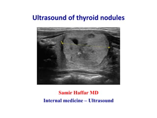
  • 1. Ultrasound of thyroid nodules Samir Haffar MD Internal medicine – Ultrasound
  • 2. Ultrasound of thyroid nodules ① Epidemiology & etiologies of thyroid nodules ② Ultrasound features of thyroid nodules ③ Ultrasound of cervical lymph nodes ④ Ultrasound systems for thyroid nodule risk stratification ⑤ Pseudo-nodules of thyroid ⑥ Fine needle aspiration of thyroid nodules
  • 3. ① Epidemiology & etiology of thyroid nodules
  • 4. Epidemiology of thyroid nodules • Very common: Palpation 4 – 8%, US 10 – 40%, autopsy 50% Increases with age • Malignancy risk: Patients younger than 20 or older than 60 years History of neck irradiation History of familial thyroid cancer • Cancer prevalence: 10-13% no matter how many nodules at US No decrease of cancer risk in multiple nodules Cancer found in dominant nodules in 2/3 of cases Frates MC et al. Ultrasound Quarterly 2006;22(4):231–238. Most thyroid nodules are benign hyperplastic nodules Thyroid cancer not common compared to very frequent benign nodules
  • 5. Indications of thyroid ultrasound • Patient with palpable thyroid nodule or with multinodular goiter • High-risk patient for thyroid malignancy: History of familial thyroid cancer Multiple endocrine neoplasia (MEN) type II Irradiated neck in childhood • Patient with palpable cervical adenopathy suspicious for malignancy • Follow-up and monitoring of thyroid nodules Haugen BR et al. Thyroid 2016;26:1–133. American thyroid association guidelines
  • 6. Etiologies of thyroid nodules Discrete lesion in thyroid distinct from surrounding thyroid parenchyma • Benign thyroid nodules: Cyst and pseudocyst Hyperplastic adenomatous nodule (adenoma) • Malignant thyroid nodules: Papillary carcinoma: 75 – 80% Follicular carcinoma: 10 – 20% Medullary carcinoma: 3 – 5% Anaplastic carcinoma: 1 – 2% Lymphomas: < 5% Metastasis: rare Dighe M et al. Med Ultrason 2017;19:195–210.
  • 7. ② Ultrasound features of thyroid nodules
  • 8. Different modes of medical ultrasonography Mode What is measured What is displayed B-mode Acoustic impedance Anatomy Doppler Motion Vascular flow Elastography Mechanical properties Tissue stiffness Elastography: A Practical Approach. Barr RG Edit, Thieme Medical Publishers, New York, 2017. The three modes are applied during ultrasound examination for a more accurate diagnosis
  • 9. Ultrasound features of thyroid nodules Nodules Ultrasound features Composition: Cystic: fluid-filled without solid component Predominantly cystic: ≥ 50% cystic Predominantly solid: ≥ 50% solid Solid: entirely solid or almost completely solid Echogenicity: Anechoic, hyperechoic, isoechoic, hypoechoic, very hypoechoic Shape: Wider-than-tall, taller-than-wide Margins: Regular, lobulated, irregular, extra-thyroidal extension Peripheral halo: No halo, thin regular vascular, thick irregular avascular Echogenic foci: Comet-tail artifact (colloid cyst) Peripheral rim calcification (eggshell) Macro-calcifications: > 1 mm Micro-calcifications: ≤ 1 mm without acoustic shadowing Vascular pattern: Peripheral, intranodular, peripheral & intranodular Tessler FN et al. J Am Coll Radiol 2017;14:587–595.
  • 10. Some ultrasound features are considered as benign Other ultrasound features are suspicious for malignancy
  • 11. Ultrasound features of thyroid nodules Category Benign features Suspicious features Composition Cystic predominantly cystic Spongiform Mixed cystic and solid Solid or almost completely solid Echogenicity Anechoic Hyperechoic Isoechoic Hypoechoic Very hypoechoic Shape Wider-than-tall Taller-than-wide Margins Smooth or regular Lobulated or irregular Extra-thyroidal extension Halo Thin hypoechoic vascular halo Lack of hypoechoic halo Thick irregular avascular halo Echogenic foci No calcifications Comet-tail artifacts (colloid cyst) Macro-calcifications Eggshell calcifications (rim) Micro-calcifications Vascularity Peripheral vascularity Intranodular vascularity Peripheral & intranodular Tessler FN et al. J Am Coll Radiol 2017;14:587–595.
  • 12. Suspicious US features don’t have the same risk of malignancy Some have lower risk of malignancy: hyperechoic, isoechoic, … Other have higher risk of malignancy: hypoechoic, irregular margin, .. Suspicious US features of thyroid nodules
  • 13. US report of thyroid nodules should include • Nodule location • Nodule size in three dimensions • Nodule sonographic features: composition, echogenicity, shape, margin, halo, calcifications, and vascularity Cosgrove D et al. Ultrasound in Med & Biol 2016;
  • 14. Drawing to locate and number thyroid nodules Russ G et al. Eur Thyroid J 2017;6:225–237. right lobe, longitudinal left lobe, longitudinal anterioranterior posterior posterior caudalcaudalcranial cranial
  • 15. Ultrasound features of thyroid nodules Composition, echogenicity, shape, margin, peripheral halo, calcifications and vascularity
  • 16. Ultrasound features of thyroid nodules Composition
  • 17. Large anechoic cyst (arrow) without calcifications or ring down artifact Most thyroid cysts are pseudocysts are hyperplastic nodules that have undergone extensive degeneration, necrosis and hemorrhage Dighe M et al. Med Ultrason 2017;19(2):195–210. 29 year old female with multiple known thyroid nodules Difficult swallows due to mass like feeling in neck Thyroid nodule Composition / Completely cystic
  • 18. Ahuja AT. Diagnostic Ultrasound: Head and Neck. 2104, Amirsys Publishing. Large cystic thyroid nodule with internal debris mobile on Doppler Posterior acoustic enhancement esophagus debris cyst Thyroid nodule Composition / Hemorrhagic thyroid cyst
  • 19. Ahuja AT. Diagnostic Ultrasound: Head and Neck. 2104, Amirsys Publishing. Large predominantly cystic nodule with internal debris and septa Perinodular vascularity with no vascularity within septa Power Doppler ultrasound of thyroid septa cyst perinodular vascularity Thyroid nodule Composition / Hemorrhagic thyroid cyst
  • 20. Ahuja AT. Diagnostic Ultrasound: Head and Neck. 2104, Amirsys Publishing. Hemorrhagic thyroid cyst Sharply defined fluid level: layering of debris in its dependent portion Marked posterior enhancement Absence of any intranodular vascularity Longitudinal power Doppler US of thyroid lobe posterior enhancement fluid level Thyroid nodule Composition / Hemorrhagic thyroid cyst
  • 21. Ahuja AT. Diagnostic Ultrasound: Head and Neck. 2104, Amirsys Publishing. Hemorrhagic thyroid nodule with multiple mural nodules within Mural nodules avascular on Doppler Representing intra-nodular blood clots Longitudinal ultrasound of thyroid lobe mural nodule Thyroid nodule Composition / Hemorrhagic thyroid cyst
  • 22. 53-year-old male with incidental mass in right lobe seen on CT Cystic mass with large ring down artifact (arrowheads) consistent with benign colloid cyst Transverse ultrasound of right lobe Dighe M et al. Med Ultrason 2017;19(2):195–210. Thyroid nodule Composition / Colloid cyst
  • 23. Multiple benign colloid cysts with ring-down artifact in several cysts Henrichsen TL et al. Radiol Clin N Am 2011;49:417–424. Thyroid nodule Composition /Colloid cyst
  • 24. Thyroid nodule Composition /Colloid cyst Comet tail artifact also referred to as a ring-down artifact, a stepladder artifact, or when a single comet tail is seen within a small colloid cyst, a cat’s eye In: Thyroid and parathyroid ultrasound and ultrasound-guided FNA. Duick DS Edit, 4th edition, 2018, Springer International Publishing, Switzerland. cat’s eye
  • 25. Thyroid nodule Composition / Spongiform nodule Spongiform benign nodule with internal microcystic appearance involving more than 50% of the lesion Classic spongiform or honeycomb pattern Head and neck ultrasonography: essential and extended applications. Second edition, 2017. Editor: LA Orloff – Plural Publishing Incorporation, San Diego, CA, USA.
  • 26. Thyroid nodule Composition / Predominantly cystic nodule Transverse US of left lobe Predominantly cystic nodule with small solid mural component Flow within mural component (arrowheads): tissue and not debris US-guided FNA directed into this area: benign lesion Corresponding color Doppler US Frates MC et al. Ultrasound Quarterly 2006;22(4):231–238.
  • 27. Thyroid nodule Composition / Predominantly solid nodule Iso- to hyperechoic thyroid nodule containing multiple cystic components Gaitini D et al. Thyroid Ultrasound. In: EFSUMB – European Course Book – Editor: Dietrich CF – 2011
  • 28. Thyroid nodule Composition / Completely solid nodule Transverse US of left lobe Completely solid thyroid nodule (calipers) Marked internal vascularity on color Doppler mode indicating increased likelihood that nodule is malignant This was a papillary thyroid carcinoma Corresponding color Doppler US Frates MC et al. Ultrasound Quarterly 2006;22(4):231–238.
  • 29. Ultrasound features of thyroid nodules Echogenicity
  • 30. Thyroid nodule Echogenicity / Hyperechoic nodule Russ G et al. Eur Thyroid J 2017;6:225–237. Hyperechoic homogenous thyroid nodule Oval shape and smooth margins – No any high-risk features Risk of malignancy decreases as the echogenicity increases Longitudinal ultrasound Transverse ultrasound
  • 31. Die C et al. Insights Imaging 2016;7:77–86. Markedly hyper-echogenic & heterogenous nodule This is considered a suspicious nodule Thyroid nodule Echogenicity / Hyperechoic nodule
  • 32. Thyroid nodule Echogenicity / Hyperechoic nodule Head and neck ultrasonography: essential and extended applications. Second edition, 2017. Editor: LA Orloff – Plural Publishing Incorporation, San Diego, CA, USA. Almost completely solid and hyperechoic nodule Some small cystic components Smooth margin – No calcifications
  • 33. Thyroid nodule Echogenicity / Isoechoic nodule Isoechoic and completely solid nodule Hypoechoic thin halo with peripheral vascularity on Doppler Benign nodule at fine needle aspiration Transverse US of right lobe Correspondent color Doppler Dighe M et al. Med Ultrason 2017;19(2):195–210.
  • 34. Thyroid nodule Echogenicity / Hypoechoic nodule Hypoechoeic nodule compared to thyroid gland (arrowheads) Asterisk: strap muscle Kwak JY et al. Radiology 2011;260(3):892–899. Transverse US of left thyroid lobe
  • 35. Suspicious hypoechoic nodule with signal lower than surrounding thyroid tissue but higher than strap muscle above Die C et al. Insights Imaging 2016;7:77–86. Thyroid nodule Echogenicity / Hypoechoic nodule nodule strap muscle normal thyroid
  • 36. Hypoechoic nodule with significant posterior acoustic enhancement Presence of intra-nodular vascularity identifies this as a solid nodule and not a cyst Thyroid nodule Echogenicity / Hypoechoic nodule or thyroid cyst Correspondent color DopplerTransverse US of right lobe In: Thyroid and parathyroid ultrasound and ultrasound-guided FNA. Duick DS Edit, 4th edition, 2018, Springer International Publishing, Switzerland.
  • 37. Thyroid nodule Echogenicity / Very hypoechoic nodule Echogenicity of nodule less than that of strap muscle Proved to be papillary thyroid carcinoma on histology In: Thyroid and parathyroid ultrasound and ultrasound-guided FNA. Duick DS Edit, 4th edition, 2018, Springer International Publishing, Switzerland.
  • 38. Thyroid nodule Echogenicity / Very hypoechoic nodule Grant EG et al. J Am Coll Radiol 2015;12:1272–1279. Very hypoechoic 10-mm nodule (N) with smooth margin Nodule less echogenic than adjacent strap muscles (S) and isoechoic to common carotid artery (C) Diagnosis: papillary thyroid carcinoma Transverse ultrasound of left thyroid lobe
  • 39. Ultrasound features of thyroid nodules Shape
  • 40. Thyroid nodule Shape / Taller-than-wide nodule Langer JE. Radiol Clin North Am. 2019;57(3):469–483. Hypoechoic solid nodule of 1.3 cm Larger in anteroposterior diameter than in width Highly predictive of malignancy & noted in smaller cancers < 1 cm Papillary thyroid carcinoma on surgery Transverse image of left lobe
  • 41. Ultrasound features of thyroid nodules Margins
  • 42. Thyroid nodule Margin / Lobular margin of nodule Die C et al. Insights Imaging 2016;7:77–86. Suspicious hypoechoic nodule with lobular margin
  • 43. Hypoechoic solid nodule with a lobulated margin (arrow) Papillary thyroid carcinoma on FNA Thyroid nodule Margin / Lobular margin of nodule In: Thyroid and parathyroid ultrasound and ultrasound-guided FNA. Duick DS Edit, 4th edition, 2018, Springer International Publishing, Switzerland.
  • 44. Grant EG et al. J Am Coll Radiol 2015;12:1272–1279. Thyroid nodule Margin / Irregular margin of nodule Heterogeneous 16-mm nodule with irregular margins Angulated borders anteriorly Diagnosis: papillary thyroid carcinoma Longitudinal image of thyroid lobe
  • 45. Markedly hypoechoic nodule which is taller than wide (arrow) Presence of speculated margin (arrowheads ) Presence of microcalcifications (open arrow ) It demonstrates most of US features associated with malignancy Thyroid nodule Margin / Irregular spiculated margin of nodule In: Ultrasound of the thyroid and parathyroid glands. RA Sofferman & AT Ahuja (Eds), Springer Science+Business Media, 2012, New York.
  • 46. Thyroid nodule Margin / Extra-thyroidal extension of nodule Longitudinal US of left thyroid lobe Hypoechoic nodule with extrathyroidal extension into strap muscle Loss of thin hyperechoic capsule (red arrow) Highly reliable sign of malignancy and unfavorable prognostic sign Papillary carcinoma on surgery Head and neck ultrasonography: essential and extended applications. Second edition, 2017. Editor: LA Orloff – Plural Publishing Incorporation, San Diego, CA, USA.
  • 47. Thyroid nodule Margin / Extra-thyroidal extension of nodule Transverse ultrasound of thyroid Extrathyroidal extension through anterior capsule (arrows) Lack of clear margin with adjacent trachea posteriorly (dashed arrow) Anaplastic carcinoma on surgery with tracheal invasion In: Thyroid and parathyroid ultrasound and ultrasound-guided FNA. Duick DS Edit, 4th edition, 2018, Springer International Publishing, Switzerland.
  • 48. Langer JE. Radiol Clin North Am 2019;57(3):469–483. Thyroid nodule Margin / Extra-thyroidal extension of nodule Sagittal ultrasound of left thyroid lobe 3.5 cm solid hypoechoic nodule with microcalcifications Along inferior aspect of lesion: normal thyroid capsule (dashed arrow) Along superior aspect lesion: extra-thyroidal extension (arrows)
  • 49. Ultrasound features of thyroid nodules Peripheral halo
  • 50. • Thin, regular and vascular halo Generally forms margin for iso- and hyperechoic nodules Found in about half of benign nodule • Thick, irregular and avascular halo Presence of fibrous capsule surrounding a neoplastic growth: Follicular carcinoma or Hürthle cell carcinoma Peripheral halo of thyroid nodules Sonolucent ring that surrounds a nodule In: Thyroid and parathyroid ultrasound and ultrasound-guided FNA. Duick DS Edit, 4th edition, 2018, Springer International Publishing, Switzerland.
  • 51. Longitudinal US of thyroid lobe Thyroid nodule Halo / Thin regular and vascular halo Halo corresponds with peripheral vascularity Coresponding color Doppler Isoechoic nodule with thin regular halo Cytology is benign In: Thyroid and parathyroid ultrasound and ultrasound-guided FNA. Duick DS Edit, 4th edition, 2018, Springer International Publishing, Switzerland.
  • 52. Thick, irregular, and incomplete halo Surrounding solid iso- to hyperechoic nodule Hürthle cell cancer on histology Thyroid nodule Halo / Thick irregular and incomplete halo Transverse ultrasound of right thyroid lobe In: Thyroid and parathyroid ultrasound and ultrasound-guided FNA. Duick DS Edit, 4th edition, 2018, Springer International Publishing, Switzerland.
  • 53. Thyroid nodule Halo / Thick avascular halo In: Ultrasound of the thyroid and parathyroid glands. RA Sofferman & AT Ahuja (Eds), Springer Science+Business Media, 2012, New York. Heterogeneous nodule with thick halo which without vascularity Avascular halo is suspicious for a malignant nodule and represents true capsule of the lesion Longitudinal power Doppler ultrasound of thyroid lobe
  • 54. Ultrasound features of thyroid nodules Echogenic foci (calcifications)
  • 55. Thyroid nodule Echogenic foci / Ring down artifact Dighe M et al. Med Ultrason 2017;19(2):195–210. Transverse ultrasound of right lobe Cystic mass with large ring down artifact (arrowheads) consistent with benign colloid cyst
  • 56. Thyroid nodule Echogenic foci / Macro-calcifications Langer JE. Radiol Clin North Am. 2019;57(3):469–483. Mixed solid & cystic nodule (calipers) Central macro-calcification (arrow) producing acoustic shadowing Benign hyperplastic nodule Sagittal US of thyroid lobe Transverse US of thyroid lobe Solid hypoechoic nodule Central dystrophic calcification outlined by calipers Medullary thyroid carcinoma
  • 57. 69-year-old woman with medullary thyroid carcinoma Hypoechoic nodule with coarse central calcification This is suggestive of medullary thyroid carcinoma Henrichsen TL et al. Radiol Clin N Am 2011;49:417–424. Thyroid nodule Echogenic foci / Macro-calcifications
  • 58. Thyroid nodule Echogenic foci / Peripheral rim calcifications (eggshell) Complete eggshell calcification Acoustic shadowing produced by calcific ring Twofold increase of cancer likelihood in case of eggshell calcification In: Ultrasound of the thyroid and parathyroid glands. RA Sofferman & AT Ahuja (Eds), Springer Science+Business Media, 2012, New York. CCA trachea eggshell Transverse ultrasound of left thyroid lobe
  • 59. Thyroid nodule Echogenic foci / Disrupted peripheral rim calcifications Die C et al. Insights Imaging 2016;7:77–86. Interrupted eggshell calcification around edges Corresponds with localized invasion into surrounding thyroid
  • 60. Coarse and disrupted calcification Posterior acoustic shadowing partially obscures the nodule Highly suspicious for malignancy – Papillary thyroid carcinoma on FNA Transverse ultrasound of right lobe Head and neck ultrasonography: essential and extended applications. Second edition, 2017. Editor: Orloff LA – Plural Publishing Incorporation, San Diego, CA, USA. Thyroid nodule Echogenic foci / Disrupted peripheral rim calcifications
  • 61. Sagittal US image of thyroid nodule Thyroid nodule (arrowheads) with micro-calcifications (arrow) Papillary thyroid carcinoma on FNA and surgery Threefold increase of cancer likelihood in case of microcalcifications Microcalcifications almost exclusively appear in hypoechoic nodules Frates MC et al. Ultrasound Quarterly 2006;22(4):231–238. Thyroid nodule Echogenic foci / Micro-calcifications (punctate echogenic foci)
  • 62. Ultrasound features of thyroid nodules Vascularity
  • 63. Vascularity pattern of thyroid nodules ACR TIRADS: American college of radiology – Thyroid imaging reporting and data system ATA: American thyroid association – BTA: British thyroid association FNA: fine needle aspiration Die C et al. Insights Imaging 2016;7:77–86. Nodular vascularity U classification of BTA FNA Peripheral vascularity Benign nodule not required Peripheral & intra-nodular vascularity Equivocal nodule required Intra-nodular vascularity Malignant nodule required Nodular vascularity included in U classification of BTA and not included in K-TI-RADS, ATA & ACR TI-RADS
  • 64. Benign nodule with peripheral vascularity on Doppler Vascular haloes in benign nodules represent compressed vessels/ thyroid tissue & such nodules do not have true capsule around them Die C et al. Insights Imaging 2016;7:77–86. Thyroid nodule Vascularity /Peripheral vascularity of nodule
  • 65. Transverse US of thyroid nodule Solid thyroid nodule with peripheral and internal vascularity This was a papillary thyroid carcinoma Increased vascularity in thyroid nodule is suggestive of malignancy but should not be considered a pathognomonic feature Corresponding color Doppler US Frates MC et al. Ultrasound Quarterly 2006;22(4):231–238. Thyroid nodule Vascularity / Peripheral and intra-nodular vascularity
  • 66. Thyroid nodule Vascularity / Intranodular vascularity Die C et al. Insights Imaging 2016;7:77–86. Thyroid nodule with intra-nodular vascularity Later confirmed to be papillary thyroid cancer
  • 67. Nodules in Hashimoto thyroiditis • Pseudo- nodules • White-knight nodule • Giraffe hide • Papillary thyroid carcinoma: increased risk in Hashimoto thyroiditis • Thyroid lymphoma: increased risk in Hashimoto thyroiditis Henrichsen TL et al. Radiol Clin N Am 2011;49:417–424.
  • 68. Hashimoto’s thyroiditis Pseudonodules Thin & thicker fibrous septa in hypoechoic thyroid Creating pseudonodules and parenchymal coarsening When it is unclear if a nodule is present, FNAC may be needed FNAC: fine needle aspiration cytology Sholosh B et al. Radiol Clin N Am 2011;49:391–416. Sagittal ultrasound of thyroid lobe
  • 69. Hashimoto’s thyroiditis white-knight nodule Background thyroid: hypoechoic with micro-nodularity features typical of diffuse Hashimoto thyroiditis Well-defined homogenous hyperechoic nodule known as white-knight Surgical removal demonstrated nodular Hashimoto thyroiditis Henrichsen TL et al. Radiol Clin N Am 2011;49:417–424. 16-year-old boy with known Hashimoto thyroiditis
  • 70. Hashimoto’s thyroiditis Giraffe hide Sholosh B et al. Radiol Clin N Am 2011;49:391–416. Background thyroid: hypoechoic with micro-nodularity features typical of diffuse Hashimoto thyroiditis Bright blocks separated by dark bands looking like a giraffe hide This nodule is variation of echogenic nodule known as “white- knight”
  • 71. Presence of multiple nodules is the norm on ultrasound examination With high-resolution ultrasound, it is quite unusual to see a truly solitary nodule
  • 72. Multiple thyroid nodules Large anechoic cyst (arrow) without calcifications or ring down artifact Two small hypoechoic nodules with multiple cystic components Dighe M et al. Med Ultrason 2017;19(2):195–210. Longitudinal ultrasound of left thyroid lobe
  • 73. Multiple thyroid nodules In: Ultrasound of the thyroid and parathyroid glands. RA Sofferman & AT Ahuja (Eds), Springer Science+Business Media, 2012, New York. Sagittal ultrasound of right thyroid lobe Multiple heterogeneous nodules in right lobe (arrows) Some nodules show cystic component with comet-tail artifacts Others are isoechoic with cystic elements None of nodules show obvious features suspicious for malignancy
  • 74. In: Ultrasound of the thyroid and parathyroid glands. RA Sofferman & AT Ahuja (Eds), Springer Science+Business Media, 2012, New York. Multiple solid isoechoic non-calcified nodules Multiple thyroid nodules Sagittal ultrasound of thyroid lobe
  • 75. Malignancy in solitary & multiple thyroid nodules 1 Cochand-Priollet B et al. Am J Med 1994; 97(2):152–157. 2 Marqusee E et al. Ann Intern Med 2000; 133(9):696–700. 3 Papini E et al. J Clin Endocrinol Metab 2002; 87(5):1941–1946. Malignancy Solitary nodules Multiple nodules Cochand-Priollet 1 13% 14% Marqusee E 2 7% 9% Papini 3 9% 6% Malignancy is similar in solitary & multiple thyroid nodules
  • 77. Thyroid cancers 1% of all malignancies Average annual incidence 5/100.000 inhabitants • Papillary thyroid carcinoma: 75 – 80% • Follicular thyroid carcinoma: 5 – 10% • Medullary thyroid carcinoma: 3 – 5% • Anaplastic thyroid carcinoma: 1 – 2% • Primary thyroid lymphoma: < 5% • Thyroid metastasis Die C et al. Insights Imaging 2016;7:77–86.
  • 78. • Ultrasound: Heterogeneous hypoechoic nodule Irregular shape Characteristic micro-calcifications • Color Doppler: Not useful to diagnose papillary cancer (useful in follicular cancer) 20% of malignant nodules have peripheral vascularity Papillary thyroid carcinoma most common thyroid cancer (75–80%) Dighe M et al. Med Ultrason 2017;19(2):195–210.
  • 79. Classic pattern of hypoechoic nodule with micro-calcifications Mild internal vascularity on color Doppler Papillary thyroid carcinoma Transverse US and color Doppler of right lobe Dighe M et al. Med Ultrason 2017;19(2):195–210.
  • 80. Papillary thyroid carcinoma Sholosh B et al. Radiol Clin N Am 2011;49:391–416. Subtle hypoechoic nodule proved to be papillary thyroid carcinoma Nodule easily blends in to adjacent hypoechoic thyroid on sagittal view Multiple tiny hypoechoic nodules consistent with Hashimoto thyroiditis Patient with long-standing Haschimoto’s thyroiditis Transverse and sagittal US of right thyroid lobe
  • 81. Langer JE. Radiol Clin North Am. 2019;57(3):469–483. Papillary thyroid carcinoma Patient with long-standing Haschimoto’s thyroiditis Sagittal ultrasound of thyroid lobe Background thyroid: micronodules of Hashimoto’s thyroiditis Papillary thyroid cancer (PTC) occurring in Haschimoto thyroiditis Echogenicity of nodule similar to background thyroid but microcalcifications within nodule facilitate its detection
  • 82. Thrombosis of internal jugular vein Patient with known papillary thyroid carcinoma Thrombosis of left internal jugular vein (IJV) Head and neck ultrasonography: essential and extended applications. Second edition, 2017. Editor: Orloff LA – Plural Publishing Incorporation, San Diego, CA, USA.
  • 83. Follicular thyroid carcinoma 5–10% of thyroid cancer • More common in females than males • Higher risk of distant metastases & mortality compared with PTC • Ultrasound: follicular carcinoma similar to follicular adenoma Adenoma: oval to round isoechoic or hypoechoic nodule Carcinoma: irregular margins, thick halo & caotic vessels • Doppler: characteristic high velocity pulsatile flow penetrating tumor • FNA insufficient to distinguish benign & malignant follicular lesions Follicular carcinoma dg by capsular or vascular invasion on histology Follicular adenoma must be excised for definitive diagnosis FNA: fine needle aspiration Dighe M et al. Med Ultrason 2017;19(2):195–210.
  • 84. Benign follicular adenoma Classic pattern of follicular benign adenoma Homogeneous hypoechoic oval nodule with thin capsule Sholosh B et al. Radiol Clin N Am 2011;49:391–416. 56-year-old woman with benign follicular adenoma
  • 85. Ovoid solid predominantly hypoechoic thyroid nodule Hypoechoic areas (arrowhead) within nodule are suspicious features Marked intranodular vascular flow on power Doppler Follicular carcinoma on histology Follicular thyroid carcinoma Longitudinal US of thyroid nodule In: Ultrasound of the thyroid and parathyroid glands. RA Sofferman & AT Ahuja (Eds), Springer Science+Business Media, 2012, New York. Correspondent color Doppler US
  • 86. • Arise from parafollicular C-cells that secrete thyrocalcitonin 10-20% of patients have familial history of pheochromocytomas Medullary carcinoma may be associated with MEN II-syndrome • Ultrasound: Solid hypoechoic nodule with central coarse calcification in 80-90% • Doppler: Chaotic intranodular vessels seen in tumor on color Doppler Medullary thyroid carcinoma 3–5 % of thyroid cancer Dighe M et al. Med Ultrason 2017;19(2):195–210.
  • 87. Medullary thyroid carcinoma Ahuja AT. Diagnostic Ultrasound: Head and Neck. 2104, Amirsys Publishing. Transverse ultrasound of left lobe Well-defined solid hypoechoic nodules in left lobe Central dense shadowing calcification in larger nodule These were confirmed to be medullary thyroid carcinoma
  • 88. Medullary thyroid carcinoma Dighe M et al. Med Ultrason 2017;19(2):195–210. 37 year old male with incidentally detected nodule on CT scan Transverse US & Doppler US of right lobe Large hypoechoic predominantly solid nodule Mild internal vascularity on color Doppler FNA suspicious for medullary carcinoma confirmed on histopathology
  • 89. • Highly aggressive form of thyroid cancer Typically in elderly women in 6th to 7th decades of life Large number of patients have history of multinodular goiter • Ultrasound: Hypoechoic nodule involving entire lobe, ill-defined margins, areas of necrosis, microcalcifications, extracapsular spread, vascular invasion, nodal or distant metastases • Doppler: multiple small intra-nodular vessels • Core biopsy may be required for diagnosis due to its fibrotic nature Anaplastic thyroid carcinoma 1–2% of thyroid cancer Dighe M et al. Med Ultrason 2017;19(2):195–210.
  • 90. Transvers ultrasound of thyroid bed ill-defined hypoechoic nodule in thyroid isthmus Tissue plane between tumor and trachea is lost (tracheal invasion) Anaplastic carcinoma revealed by core needle biopsy Anaplastic thyroid carcinoma 1–2% of thyroid cancer Ahuja AT. Diagnostic Ultrasound: Head and Neck. 2104, Amirsys Publishing.
  • 91. ill-defined heterogeneous hypoechoic nodule (arrows) encasing left CCA (open arrow) Anaplastic carcinoma on histology Extrathyroid extension is better evaluated with CT or MR Transverse ultrasound of left thyroid lobe Anaplastic thyroid carcinoma 1–2% of thyroid cancer Ahuja AT. Diagnostic Ultrasound: Head and Neck. 2104, Amirsys Publishing.
  • 92. Thyroid lymphoma • Uncommon: < 5% of all thyroid malignancies • Almost always in patients with underlying Hashimoto’s thyroiditis • Classically presents with rapidly enlarging thyroid gland • Ultrasound features: Markedly hypoechoic nodule in background of chronic thyroiditis Enhanced through transmission posterior to the lesion • Treatment: Chemotherapy and external beam radiation Surgery only if trachea markedly compressed by tumor • Good prognosis when disease confined to thyroid gland Sholosh B et al. Radiol Clin N Am 2011;49:391–416.
  • 93. CECT: contrast enhanced computed tomography – PET: positron emission tomography Sholosh B et al. Radiol Clin N Am 2011;49:391–416. Thyroid lymphoma Markedly hypoechoic large expansile masses in both lobes on US Low density masses on CT scan Intensely hypermetabolic masses on PET scan Patient with Hashimoto thyroiditis & rapidly enlarging thyroid mass Transverse US of thyroid CECT PET scan
  • 94. Thyroid lymphoma almost always in patients with underlying Hashimoto’s thyroiditis Sholosh B et al. Radiol Clin N Am 2011;49:391–416. Markedly hypoechoic nodule in isthmus Increased through-transmission: finding suggesting lymphoma Relative hypovascularity on color Doppler Background thyroid: micronodules of Hashimoto’s thyroiditis
  • 95. Asymmetrically enlargement of left lobe with preserved contour Homogeneous & hypoechoic parenchyma of both lobes with increased through-transmission (features suggestive of lymphoma) Haschimoto thyroiditis: heterogeneous hypoechoic regions that attenuate sound Thyroid lymphoma Patient with long-standing Haschimoto thyroiditis & new neck swelling Transverse ultrasound of thyroid Langer JE. Radiol Clin North Am. 2019;57(3):469–483.
  • 96. Thyroid metastasis rare in daily clinical practice • Generally associated with advanced stage of malignancy • Main primary tumors spreading to thyroid gland: malignant melanoma, breast carcinoma, renal cell carcinoma • Difficult to distinguish from primary thyroid lesion • No specific features on US: Solitary/multiple hypoechoic nodules without calcifications No specific information about color Doppler of metastases Dighe M et al. Med Ultrason 2017;19(2):195–210.
  • 97. Transverse US of right and left lobe Several small hypoechoic nodules in right lobe Large hypoechoic nodule in left lobe Adenocarcinoma most probably from gastrointestinal origin on FNA Thyroid metastasis Gaitini D et al. Thyroid Ultrasound. In: EFSUMB – European Course Book – Editor: Dietrich CF – 2011
  • 98. In: Ultrasound of the thyroid and parathyroid glands. RA Sofferman & AT Ahuja (Eds), Springer Science+Business Media, 2012, New York. Thyroid metastasis Longitudinal in a patient with a known lung cancer Solid hypoechoic noncalcified nodule (arrow) in lower thyroid pole Lower pole is common location for large thyroid metastases Presence of such a nodule in a patient with known cancer suggests thyroid metastases unless proven otherwise
  • 99. ③ Ultrasound of cervical lymph nodes
  • 100. Six zones of cervical lymph nodes Zone I Submental triangle Zone II Upper internal jugular chain nodes Zone III Middle internal jugular chain nodes between hyoid bone & cricoid cartilage Zone IV Lower internal jugular chain nodes from zone II to clavicle Zone V Entire posterior triangle between SCM, trapezius and clavicle Zone VI hyoid bone superiorly sternal manubrium inferiorly carotid arteries laterally Head and neck ultrasonography: essential and extended applications. Second edition, 2017. Editor: Orloff LA – Plural Publishing Incorporation, San Diego, CA, USA.
  • 101. Zones in dark blue are most common sites of metastatic papillary carcinoma Zones in lighter blue are less frequent areas of metastatic papillary carcinoma Six zones of cervical lymph nodes In: Ultrasound of the thyroid and parathyroid glands. RA Sofferman & AT Ahuja (Eds), 2012, Springer Science+Business Media, New York.
  • 102. Pretracheal and paratracheal lymph nodes Schematic transverse section at level of thyroid gland In: Ultrasound of the thyroid and parathyroid glands. RA Sofferman & AT Ahuja (Eds), 2012, Springer Science+Business Media, New York. Relationship of pretracheal and paratracheal lymph nodes to adjacent structures
  • 103. • Small: short axis < 5 – 8 mm • Oval: short axis/long axis < 0.5 • Echogenic hilum • Hilar vascularity or avascular hilum on Doppler Ultrasound features of benign lymph nodes At least 5–6 normal cervical nodes identified routinely by US Ting M et al. Clinical Radiology 2003;58:351–358.
  • 104. Normal cervical lymph node Oval shape and echogenic hilum Ahuja AT et al. AJR 2005;184:1691–1699. Hypoechoic, elliptic, and elongated lymph node (arrows) Arrowheads indicate echogenic hilus continuous with adjacent soft tissue
  • 105. Richards PS et al. Cancer Imaging 2007;7:167–178. Normal cervical lymph node oval shape & echogenic hilum Normal elliptical node with echogenic hilum
  • 106. Normal cervical lymph node Hilar vascularity Vessels radiate out from the hilum Richards PS et al. Cancer Imaging 2007;7:167–178.
  • 107. Ultrasound features of malignant lymph nodes • Round shape • Loss of echogenic hilum • Cystic change in lymph node • Microcalcifications • Non-hilar vascularity • Extracapsular spread Sholosh B et al. Radiol Clin N Am 2011;49:391–416.
  • 108. Ahuja AT et al. Cancer Imaging 2008;8:48–56. Malignant lymph node Round shape Metastatic lymph node which is enlarged, hypoechoic, well-defined and without echogenic hilum (arrows)
  • 109. Enlarged node (calipers) with cystic component Node proved to be metastatic papillary carcinoma Frates MC et al. Ultrasound Quarterly 2006;22(4):231–238. Malignant lymph node Cystic change
  • 110. Metastatic cervical node (arrows) with intra-nodal cystic necrosis which appears ill-defined and echolucent (arrowheads) Ahuja AT et al. Cancer Imaging 2008;8:48–56. Malignant lymph node Cystic change
  • 111. Malignant lymph node Cystic change In: Ultrasound of the thyroid and parathyroid glands. RA Sofferman & AT Ahuja (Eds), Springer Science+Business Media, 2012, New York. Cystic metastatic lymph node (arrow) from papillary carcinoma Microcalcifications (arrowhead) within solid component (open arrow) of metastatic node Transverse ultrasound of left neck CCA
  • 112. Ahuja AT et al. Cancer Imaging 2008;8:48–56. Metastatic lymph node from papillary thyroid carcinoma (arrows) Hyperechoic component within node which may be related to intra-nodal deposition of thyroglobulin (arrowheads) Malignant lymph node Hyperechoic component
  • 113. Malignant lymph node Micro-calcifications Ahuja AT. Diagnostic Ultrasound: Head and Neck. 2104, Amirsys Publishing. Round heterogeneous hyperechoic node – Loss of echogenic hilum Characteristic punctate calcification with fine acoustic shadowing Compressed internal jugular vein (IJV) Patient with known papillary thyroid carcinoma CCA collapsed IJV lymph node punctate calcifications
  • 114. Ahuja AT et al. Cancer Imaging 2008;8:48–56. Metastatic lymph node with peripheral vascularity (arrowheads) Power Doppler ultrasound Malignant lymph node Peripheral vascularity
  • 115. Malignant lymph node with both peripheral (arrowheads) and hilar vascularity (arrows) Power Doppler ultrasound Ahuja AT et al. Clinical Radiology 2003:58:359–366. Malignant lymph node Peripheral & hilar vascularity
  • 116. Head and neck ultrasonography: essential and extended applications. Second edition, 2017. Editor: Orloff LA – Plural Publishing Incorporation, San Diego, CA, USA. Metastatic lymph node Mixed (peripheral and hilar) vascularity on power Doppler Patient with known papillary thyroid carcinoma Sagittal gray scale & power Doppler ultrasound Malignant lymph node Peripheral & hilar vascularity
  • 117. Richards PS et al. Cancer Imaging 2007;7:167–178. Malignant lymph node Extracapsular spread
  • 118. ④ Ultrasound systems for thyroid nodule risk stratification
  • 119. Reliable US systems are necessary to specifically target nodules that require biopsy Several published US systems for risk stratification
  • 120. Some US systems for thyroid nodule risk stratification 2009 First TI-RADS Horvath E et al. Clin Endocrinol Metab 2009;94(5):1748–51. 2011 Korean TI-RADS Kwak JY et al. Radiology 2011;260(3):892–899. 2016 U classification of British Thyroid Association (BTA) Die C et al. Insights Imaging 2016;7:77–86. 2016 American Thyroid Association (ATA) Haugen BR et al. Thyroid 2016;26(1):1–133. 2017 ACR TI-RADS Tessler FN et al. J Am Coll Radiol 2017;14:587–595. ACR: American college of radiology TI-RADS: Thyroid imaging reporting and data system No one system has achieved universal acceptance This leads to some confusion for practitioners of sonography
  • 121. Korean TIRADS Analysis of 1658 nodules > 1 cm that underwent FNA Category Definition Ultrasound features FNA 1 Normal thyroid gland follow-up 2 Benign: 0% Cyst & colloid cysts Spongiform grid nodule follow-up follow-up 3 Probably benign: < 5% No suspicious feature follow-up 4 4 a 4 b 4 c Suspicious nodules: 5-80% Low suspicion: Intermediate suspicion Moderate suspicion 1 suspicious feature 2 suspicious features 3-4 suspicious features ≥ 1.0 cm ≥ 1.0 cm ≥ 1.0 cm 5 Probably malignant: >80% 5 suspicious features ≥ 1.0 cm 6 Biopsy proven malignancy Less complex TI-RADS stratifying malignancy risk Kwak JY et al. Radiology 2011;260(3):892–899.
  • 122. Suspicious ultrasound features in Korean TI-RADS • Solid component • Hypoechogenicity • Marked hypoechogenicity • Microlobulated or irregular margins • Microcalcifications • Taller-than-wide shape Kwak JY et al. Radiology 2011;260(3):892–899. Six ultrasound features
  • 123. U classification of British Thyroid Association Classification US features of thyroid nodule FNA U1: benign thyroid not required U2: indeterminate nodule Iso- or slightly hyperechoic nodule Hypoechoic halo around nodule Anechoic with ring-down sign (colloid) Spongiform/honeycomb nodule Complete eggshell calcification Peripheral vascularity of nodule not required U3: equivocal nodule Markedly hyperechoic nodule Nodule with cystic change Peripheral & intra-nodular vascularity required U4: suspicious nodule Hypoechoic nodule Disrupted eggshell calcification Nodule with lobular margin required U5: malignant nodule Taller than wide nodule Micro-calcifications Intra-nodular vascularity required Die C et al. Insights Imaging 2016;7:77–86.
  • 124. American Thyroid Association (ATA) guidelines for adult patients with thyroid nodules FNAC: fine needle aspiration cytology – ETE: extra-thyroidal extension – F/U: follow-up Haugen BR et al. Thyroid 2016;26(1):1–133. Category Risk of malignancy US features FNAC High suspicion > 70 – 90% Microcalcifications, hypoechoic, irregular Hypoechoic, irregular margin Hypoechoic, taller-than-wide Hypoechoic, irregular margin, ETE Hypoechoic, interrupted rim calcifications Irregular, suspicious lateral lymph node ≥ 1 cm Intermediate suspicion 10 – 20% hypoechoic solid, regular margin ≥ 1 cm Low suspicion 5 – 10% Hyperechoic solid, regular margin Isoechoic solid, regular margin Partially cystic, eccentric solid area ≥ 1.5 cm Very low suspicion < 3% Spongiform Partially cystic, no suspicious features ≥ 2 cm or F/U Benign < 1% Cyst no FNA
  • 125. ACR TI-RADS Tessler FN et al. J Am Coll Radiol 2017;14:587–595.
  • 126. ACR TI-RADS • Less concerning features are awarded less or no points • More suspicious features are awarded higher points • Add points of all categories to determine ACR TI-RADS level ACR TI-RADS: American college of radiology – Thyroid imaging reporting and data system Langer JE. Radiol Clin North Am. 2019;57(3):469–483.
  • 127. Categories of nodule features in ACR TI-RADS Category Ultrasound features Points Composition: choose only one Cystic or almost completely cystic Spongiform: > 50% of small cystic spaces Mixed cystic and solid Solid or almost completely solid 0 point 0 point 1 point 2 points Echogenicity: choose only one Anechoic Hyperechoic or isoechoic Hypoechoic Very hypoechoic 0 point 1 point 2 points 3 points Shape: choose only one Wider-than-tall Taller-than-wide 0 point 3 points Margins: choose only one Smooth or ill-defined Lobulated or irregular Extra-thyroidal extension 0 point 2 points 3 points Echogenic foci: all that apply No echogenic foci or large comet-tail artifacts Macrocalcifications Peripheral (rim) calcifications Punctate echogenic foci (microcalcifications) 0 point 1 point 2 points 3 points Tessler FN et al. J Am Coll Radiol 2017;14:587–595.
  • 128. ACR TI-RADS ACR: American college of radiology TIRADS: Thyroid imaging reporting and data system FNA: fine needle aspiration Tessler FN et al. J Am Coll Radiol 2017;14:587–595. TI-RADS Description Points FNA TR1 benign 0 points no FNA TR2 not suspicious 2 points no FNA TR3 mildly suspicious 3 points FNA if ≥ 2.5 cm TR4 moderately suspicious 4 – 6 points FNA if ≥ 1.5 cm TR5 highly suspicious ≥ 7 points FNA if ≥ 1 cm
  • 129. Advantages and disadvantages of ARC TI-RADS • Advantages: Standard terms (lexicon) for ultrasound reporting Able to classify almost all thyroid nodules Evidence based to the greatest extent possible • Disadvantages: High size threshold for FNA in mild & moderate suspicious lesions Doesn't take into consideration thyroid nodule vascularity Doesn't take into consideration elastography ACR TI-RADS: American college of radiology – Thyroid imaging reporting and data system FNA: fine needle aspiration
  • 130. Head and neck ultrasonography: essential and extended applications. Second edition, 2017. Editor: LA Orloff – Plural Publishing Incorporation, San Diego, CA, USA. Almost completely solid (2 points) – Hyperechoic nodule (1 point) Smooth margin (0 point) – No calcifications (0 point) Total: 3 points – ACR TR3: mildly suspicious – FNA if ≥ 2.5 cm Classification of thyroid nodule by ACR TI-RADS
  • 131. Completely solid (2 points) – hyperechoic (1 point) Smooth margin (0 point) – No echogenic foci (0 point) Total: 3 points – ACR TR 3: mildly suspicious – FNA if ≥ 2.5 cm Classification of thyroid nodule by ACR TI-RADS In: Thyroid and parathyroid ultrasound and ultrasound-guided FNA. Duick DS Edit, 4th edition, 2018, Springer International Publishing, Switzerland.
  • 132. Longitudinal ultrasound of thyroid Completely solid nodule (2 points) – Hypoechoic (2 points) Lobulated margin (2 points) – Microcalcifications (arrows, 3 points) Total: 9 points – ACR TR 5: highly suspicious – FNA if ≥ 1 cm Papillary carcinoma on FNA ACR: American college of radiology – FNA: fine needle aspiration – TR: TIRADS In: Ultrasound of the thyroid and parathyroid glands. RA Sofferman & AT Ahuja (Eds), Springer Science+Business Media, 2012, New York. Classification of thyroid nodule by ACR TI-RADS
  • 134. Pseudo-nodules of thyroid • Pyramidal lobe • Zuckerkandl tubercle • Pseudo-nodules in Hashimoto thyroiditis • Parathyroid adenoma • Osteophytes Germano A et al. Clinical imaging 2019;58:114–128.
  • 135. • Frequency: 10–30% of individuals • Remnant of thyroglossal tract Arises from isthmus & ascends towards hyoid bone Anterior to thyroid cartilage on midline or slightly to left • Rarely visualized on ultrasound: small anteroposterior diameter Can be seen when hypertrophied such as in case of Graves disease • More commonly detected in young: progressive atrophy in adulthood In: Ultrasound of the thyroid and parathyroid glands. RA Sofferman & AT Ahuja (Eds), 2012, Springer Science+Business Media, New York. Pyramidal lobe (Lalouette lobe)
  • 136. Pseudo-nodules of thyroid pyramidal lobe Germano A et al. Clinical imaging 2019;58:114–128. Transverse ultrasound: pyramidal lobe simulates a nodule Longitudinal ultrasound: pyramidal shape can be assessed Transverse US of thyroid Sagital US of thyroid
  • 137. • Lateral ingress of embryologic thyroid tissue • Located along posterior midportion of thyroid lobe • Ranges from small round fragment to 2–3 cm oval structure • Becomes prominent in the setting of diffuse thyroid disease • It may be misinterpreted as a nodule & unnecessarily biopsied • Surgically, the tubercule can be a marker of nearby trajectory of recurrent laryngeal nerve Tubercle of Zuckerkandl Embryologic remnant in posterior aspect of thyroid lobe In: Thyroid and parathyroid ultrasound and ultrasound-guided FNA. Duick DS Edit, 4th edition, 2018, Springer International Publishing, Switzerland.
  • 138. Hyperechoic thyroid capsule separates body of thyroid from tubercle of Zuckerkandl Misinterpreted as thyroid nodule which does not have capsule Transverse US of thyroid In: Atlas of Thyroid Ultrasonography. Halenka M & Fryšák Z (Edits), Springer International Publishing AG, Switzerland, 2017. Sagittal US of left lobe Pseudo-nodules of thyroid Zuckerkandl tubercle
  • 139. Pseudo-nodules of thyroid Zuckerkandl tubercle Germano A et al. Clinical imaging 2019;58:114–128. Zuckerkandl tubercle (arrows) Longitudinal ultrasound of right lobe
  • 140. Thin & thicker fibrous septa in hypoechoic thyroid Creating pseudonodules and parenchymal coarsening When it is unclear if a nodule is present, FNAC may be needed FNAC: fine needle aspiration cytology Sholosh B et al. Radiol Clin N Am 2011;49:391–416. Sagittal ultrasound of thyroid lobe Pseudo-nodules of thyroid Hashimoto thyroiditis
  • 141. Germano A et al. Clinical imaging 2019;58:114–128. Pseudo-nodules of thyroid parathyroid adenoma Histology-proven parathyroid adenoma (arrow) located posteriorly to thyroid, compressing thyroid parenchyma and mimicking a thyroid nodule Transverse ultrasound of right lobe
  • 142. Pseudo-nodules of thyroid osteophytes Osteophytes posterior to lateral lobes of thyroid gland Mimicking calcified thyroid nodules (arrows) Can be easily ruled-out with neck X-ray Longitudinal ultrasound of thyroid Germano A et al. Clinical imaging 2019;58:114–128.
  • 143. ⑥ Fine needle aspiration of thyroid nodules
  • 144. Indications for FNA according to US findings FNA: fine needle aspiration – US: ultrasound Gharib H et al. Endocrine Practice 2016;22 (Suppl 1):1–60. FNA Ultrasound findings
  • 145. Indications of FNA in ACR TI-RADS • TR3 (mildly suspicious nodules): if they are ≥ 2.5 cm • TR4 (moderately suspicious nodules): if they are ≥ 1.5 cm • TR5 (highly suspicious nodules): if they are ≥ 1 cm • No FNA of nodules < 1 cm even if they are highly suspicious This is in concordance with other guidelines FNA of 5-9 mm nodules may be done in certain conditions: Shared decision making between referring physician and patient FNA: fine needle aspiration ACR TI-RADS: American college of radiology – Thyroid imaging reporting and data system Tessler FN et al. J Am Coll Radiol 2017;14:587–595.
  • 146. Fine needle aspiration of thyroid nodules Advantages Inexpensive, widely available, easy to perform, accurate (>90%) and cost-effective Disadvantages Depends on skill of operator & cytopathologist Complications Rare – no reported case of cutaneous implantation of malignancy following FNA False negative rate 0.5 – 11.8% (pooled rate 2.4%) False positive rate 0 – 7.1% (pooled rate of 1.2%) Non-diagnostic rate Vary among different centers 5% is the maximum acceptable limit FNA: fine needle aspiration In: Ultrasound of the thyroid and parathyroid glands. RA Sofferman & AT Ahuja (Eds), Springer Science+Business Media, 2012, New York.
  • 147. Bethesda system for reporting thyroid cytopathology BSRTC Category Management I Nondiagnostic or unsatisfactory Repeat FNAC in 3 m II Benign Ultrasound follow-up III Atypia of undetermined significance or Follicular lesion of undetermined significance Repeat FNAC in 3 m or surgery w/o repeat FNAC IV Follicular neoplasm or Suspicious for follicular neoplasm Lobectomy or Thyroidectomy if proven malig. V Suspicious for malignancy Lobectomy or Thyroidectomy VI Malignant Thyroidectomy FNAC: fine needle aspiration cytology Haugen BR et al. Thyroid 2016;26(1):1–133.
  • 148. Complication of thyroid fine needle aspiration 1–12% of patients In: Thyroid ultrasound – From simple to complex. Editor: Sencha AN, Springer AG, 2019. Hematoma Sub-capsular, interfacial, inter-muscular, subcutaneous Pain in puncture region Usually disappears within 2 – 5 days Injury of CCA Hematoma in CCA wall with stenosis Injury of IJV Partial or complete thrombosis of IJV Nerve puncture Cervical plexus: neck, shoulder & upper extremity pain Superior laryngeal nerve: choking specially with liquids Inferior laryngeal nerve: hoarse voice Trachea puncture Instant dry hoarse cough for 1 – 5 min Insignificant subcutaneous emphysema possible Esophageal puncture Oval lesion in posterior left lobe on transverse scan Can be misinterpreted as nodule
  • 149. In: Thyroid ultrasound – From simple to complex. Editor: Sencha AN, Springer AG, 2019. Complications of fine needle aspiration Subcapsular hematoma of thyroid
  • 150. In: Thyroid ultrasound – From simple to complex. Editor: Sencha AN, Springer AG, 2019. Complications of fine needle aspiration interfascial hematoma
  • 151. Complications of fine needle aspiration hematoma within common carotid artery wall CCA: common carotid artery In: Thyroid ultrasound – From simple to complex. Editor: Sencha AN, Springer AG, 2019. Transverse US of CCA Longitudinal color Doppler of CCA
  • 152. Complications of fine needle aspiration thrombosis of internal jugular vein In: Thyroid ultrasound – From simple to complex. Editor: Sencha AN, Springer AG, 2019. Transverse US of left thyroid lobe Partial thrombosis of left internal jugular vein (arrow)
  • 153. Conclusion • Thyroid nodules are very common • Vast majority of nodules are benign, hyperplastic regions of thyroid or benign adenomas • Minority of nodules representing a malignancy • Sonography serves as an effective tool in determining the need for FNA to diagnose or exclude thyroid malignancy especially in low-risk patients
  • 155. Benign features of thyroid nodules in ACR TI-RADS ACR TI-RADS: American college of radiology – Thyroid imaging reporting and data system Langer JE. Radiol Clin North Am 2019;57(3):469–483. Nodule Ultrasound features Points Composition Cystic or almost completely cystic Spongiform: > 50% of small cystic spaces add 0 point add 0 point Echogenicity Anechoic: cystic or almost completely cystic add 0 point Shape Taller-than-wide: assessed by visual inspection add 0 point Margin Smooth add 0 point Echogenic foci None Large comet-tail artifacts: >1 mm in cystic parts add 0 point
  • 156. Suspicious features of thyroid nodules in ACR TI-RADS Tessler FN et al. J Am Coll Radiol 2017;14:587–595. Nodule Ultrasound features Points Choose only one Composition: Mixed cystic and solid & predominantly solid Solid or almost completely solid add 1 point add 2 points Echogenicity: Hyperechoic or isoechoic: compared to thyroid Hypoechoic: compared to thyroid Very hypoechoic: compared to strap muscle add 1 point add 2 points add 3 points Shape: Taller-than-wide: assessed by visual inspection add 3 points Margin: Lobulated or irregular Extra-thyroidal extension: obvious malignancy add 2 points add 3 points Choose all that apply Echogenic foci: Macrocalcifications: causing acoustic shadowing Peripheral rim calcifications: eggshell Punctate echogenic foci: mirocalcifications add 1 point add 2 points add 3 points Add points from all categories to determine ACR TI-RADS level