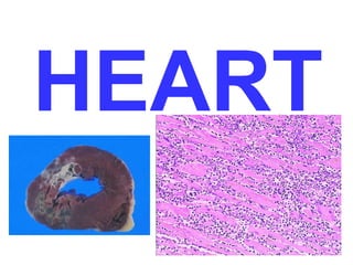
2. THE HEART
• Normal
• Pathology
– Heart Failure: L, R
– Heart Disease
• Congenital: LR shunts, RL shunts, Obstructive
• Ischemic: Angina, Infarction, Chronic Ischemia, Sudden Death
• Hypertensive: Left sided, Right sided
• Valvular: AS, MVP, Rheumatic, Infective, Non-Infective,
Carcinoid, Artificial Valves
• Cardiomyopathy: Dilated, Hypertrophic, Restrictive, Myocarditis,
Other
• Pericardium: Effusions, Pericarditis
• Tumors: Primary, Effects of Other Primaries
• Transplants
3. NORMAL Features
• 6000 L/day
• 250-300 grams
• 40% of all deaths (2x cancer)
• Wall thickness ~ pressure
• (i.e., a wall is only as thick as it has to be)
– LV=1.5 cm
– RV= 0.5 cm
– Atria =.2 cm
• Systole/Diastole
• Starling’s Law
4. TERMS
• CARDIOMEGALY
• DILATATION, any chamber, or all
• HYPERTROPHY, and chamber, or all
21. Left Sided Failure
• Low output vs. congestion
• Lungs
– pulmonary congestion and edema
– heart failure cells
• Kidneys
– pre-renal azotemia
– salt and fluid retention
• renin-aldosterone activation
• natriuretic peptides
• Brain: Irritability, decreased attention,
stuporcoma
22. Left Heart Failure Symptoms
• Dyspnea
– on exertion
– at rest
• Orthopnea
– redistribution of peripheral edema fluid
– graded by number of pillows needed
• Paroxysmal Nocturnal Dyspnea (PND)
28. CONGENITAL HEART
DEFECTS
• Faulty embryogenesis (week 3-8)
• Usually MONO-morphic (ASD, VSD,
hypo-RV, hypo-LV)
• May not be evident until adult life
(Coarctation, ASD)
• Overall incidence 1% of USA births
• INCREASED simple early detection via
non invasive methods, e.g., US, MRI,
CT, etc.
29. Malformation Births
Ventricular septal defect 4482 42
Atrial septal defect 1043 10
Pulmonary stenosis 836 8
Patent ductus arteriosus 781 7
Tetralogy of Fallot 577 5
Coarctation of aorta 492 5
Atrioventricular septal defect 396 4
Aortic stenosis 388 4
Transposition of great arteries 388 4
Truncus arteriosus 136 1
Total anomalous pulmonary venous connection 120 1
Tricuspid atresia
%
Incidence per Million Live
30. GENETICS
• Gene abnormalities in only 10% of CHD
• Trisomies 21, 13, 15, 18, XO
• Mutations of genes which encode for
transcription factorsTBX5ASD,VSD
• NKX2.5ASD
• Region of chromosome 22 important in
heart development, 22q11.2
deletionconotruncus, branchial arch,
face
32. CHD • LR SHUNTS: all “D’s” in their names
– NO cyanosis
– Pulmonary hypertension
– SIGNIFICANT pulmonary hypertension is
IRREVERSIBLE
• RL SHUNTS: all “T’s” in their names
– CYANOSIS
– VENOUS EMBOLI become SYSTEMIC
• OBSTRUCTIONS
33. LR
• ASD
• VSD
• ASVD
• PDA
NON CYANOTIC
IRREVERSIBLE
PULMONARY
HYPERTENSION
IS THE MOST
FEARED
CONSEQUENCE
34.
35. ASD
• NOT patent foramen ovale
• Usually asymptomatic until adulthood
• SECUNDUM (90%): Defective fossa
ovalis
• PRIMUM (5%): Next to AV valves, mitral
cleft
• SINUS VENOSUS (5%): Next to SVC
with anomalous pulmonary veins
draining to SVC or RA
36. VSD
• By far, most common CHD defect
• Only 30% are isolated
• Often with TETRALOGY of FALLOT
• 90% involve the membranous septum
• If muscular septum is involved, likely to
have multiple holes
• SMALL ones often close spontaneously
• LARGE ones progress to pulmonary
hypertension
37.
38. PDA
• 90% isolated
• HARSH, machinery-like murmur
• LR, possibly RL as pulmonary
hypertension approaches systemic
pressure
• Closing the defect may be life saving
• Keeping it open may be life saving
(Prostaglandin E). Why?
39. AVSD • Associated with defective,
inadequate AV valves
• Can be partial, or COMPLETE
(ALL 4 CHAMBERS FREELY
COMMUNICATE)
40. RL SHUNTS
• TETRALOGY of FALLOT most COMMON
– 1) VSD, large
– 2) OBSTRUCTION to RV flow
– 3) Aorta OVERRIDES the VSD
– 4) RVH
– SURVIVAL DEPENDS on SEVERITY of
SUBPULMONIC STENOSIS
– Can be a “PINK” tetrology if pulmonic
obstruction is small, but the greater the
obstruction, the greater is the RL shunt
41.
42. TGA (TRANSPOSITION
of GREAT ARTERIES)
• NEEDS a SHUNT for
survival
–PDA or PFO (65%),
“unstable” shunt
–VSD (35%), “stable” shunt
–RV>LV in thickness
–Fatal in first few months
–Surgical “switching”
44. TRICUSPID ATRESIA
• Hypoplastic RV
• Needs a shunt, ASD, VSD, or PDA
• High mortality
45. Total Anomalous Pulmonary
Venous Connection (TAPVC)
• PULMONARY VEINS do NOT go into
LA, but into L. innominate v. or
coronary sinus
• Needs a PFO or a VSD
• HYPOPLASTIC LA
47. COARCTATION of AORTA
• M>F
• But XO’s frequently have it
• INFANTILE FORM (proximal to PDA) (SERIOUS)
• ADULT FORM (CLOSED DUCTUS)
• Bicuspid aortic valve 50% of the time
48. PULMONIC
STENOSIS/ATRESIA
• If 100% atretic, hypoplastic RV with ASD
• Clinical severity ~ stenosis severity
52. IHD RISK
• Number of plaques
• Distribution of plaques
• Size, structure of plaques
53. ACUTE CORONARY SYNDROMES
• “The acute coronary syndromes are
frequently initiated by an
unpredictable and abrupt conversion
of a stable atherosclerotic plaque to
an unstable and potentially life-threatening
atherothrombotic lesion
through superficial erosion,
ulceration, fissuring, rupture, or deep
hemorrhage, usually with
superimposed thrombosis.”
54. EPIDEMIOLOGY
• ½ million die of IHD yearly in USA
• 1 million in 1963. Why?
– Prevention of control controllable risk factors
– Earlier, better diagnostic methods
– PTCA, CABG, arrythmia control
• 90% of IHD patients have
ATHEROSCLEROSIS
55. ACUTE CORONARY
SYNDROME FACTORS
• ACUTE PLAQUE CHANGE *******
• Inflammation
• Thrombus
• Vasoconstriction
******* MOST IMPORTANT
56. ACUTE PLAQUE CHANGE
• Rupture/Refissuring
• Erosion/Ulceration, exposing ECM
• Acute Hemorrhage
NB: Plaques do NOT have to be severely stenotic to
cause acute changes, i.e., 50% of AMI results from
thromboses of plaques showing LESS THAN 50%
stenosis
57.
58. INFLAMMATION
• Endothelial cells release CAMs,
selectins
• T-cells release TNF, IL-6, IFN-gamma to
stimulate and activate endothelial cells
and macrophages
• CRP predicts the probability of damage
in angina patients
60. VASOCONSTRICTION
• Circulating adrenergic agonists
• Platelet release products
• Endothelially released factors, such as
endothelin
61.
62. Coronary Artery Pathology in Ischemic Heart Disease
Plaque Plaque-Associated Thrombus
Syndrome Stenoses Disruption
Stable angina >75% No No
Unstable angina Variable Frequent Nonocclusive, often with thromboemboli
Transmural Variable Frequent Occlusive
myocardial infarction
Often small platelet aggregates or thrombi
and/or thromboemboli
Usually Frequent
severe
Sudden death
Widely variable, may be absent,
partial/complete, or lysed
Subendocardial Variable Variable
myocardial infarction
63. ANGINA PECTORIS
• Paroxysmal (sudden)
• Recurrent
• 15 sec.15 min.
• Reduced perfusion, but NO infarction
• THREE TYPES
– STABLE: relieved by rest or nitro
– PRINZMETAL: SPASM is main feature, responds to
nitro, S-T elevation
– UNSTABLE (crescendo, PRE-infarction, Q-wave
angina): perhaps some thrombosis, perhaps some
non transmural necrosis, perhaps some
embolization, but DISRUPTION of PLAQUE is
universally agreed upon
64. MYOCARDIAL INFARCTION
• Transmural vs. Subendocardial (inner 1/3)
• DUH! EXACT SAME risk factors ar
atherosclerosis
• Most are TRANSMURAL, and MOST are
caused by coronary artery occlusion
• In the 10% of transmural MIs NOT associated
with atherosclerosis:
– Vasospasm
– Emboli
– UNexplained
65. MYOCARDIAL RESPONSE
Feature Time
Onset of ATP depletion Seconds
Loss of contractility <2 min
ATP reduced
to 50% of normal 10 min
to 10% of normal 40 min
Irreversible cell injury 20–40 min
Microvascular injury >1 hr
67. TIMING of Gross and
Microscopic Findings
½–4 hr None Usually none; variable waviness of fibers at border
4–12 hr Occasionally dark mottling Beginning coagulation necrosis; edema; hemorrhage
Ongoing coagulation necrosis; pyknosis of nuclei;
myocyte hypereosinophilia; marginal contraction
band necrosis; beginning neutrophilic infiltrate
Coagulation necrosis, with loss of nuclei and striations;
interstitial infiltrate of neutrophils
Beginning disintegration of dead myofibers, with dying
neutrophils; early phagocytosis of dead cells by
macrophages at infarct border
Well-developed phagocytosis of dead cells; early
formation of fibrovascular granulation tissue at
margins
Well-established granulation tissue with new blood
vessels and collagen deposition
Increased collagen deposition, with decreased
cellularity
12–24 hr Dark mottling
Mottling with yellow-tan
infarct center
Hyperemic border; central
yellow-tan softening
Maximally yellow-tan and
soft, with depressed red-tan
margins
Red-gray depressed infarct
borders
Gray-white scar, progressive
from border toward core
of infarct
1–3 days
3–7 days
7–10
days
10–14
days
2–8 wk
>2 mo Scarring complete Dense collagenous scar
69. RE-PERFUSION
• Thrombolysis
• PTCA
• CABG
• Reperfusion CANNOT restore necrotic
or dead fibers, only reversibly injured
ones
• REPERFUSION “INJURY”
– Free radicals
– Interleukins
70. AMI DIAGNOSIS
• SYMPTOMS
• EKG
• DIAPHORESIS
• (10% of MIs are “SILENT” with Q-waves)
• CKMB gold standard enzyme
• Troponin-I, Troponin-T better
• CRP predicts risk of AMI in angina
patients
72. CIHD, aka, ischemic
“cardiomyopathy”
• Progress to CHF often with no
pathologic or clinical evidence of
localized infarction
–Extensive atherosclerosis
–No infarct
–H&D present
73. SUDDEN CARDIAC DEATH
• 350,000 in USA yearly from atherosclerosis
• NON-atherosclerotic sudden cardiac death includes:
– Congenital coronary artery disease
– Aortic stenosis
– MVP
– Myocarditis
– Cardiomyopathy (sudden death in young athletes)
– Pulmonary hypertension
– Conduction defects
– HTN, hypertrophy of UNKNOWN etiology
74. AUTOPSY findings in SCD
• >75% narrowing of 1-3 vessels
• Healed infarcts 40%
• “ARRHYTHMIA” is often a very
convenient conclusion when no
anatomic findings are present
76. HHD (Left)
• DEFINITION: Hypertrophic
adaptive response of the heart,
which can progress:
–Myocardial dysfunction
–Cardiac dilatation
–CHF
–Sudden death
88. 70% of all VHD
• AS
–Calcification of a deformed valve
– “Senile” calcific AS
– Rheum, Heart Dis.
• MS
–Rheumatic Heart Disease
89. AORTIC STENOSIS
2X gradient pressure
LVH, ischemia
Cardiac decompensation, angina, CHF
50% die in 5 years if angina present
50% die in 2 years if CHF present
90. MITRAL ANNULAR
CALCIFICATION
• Calcification of the
mitral “skeleton”
• Usually NO
dysfunction
• Regurgitation or
Stenosis possible
• F>>M
102. DIAGNOSIS=MMm, Mmmm, mmmmm
• MAJOR
• Positive blood culture(s) indicating characteristic organism or persistence of unusual organism
• Echocardiographic findings, including valve-related or implant-related mass or abscess, or
partial separation of artificial valve
• New valvular regurgitation
• minor
• Predisposing heart lesion or intravenous drug use
• Fever
• Vascular lesions, including arterial petechiae, subungual/splinter hemorrhages, emboli, septic
infarcts, mycotic aneurysm, intracranial hemorrhage, Janeway lesions
• Immunologic phenomena, including glomerulonephritis, Osler nodes, Roth spots, rheumatoid
factor
• Microbiologic evidence, including single culture showing uncharacteristic organism
• Echocardiographic findings consistent with but not diagnostic of endocarditis, including new
valvular regurgitation, pericarditis
103.
104. NON-infective VEGETATIONS
• <5 mm
• PE
• Trousseau syndrome (migratory
thrombophlebitis with malignancies)
• s/p Swan-Ganz
• Libman-Saks with SLE (both sides of
valve)
105. Carcinoid Syndrome
• Episodic skin flushing
• Cramps
• Nausea & Vomiting
• Diarrhea
• ↑serotonin, ↑ 5HIAA in urine
• FIBROUS INTIMAL THICKENING
– RV, Tricuspid valve, Pulmonic valve (all RIGHT side)
– Similar to what Fen-Phen does on the LEFT side
106.
107. ARTIFICIAL VALVES
• Mechanical
• Xenografts (porcine)
• 60% have complications within
10 years
115. Arrhythmogenic Right Ventricular Cardiomyopathy
(Arrhythmogenic Right Ventricular Dysplasia)
This is an uncommon dilated cardiomyopathy predominantly RIGHT ventricle.
So is NAXOS syndrome.
116. HYPERTROPHIC cardiomyopathy
• Also called IHSS, (Idiopathic Hypertrophic
Subaortic Stenosis)
– GENETIC defects involving:
• Beta-myosin heavy chain
• Troponin T
• Alpha-tropomyosin
• Myosin binding protein C
– PATHOLOGY: Massive hypertrophy, Asymmetric
septum, DISARRAY of myocytes, INTERSTITIAL
fibrosis
– CLINICAL: ↓chamber volume, ↓SV, ↓ diastolic
filling
117. RESTRICTIVE cardiomyopathy
• (idiopathic)
• ↓ ventricular compliance
• Chiefly affects DIASTOLE
• NORMAL chamber size and wall thickness
• THREE similar diseases affecting
predominantly the SUBENDOCARDIAL area:
– Endomyocardial Fibrosis (African children)
– Loeffler Endomyocarditis (eosinophilic leukemia)
– Endocardial Fibroelastosis (infants)
118. MYOCARDITIS
• INFLAMMATION of MYOCARDIUM
• Chiefly microbial
–COXACKIE A & B, CMV, HIV
– Trypanosoma cruzi (Chagas dis.), 80%
– Trichinosis
– Toxoplasmosis
– Lyme disease (5%)
– Diphtheria
• IMMUNE: Post-viral, rheumatic, SLE, drug
hypersensitivityalpha-methyl dopa, sulfas
119. LYM PHOCYTIC INFILTR ATES are the USUAL pattern of ALL myocarditis, but
eosinophils, giant cells, and even trypanosomes can be seen occasionally
120. OTHER Myocarditides
• Adriamycin
• Cyclophosphamide
• Catecholamines (Pheochromocytomas)
• Amyloid, systemic or primary cardiac
– Congo red stain: green birefringence with
polarization
• Amyloid, aging
– Congo red stain: green birefringence with
polarization
• Hemochromatosis (Prussian Blue)
• BOTH HYPER-, HYPO- -thyroidism
Whichever artery winds up supplying the posterior interventricular septum is said to be “DOMINANT”
The pigment which accumulates with age is called lipofucsin, and caused the heart to appear “browner” than normal. This is called “brown” atrophy of the heart.
Note that not only is the FIBER thick, but so are the nuclei. Note squaring off of the nuclei, so called “BOXCAR” effect.
LEFT to RIGHT SHUNTS, NON-cyanotic
CLASSICAL TETROLOGY of FALLOT
NOTE: In sichemia, NO gross or microscopic findings are seen, visible changes are seen only with INFARCTION. You cannoe see ISCHEMIA!!!
Answer: owing to left atrial enlargement
Answer: lenticulostriate in basal ganglia most susceptible to hypertensive CVA
Why do BOTH stenosis and regurgitation cause hypertrophy of the chamber proximal to the valve?