SlideShare a Scribd company logo
1 of 67
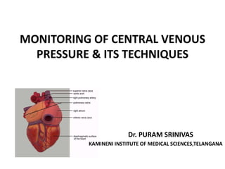
MONITORING OF CENTRAL VENOUS
PRESSURE & ITS TECHNIQUES
Dr. PURAM SRINIVAS
KAMINENI INSTITUTE OF MEDICAL SCIENCES,TELANGANA
OVERVIEW
• Introduction
• Types Of Central Line
• Indications & Relative Contraindications Of
Central Venus Line (CVL)
• PICC Line Indications & Contraindications
• CVL Insertion
• Factors Affecting CVP
• Central Venous Pressure
Monitoring
• Interpretation Of Waveforms
• Summary
INTRODUCTION
• The central venous pressure (CVP) is the pressure
measured in the central veins close to the heart.
• It indicates mean right atrial pressure and is
frequently used as an estimate of right ventricular
preload.
• CVP reflects the amount of blood returning to the
heart and the ability of the heart to pump the blood
into the arterial system
INTRODUCTION Cont’
• It is the pressure measured at the junction of the
superior vena cava and the right atrium.
• It reflects the driving force for filling of the right
atrium & ventricle.
• Normal CVP in an awake spontaneously breathing
patient : 1-7 mmHg or 5-10 cm H2O.
• Mechanical ventilation : 3-5 cm H2O higher
TYPES OF CENTRAL LINE
• SINGLE LUMEN
• DOUBLE LUMEN
• TRIPLE LUMEN
• QUADRUPLE LUMEN
• QUINTUPLE LUMEN
• PERIPHERALLY INSERTED CENTRAL CATHETERS
(PICCS)
Single, Double, and Triple Lumen
Central Lines
Indications Central Venus Line (CVL)
• Major operative procedures involving large fluid
shifts or blood loss
• Intravascular volume assessment when urine output
is not reliable or unavailable
• Temporary Hemodialysis
• Surgical procedures with a high risk for air embolism,
CVP catheter may be used to aspirate intracardiac air
Indications Central Venus Line (CVL) CONT’
• Frequent venous blood sampling, Inadequate
peripheral intravenous access
• Temporary pacing
• Venous access for vasoactive or irritating drugs &
Chronic drug administration
• Rapid infusion of intravenous fluids (using large
cannulae)
• Total parenteral nutrition
Relative Contraindications
• Bleeding disorders (platelet counts <50,000,
bleeding is uncommon and easily managed).
• Anticoagulation or thrombolytic therapy.
• Combative patients.
• Distorted local anatomy.
• Cellulitis, burns, severe dermatitis at site.
• Vasculitis.
Peripherally Inserted Central
Catheters (PICCs)
• LOCATION OR SITE OF
INSERTION
• INDICATIONS
• CONTRAINDICATIONS
• BENEFITS AND RISKS
PICC LINE INTRODUCTION
• A Peripherally Inserted Central Catheter (PICC)
is a small gauge catheter that is inserted
peripherally but the tip sits in the central
venous circulation in the lower 1/3 of the
superior vena cava.
• It is suitable for long term use and there are
no restrictions for age, or gender.
SITE’S OF INSERTION OF PICC LINE
• PICCs are commonly placed at or above the
antecubital space in the following veins;
 Cephalic vein
 Basilic vein
 Medial-cubital vein
INDICATIONS FOR PICC LINE
INSERTION
• PICC lines are suitable for many situations when
access is limited or expected to last longer than 2
weeks.
• Compromised/Inadequate peripheral access
• Infusion of hyperosmolar solutions or solutions with
high acidity or alkalinity
(e.g. Total Parenteral Nutrition)
• Infusion of vesicant or irritant agents
(Inotropes, Chemotherapy)
• Short or long term intravenous therapy
(e.g. Antibiotics)
CONTRAINDICATIONS FOR PICC
INSERTION
• Previous upper extremity venous thrombosis (DVT)
• Trauma or vascular surgeries at or near the site of
insertion
• Presence of a device related infection, cellulitis, or
bacteremia at or near the insertion site
• Lymphedema.
• Mastectomy surgery with axillary dissection +/-
lymphedema on affected side (unless urgent
condition requires it)
• Allergy to materials
• Irradiation of insertion site
Sites for insertion of CVL
• Internal Jugular
• Subclavian
• Femoral
• External Jugular
• Basilic
• Axillary
Right IJV is Preferred
• Consistent, predictable anatomy
• Alignment with RA
• Palpable landmark and high success rate
• No thoracic duct injury
CVL Insertion
• Equipment.
• Patient position.
• Procedure.
• After insertion
Equipment
• Sterile gloves, gown, suture pack.
• Iodine solution.
• 10 ml syringe, 2% lidocaine, 10 ml N.S.
• Catheter special size.
• H2O manometer.
• Flush solution with complete CVP line.
• Dressing set.
Patient Position
• Patient is moved to the side of the bed so physician
would not lean over.
• The bed is high enough so physician would not have
to stoop over.
• Patient should be flat without a pillow,
Trendelenburg position if patient is hypovolemic.
• The head is turned away from the side of the
procedure.
• Wrist restraints if necessary.
Procedure
Skin preparation:
• Prepare before putting sterile gloves
• Allow time for the sterilizing agent to dry
Drape:
• Large enough and Handed sterilely by the assistant.
• Hole in the area of placement.
Prepare the tray:
• Prepare the equipment before starting.
Anesthesia:
• Use local anesthesia with lidocaine
USING THE CENTRAL LINE
• Flush it, before and after use( with NS).
• Some places also require heparin flush.
• Close clamps when not in use.
• Dressing is usually changed every days.
• Line can be used for blood drawing –withdraw
and waste 10 cc, then withdraw blood for
samples.
• If port becomes clotted, do not use – sometimes
ports can be opened up.
Immediately Complications of
Insertion CVL
• Hemothorax.
• Pneumothorax (most common).
• Bleeding
• Arterial puncture.
• Vessel erosion
• Nerve Injury.
• Dysrhythmias.
• Catheter malplacement.
• Embolus.
• Cardiac tamponade.
Delayed Complications
• Dysrhythmias
• Infection (“Femoral > IJ > subclavian”)
• Catheter malplacement.
• Vessel erosion.
• Embolus.
• Cardiac tamponade.
• Thrombosis
Factors Affecting CVP
•Cardiac Function
•Blood Volume
•Capacitance of vessel
•Intrathoracic Pressure
•Intraperitoneal
pressure
Causes for Increase in CVP
• Over hydration.
• Right-sided heart failure.
• Cardiac tamponade.
• Constrictive pericarditis.
• Pulmonary hypertension.
• Tricuspid stenosis and regurgitation.
• Stroke volume is high.
Causes for Increase in CVP CONT’
Decrease of CVP
• Hypovolemia.
• Decreased venous return.
• Excessive veno or vasodilation.
• Shock.
• If the measure is less than 5 cm water that
mean that the circulating volume is decrease.
Decrease of CVP CONT
CENTRAL VENOUS PRESSURE
MONITORING
Methods to measure CVP
Indirect assessment:
• Inspection of jugular venous pulsations in the
neck.
Direct assessment:
• Fluid filled manometer connected to central
venous catheter.
• Calibrated transducer.
Inspection of jugular venous pulsations in the
neck.
• No valve between Right atrium & Internal
Jugular Vein.
• Degree of distention & venous wave form
reflects information about cardiac function
Measuring central venous pressure
Using a manometer
• Line up the manometer
arm with the
phlebostatic axis
ensuring that the
bubble is between the
two lines of the spirit
level
Phlebostatic Axis
4th intercostal space, mid-
axillary line
Level of the atria
• Move the manometer
scale up and down to
allow the bubble to be
aligned with zero on the
scale. This is referred to
as 'zeroing the
manometer
• Turn the three-way tap
off to the patient and
open to the manometer
• Open the IV fluid bag
and slowly fill the
manometer to a level
higher than the
expected CVP
• Turn off the flow from
the fluid bag and open
the three-way tap from
the manometer to the
patient
The fluid level inside the
manometer should fall
until gravity equals the
pressure in the central
veins
• When the fluid stops
falling the CVP
measurement can be
read. If the fluid moves
with the patient's
breathing, read the
measurement from the
lower number.
• Turn the tap off to the
manometer veins
• Document the
measurement and
report any changes or
abnormalities
Measuring central venous pressure
Using a transducer
• Turn the tap off to the
patient and open to the
air by removing the cap
from the three-way port
opening the system to
the atmosphere.
• Press the zero button
on the monitor and
wait while calibration
occurs.
• When 'zeroed' is
displayed on the
monitor, replace the
cap on the three-way
tap and turn the tap on
to the patient.
• Observe the CVP trace
on the monitor. The
waveform undulates as
the right atrium
contracts and relaxes,
emptying and filling
with blood. (light blue
in this image)
Interpretation from Waveform
The CVP waveform consists of five
phasic events,
three peaks (a, c, v) and two
descents (x, y)
Mechanical Events
‘a’ wave
• Atrial Contraction(after P wave)
• Prominent a wave: resistance in
RV filling- RVH, TS,
Temponade,PS, Pulmonary
hypertension.
• Cannon A waves occur as the
RA contracts against a closed
TV: junctional rhythm,
CHB,ventricular arrhythmias
• Absent a wave: Atrial
fibrillation or
• • flutter
‘c’ wave
• Isovolumic right
ventricle contraction,
TV bow in RA(after QRS)
• Early Systole
• TR: Tall Systolic c-v wave
• It is call holosystolic
cannon v waves
‘x’ descent
• Atrial Relaxation
• Mid Systole
• Dominant x descent –
good RV function and
vice versa
• Cardiac Tamponade- X
descent is steep & Y
descent is diminished
‘v’ wave
• Filing of RA with venous
blood(just after T wave)
• Late Systole
• Prominent v wave with
increase venous return. ASD,
PAPVC or TAPVC, A-V
malformation
• Large V waves may also
appear later in systole if the
ventricle becomes
noncompliant because of
ischemia or RV failure.
• Decrease in RA emptying. TS
‘y’ descent
• Early ventricular filling,
opening of TV
• Early Diastole
• Attentuation of y
descent: TS,
Tachycardia, RVF,
Tamponade,PS
CVP Changes with Respiration
• A, During spontaneous
ventilation, the onset of
inspiration (arrows) causes a
reduction in intrathoracic
pressure, which is transmitted
to both the CVP and
pulmonary artery pressure
(PAP) waveforms. CVP should
be recorded at end-expiration.
• B, During positive-pressure
ventilation, the onset of
inspiration (arrows) causes an
increase in intrathoracic
pressure. CVP is still recorded
at end-expiration.
• Kussmaul sign is a paradoxical rise in jugular venous
pressure (JVP) on inspiration, or a failure in the
appropriate fall of the JVP with inspiration.
• It can be seen in some forms of heart disease and is
usually indicative of limited right ventricular filling
due to right heart dysfunction.
• Hepatojugular Reflex: A positive result is variously
defined as either a sustained rise in the JVP of at
least 3 cm or more or a fall of 4 cm or more after the
examiner releases pressure
REMOVAL OF CENTRAL LINE
• This is an aseptic procedure.
• The patient should be supine with head tilted
down.
• Ensure no drugs are attached and running via the
central line.
• Remove dressing.
• Cut the stitches.
• If there is resistant then call for assistance.
• Apply digital pressure with gauze until bleeding
stops.
• Dress with gauze and clear dressing.
SUMMARY
• Central Venous Line becomes the key element
in managing critically ill patients
• One should have decent amount of
knowledge & Skill about insertion and
maintanance of central lines.
REFERENCES
• Millar’s Anesthesia 8th Edition
• Samson Wrights Textbook of Applied
Physiology 13th Edition
• Marino’s The ICU Book 4th Edition
• Measuring central venous pressure Elaine Cole
Senior lecturer ED/Trauma, City University
Bartsand the London NHS Trust.
•THANK YOU

More Related Content

What's hot

Central line
Central line Central line
Central line Irfan Munna
 
CENTRAL VENOUS PRESSURE MONITORING- IACTACON 2018
CENTRAL VENOUS PRESSURE MONITORING- IACTACON 2018CENTRAL VENOUS PRESSURE MONITORING- IACTACON 2018
CENTRAL VENOUS PRESSURE MONITORING- IACTACON 2018Dr. Harshil Joshi
 
Intercostal drainage tube insertion
Intercostal drainage tube insertionIntercostal drainage tube insertion
Intercostal drainage tube insertionMahesh Chand
 
Endotracheal intubation nursing procedure
Endotracheal intubation nursing procedureEndotracheal intubation nursing procedure
Endotracheal intubation nursing procedureABHIJIT BHOYAR
 
Defibrillation
DefibrillationDefibrillation
Defibrillationresmigs
 
Venturi Mask for Oxygen Delivery Administration
Venturi Mask for Oxygen Delivery AdministrationVenturi Mask for Oxygen Delivery Administration
Venturi Mask for Oxygen Delivery AdministrationSmart Medical Buyer
 
Mechanical Ventilator by AJ
Mechanical Ventilator by AJMechanical Ventilator by AJ
Mechanical Ventilator by AJAbhishek Joshi
 
ENDOTRACHEAL TUBE INTUBATION II Parts II Details II Clinical Discussion
ENDOTRACHEAL TUBE INTUBATION II Parts II Details II Clinical DiscussionENDOTRACHEAL TUBE INTUBATION II Parts II Details II Clinical Discussion
ENDOTRACHEAL TUBE INTUBATION II Parts II Details II Clinical DiscussionSwatilekha Das
 
Intravenous cannulation
Intravenous cannulationIntravenous cannulation
Intravenous cannulationAYM NAZIM
 
Endotracheal tubes
Endotracheal tubesEndotracheal tubes
Endotracheal tubesPratik Kumar
 
Mechanical ventilator for nurses 08.02.19
Mechanical ventilator for nurses 08.02.19Mechanical ventilator for nurses 08.02.19
Mechanical ventilator for nurses 08.02.19Johny Wilbert
 
Cardiac monitoring
Cardiac monitoringCardiac monitoring
Cardiac monitoringRajee Ravindran
 
Cardioversion
Cardioversion Cardioversion
Cardioversion Pranav Bansal
 
Cardiac monitoring & ECG
Cardiac monitoring & ECGCardiac monitoring & ECG
Cardiac monitoring & ECGMathew Varghese V
 
Central Venous Catheter Care- A Nursing skill
Central Venous Catheter Care- A Nursing skill Central Venous Catheter Care- A Nursing skill
Central Venous Catheter Care- A Nursing skill Tse Sona
 

What's hot (20)

Central line
Central line Central line
Central line
 
CENTRAL VENOUS PRESSURE MONITORING- IACTACON 2018
CENTRAL VENOUS PRESSURE MONITORING- IACTACON 2018CENTRAL VENOUS PRESSURE MONITORING- IACTACON 2018
CENTRAL VENOUS PRESSURE MONITORING- IACTACON 2018
 
Intercostal drainage tube insertion
Intercostal drainage tube insertionIntercostal drainage tube insertion
Intercostal drainage tube insertion
 
Cvp line
Cvp lineCvp line
Cvp line
 
ACLS & BLS
ACLS & BLSACLS & BLS
ACLS & BLS
 
Endotracheal intubation nursing procedure
Endotracheal intubation nursing procedureEndotracheal intubation nursing procedure
Endotracheal intubation nursing procedure
 
Defibrillation
DefibrillationDefibrillation
Defibrillation
 
Venturi Mask for Oxygen Delivery Administration
Venturi Mask for Oxygen Delivery AdministrationVenturi Mask for Oxygen Delivery Administration
Venturi Mask for Oxygen Delivery Administration
 
Mechanical Ventilator by AJ
Mechanical Ventilator by AJMechanical Ventilator by AJ
Mechanical Ventilator by AJ
 
ENDOTRACHEAL TUBE INTUBATION II Parts II Details II Clinical Discussion
ENDOTRACHEAL TUBE INTUBATION II Parts II Details II Clinical DiscussionENDOTRACHEAL TUBE INTUBATION II Parts II Details II Clinical Discussion
ENDOTRACHEAL TUBE INTUBATION II Parts II Details II Clinical Discussion
 
Intravenous cannulation
Intravenous cannulationIntravenous cannulation
Intravenous cannulation
 
Endotracheal tubes
Endotracheal tubesEndotracheal tubes
Endotracheal tubes
 
Mechanical ventilator for nurses 08.02.19
Mechanical ventilator for nurses 08.02.19Mechanical ventilator for nurses 08.02.19
Mechanical ventilator for nurses 08.02.19
 
Cardiac monitoring
Cardiac monitoringCardiac monitoring
Cardiac monitoring
 
Cardioversion
Cardioversion Cardioversion
Cardioversion
 
Intravenous Cannulation
Intravenous CannulationIntravenous Cannulation
Intravenous Cannulation
 
Cardiac monitoring & ECG
Cardiac monitoring & ECGCardiac monitoring & ECG
Cardiac monitoring & ECG
 
ICU Care Bundles
ICU Care BundlesICU Care Bundles
ICU Care Bundles
 
Central Venous Catheter Care- A Nursing skill
Central Venous Catheter Care- A Nursing skill Central Venous Catheter Care- A Nursing skill
Central Venous Catheter Care- A Nursing skill
 
Central Venous Access
Central Venous AccessCentral Venous Access
Central Venous Access
 

Similar to CVP Monitoring Techniques

MONITORING HEMODYNAMIC STATUS-1.pptx
MONITORING HEMODYNAMIC STATUS-1.pptxMONITORING HEMODYNAMIC STATUS-1.pptx
MONITORING HEMODYNAMIC STATUS-1.pptxRizwanUllah151255
 
Cardiac diagnosis.pptx
Cardiac diagnosis.pptxCardiac diagnosis.pptx
Cardiac diagnosis.pptxpoojaprakash50
 
Ccpa catheter basics07medicine
Ccpa catheter basics07medicineCcpa catheter basics07medicine
Ccpa catheter basics07medicineanjika
 
Invasive procedures
Invasive proceduresInvasive procedures
Invasive proceduresSamir Elkafrawy
 
Hemodynamic monitorig
Hemodynamic monitorigHemodynamic monitorig
Hemodynamic monitorigStephi Poulose
 
Central venous catheterization
Central venous catheterizationCentral venous catheterization
Central venous catheterizationsuji kalai
 
Basic haemodynamic monitoring
Basic haemodynamic monitoringBasic haemodynamic monitoring
Basic haemodynamic monitoringDeepankar Mishra
 
LV angiography.pptx
LV angiography.pptxLV angiography.pptx
LV angiography.pptxravitulluru1
 
central venous pressure and intra-arterial blood pressure monitoring. invasiv...
central venous pressure and intra-arterial blood pressure monitoring. invasiv...central venous pressure and intra-arterial blood pressure monitoring. invasiv...
central venous pressure and intra-arterial blood pressure monitoring. invasiv...prateek gupta
 
centrallineaig-180518100313 (1).pdf download link
centrallineaig-180518100313 (1).pdf download linkcentrallineaig-180518100313 (1).pdf download link
centrallineaig-180518100313 (1).pdf download linkGokulnathMbbs
 
Cardiovascular monitoring Part II
Cardiovascular monitoring Part IICardiovascular monitoring Part II
Cardiovascular monitoring Part IISiddhanta Choudhury
 
CLASS 7 - CABG.pptx
CLASS 7 - CABG.pptxCLASS 7 - CABG.pptx
CLASS 7 - CABG.pptxDaisy Thomas
 
3. CVS monitoring.pptx
3. CVS monitoring.pptx3. CVS monitoring.pptx
3. CVS monitoring.pptxFLOWERSOFPAKISTAN
 
Basic hemodynamic monitoring for nurses
Basic hemodynamic monitoring for nurses Basic hemodynamic monitoring for nurses
Basic hemodynamic monitoring for nurses Muhammad Asim Rana
 
Swan-Ganz-catheterisation_amit-panjwani.pdf
Swan-Ganz-catheterisation_amit-panjwani.pdfSwan-Ganz-catheterisation_amit-panjwani.pdf
Swan-Ganz-catheterisation_amit-panjwani.pdframbhoopal1
 
Invasive_Cardio-Devices_procedures[1].pdf
Invasive_Cardio-Devices_procedures[1].pdfInvasive_Cardio-Devices_procedures[1].pdf
Invasive_Cardio-Devices_procedures[1].pdfBatMan752678
 
centralvenouscatheter-1.pdf
centralvenouscatheter-1.pdfcentralvenouscatheter-1.pdf
centralvenouscatheter-1.pdfisha sharma
 
CENTRAL VENOUS CATHETER
CENTRAL VENOUS CATHETERCENTRAL VENOUS CATHETER
CENTRAL VENOUS CATHETERAvijit Prusty
 
Cardiac catheterization123
Cardiac catheterization123Cardiac catheterization123
Cardiac catheterization123TriXie Sorrilla
 

Similar to CVP Monitoring Techniques (20)

MONITORING HEMODYNAMIC STATUS-1.pptx
MONITORING HEMODYNAMIC STATUS-1.pptxMONITORING HEMODYNAMIC STATUS-1.pptx
MONITORING HEMODYNAMIC STATUS-1.pptx
 
CVP.pptx
CVP.pptxCVP.pptx
CVP.pptx
 
Cardiac diagnosis.pptx
Cardiac diagnosis.pptxCardiac diagnosis.pptx
Cardiac diagnosis.pptx
 
Ccpa catheter basics07medicine
Ccpa catheter basics07medicineCcpa catheter basics07medicine
Ccpa catheter basics07medicine
 
Invasive procedures
Invasive proceduresInvasive procedures
Invasive procedures
 
Hemodynamic monitorig
Hemodynamic monitorigHemodynamic monitorig
Hemodynamic monitorig
 
Central venous catheterization
Central venous catheterizationCentral venous catheterization
Central venous catheterization
 
Basic haemodynamic monitoring
Basic haemodynamic monitoringBasic haemodynamic monitoring
Basic haemodynamic monitoring
 
LV angiography.pptx
LV angiography.pptxLV angiography.pptx
LV angiography.pptx
 
central venous pressure and intra-arterial blood pressure monitoring. invasiv...
central venous pressure and intra-arterial blood pressure monitoring. invasiv...central venous pressure and intra-arterial blood pressure monitoring. invasiv...
central venous pressure and intra-arterial blood pressure monitoring. invasiv...
 
centrallineaig-180518100313 (1).pdf download link
centrallineaig-180518100313 (1).pdf download linkcentrallineaig-180518100313 (1).pdf download link
centrallineaig-180518100313 (1).pdf download link
 
Cardiovascular monitoring Part II
Cardiovascular monitoring Part IICardiovascular monitoring Part II
Cardiovascular monitoring Part II
 
CLASS 7 - CABG.pptx
CLASS 7 - CABG.pptxCLASS 7 - CABG.pptx
CLASS 7 - CABG.pptx
 
3. CVS monitoring.pptx
3. CVS monitoring.pptx3. CVS monitoring.pptx
3. CVS monitoring.pptx
 
Basic hemodynamic monitoring for nurses
Basic hemodynamic monitoring for nurses Basic hemodynamic monitoring for nurses
Basic hemodynamic monitoring for nurses
 
Swan-Ganz-catheterisation_amit-panjwani.pdf
Swan-Ganz-catheterisation_amit-panjwani.pdfSwan-Ganz-catheterisation_amit-panjwani.pdf
Swan-Ganz-catheterisation_amit-panjwani.pdf
 
Invasive_Cardio-Devices_procedures[1].pdf
Invasive_Cardio-Devices_procedures[1].pdfInvasive_Cardio-Devices_procedures[1].pdf
Invasive_Cardio-Devices_procedures[1].pdf
 
centralvenouscatheter-1.pdf
centralvenouscatheter-1.pdfcentralvenouscatheter-1.pdf
centralvenouscatheter-1.pdf
 
CENTRAL VENOUS CATHETER
CENTRAL VENOUS CATHETERCENTRAL VENOUS CATHETER
CENTRAL VENOUS CATHETER
 
Cardiac catheterization123
Cardiac catheterization123Cardiac catheterization123
Cardiac catheterization123
 

Recently uploaded

Full Stack Web Development Course for Beginners
Full Stack Web Development Course  for BeginnersFull Stack Web Development Course  for Beginners
Full Stack Web Development Course for BeginnersSabitha Banu
 
Incoming and Outgoing Shipments in 3 STEPS Using Odoo 17
Incoming and Outgoing Shipments in 3 STEPS Using Odoo 17Incoming and Outgoing Shipments in 3 STEPS Using Odoo 17
Incoming and Outgoing Shipments in 3 STEPS Using Odoo 17Celine George
 
Grade 9 Q4-MELC1-Active and Passive Voice.pptx
Grade 9 Q4-MELC1-Active and Passive Voice.pptxGrade 9 Q4-MELC1-Active and Passive Voice.pptx
Grade 9 Q4-MELC1-Active and Passive Voice.pptxChelloAnnAsuncion2
 
ECONOMIC CONTEXT - PAPER 1 Q3: NEWSPAPERS.pptx
ECONOMIC CONTEXT - PAPER 1 Q3: NEWSPAPERS.pptxECONOMIC CONTEXT - PAPER 1 Q3: NEWSPAPERS.pptx
ECONOMIC CONTEXT - PAPER 1 Q3: NEWSPAPERS.pptxiammrhaywood
 
Visit to a blind student's school🧑‍🦯🧑‍🦯(community medicine)
Visit to a blind student's school🧑‍🦯🧑‍🦯(community medicine)Visit to a blind student's school🧑‍🦯🧑‍🦯(community medicine)
Visit to a blind student's school🧑‍🦯🧑‍🦯(community medicine)lakshayb543
 
INTRODUCTION TO CATHOLIC CHRISTOLOGY.pptx
INTRODUCTION TO CATHOLIC CHRISTOLOGY.pptxINTRODUCTION TO CATHOLIC CHRISTOLOGY.pptx
INTRODUCTION TO CATHOLIC CHRISTOLOGY.pptxHumphrey A BeĂąa
 
Difference Between Search & Browse Methods in Odoo 17
Difference Between Search & Browse Methods in Odoo 17Difference Between Search & Browse Methods in Odoo 17
Difference Between Search & Browse Methods in Odoo 17Celine George
 
Earth Day Presentation wow hello nice great
Earth Day Presentation wow hello nice greatEarth Day Presentation wow hello nice great
Earth Day Presentation wow hello nice greatYousafMalik24
 
MULTIDISCIPLINRY NATURE OF THE ENVIRONMENTAL STUDIES.pptx
MULTIDISCIPLINRY NATURE OF THE ENVIRONMENTAL STUDIES.pptxMULTIDISCIPLINRY NATURE OF THE ENVIRONMENTAL STUDIES.pptx
MULTIDISCIPLINRY NATURE OF THE ENVIRONMENTAL STUDIES.pptxAnupkumar Sharma
 
How to do quick user assign in kanban in Odoo 17 ERP
How to do quick user assign in kanban in Odoo 17 ERPHow to do quick user assign in kanban in Odoo 17 ERP
How to do quick user assign in kanban in Odoo 17 ERPCeline George
 
Procuring digital preservation CAN be quick and painless with our new dynamic...
Procuring digital preservation CAN be quick and painless with our new dynamic...Procuring digital preservation CAN be quick and painless with our new dynamic...
Procuring digital preservation CAN be quick and painless with our new dynamic...Jisc
 
Keynote by Prof. Wurzer at Nordex about IP-design
Keynote by Prof. Wurzer at Nordex about IP-designKeynote by Prof. Wurzer at Nordex about IP-design
Keynote by Prof. Wurzer at Nordex about IP-designMIPLM
 
Karra SKD Conference Presentation Revised.pptx
Karra SKD Conference Presentation Revised.pptxKarra SKD Conference Presentation Revised.pptx
Karra SKD Conference Presentation Revised.pptxAshokKarra1
 
How to Add Barcode on PDF Report in Odoo 17
How to Add Barcode on PDF Report in Odoo 17How to Add Barcode on PDF Report in Odoo 17
How to Add Barcode on PDF Report in Odoo 17Celine George
 
Inclusivity Essentials_ Creating Accessible Websites for Nonprofits .pdf
Inclusivity Essentials_ Creating Accessible Websites for Nonprofits .pdfInclusivity Essentials_ Creating Accessible Websites for Nonprofits .pdf
Inclusivity Essentials_ Creating Accessible Websites for Nonprofits .pdfTechSoup
 
ANG SEKTOR NG agrikultura.pptx QUARTER 4
ANG SEKTOR NG agrikultura.pptx QUARTER 4ANG SEKTOR NG agrikultura.pptx QUARTER 4
ANG SEKTOR NG agrikultura.pptx QUARTER 4MiaBumagat1
 

Recently uploaded (20)

Full Stack Web Development Course for Beginners
Full Stack Web Development Course  for BeginnersFull Stack Web Development Course  for Beginners
Full Stack Web Development Course for Beginners
 
Incoming and Outgoing Shipments in 3 STEPS Using Odoo 17
Incoming and Outgoing Shipments in 3 STEPS Using Odoo 17Incoming and Outgoing Shipments in 3 STEPS Using Odoo 17
Incoming and Outgoing Shipments in 3 STEPS Using Odoo 17
 
Grade 9 Q4-MELC1-Active and Passive Voice.pptx
Grade 9 Q4-MELC1-Active and Passive Voice.pptxGrade 9 Q4-MELC1-Active and Passive Voice.pptx
Grade 9 Q4-MELC1-Active and Passive Voice.pptx
 
FINALS_OF_LEFT_ON_C'N_EL_DORADO_2024.pptx
FINALS_OF_LEFT_ON_C'N_EL_DORADO_2024.pptxFINALS_OF_LEFT_ON_C'N_EL_DORADO_2024.pptx
FINALS_OF_LEFT_ON_C'N_EL_DORADO_2024.pptx
 
ECONOMIC CONTEXT - PAPER 1 Q3: NEWSPAPERS.pptx
ECONOMIC CONTEXT - PAPER 1 Q3: NEWSPAPERS.pptxECONOMIC CONTEXT - PAPER 1 Q3: NEWSPAPERS.pptx
ECONOMIC CONTEXT - PAPER 1 Q3: NEWSPAPERS.pptx
 
Visit to a blind student's school🧑‍🦯🧑‍🦯(community medicine)
Visit to a blind student's school🧑‍🦯🧑‍🦯(community medicine)Visit to a blind student's school🧑‍🦯🧑‍🦯(community medicine)
Visit to a blind student's school🧑‍🦯🧑‍🦯(community medicine)
 
INTRODUCTION TO CATHOLIC CHRISTOLOGY.pptx
INTRODUCTION TO CATHOLIC CHRISTOLOGY.pptxINTRODUCTION TO CATHOLIC CHRISTOLOGY.pptx
INTRODUCTION TO CATHOLIC CHRISTOLOGY.pptx
 
Difference Between Search & Browse Methods in Odoo 17
Difference Between Search & Browse Methods in Odoo 17Difference Between Search & Browse Methods in Odoo 17
Difference Between Search & Browse Methods in Odoo 17
 
Earth Day Presentation wow hello nice great
Earth Day Presentation wow hello nice greatEarth Day Presentation wow hello nice great
Earth Day Presentation wow hello nice great
 
MULTIDISCIPLINRY NATURE OF THE ENVIRONMENTAL STUDIES.pptx
MULTIDISCIPLINRY NATURE OF THE ENVIRONMENTAL STUDIES.pptxMULTIDISCIPLINRY NATURE OF THE ENVIRONMENTAL STUDIES.pptx
MULTIDISCIPLINRY NATURE OF THE ENVIRONMENTAL STUDIES.pptx
 
How to do quick user assign in kanban in Odoo 17 ERP
How to do quick user assign in kanban in Odoo 17 ERPHow to do quick user assign in kanban in Odoo 17 ERP
How to do quick user assign in kanban in Odoo 17 ERP
 
Procuring digital preservation CAN be quick and painless with our new dynamic...
Procuring digital preservation CAN be quick and painless with our new dynamic...Procuring digital preservation CAN be quick and painless with our new dynamic...
Procuring digital preservation CAN be quick and painless with our new dynamic...
 
Model Call Girl in Tilak Nagar Delhi reach out to us at 🔝9953056974🔝
Model Call Girl in Tilak Nagar Delhi reach out to us at 🔝9953056974🔝Model Call Girl in Tilak Nagar Delhi reach out to us at 🔝9953056974🔝
Model Call Girl in Tilak Nagar Delhi reach out to us at 🔝9953056974🔝
 
YOUVE_GOT_EMAIL_PRELIMS_EL_DORADO_2024.pptx
YOUVE_GOT_EMAIL_PRELIMS_EL_DORADO_2024.pptxYOUVE_GOT_EMAIL_PRELIMS_EL_DORADO_2024.pptx
YOUVE_GOT_EMAIL_PRELIMS_EL_DORADO_2024.pptx
 
OS-operating systems- ch04 (Threads) ...
OS-operating systems- ch04 (Threads) ...OS-operating systems- ch04 (Threads) ...
OS-operating systems- ch04 (Threads) ...
 
Keynote by Prof. Wurzer at Nordex about IP-design
Keynote by Prof. Wurzer at Nordex about IP-designKeynote by Prof. Wurzer at Nordex about IP-design
Keynote by Prof. Wurzer at Nordex about IP-design
 
Karra SKD Conference Presentation Revised.pptx
Karra SKD Conference Presentation Revised.pptxKarra SKD Conference Presentation Revised.pptx
Karra SKD Conference Presentation Revised.pptx
 
How to Add Barcode on PDF Report in Odoo 17
How to Add Barcode on PDF Report in Odoo 17How to Add Barcode on PDF Report in Odoo 17
How to Add Barcode on PDF Report in Odoo 17
 
Inclusivity Essentials_ Creating Accessible Websites for Nonprofits .pdf
Inclusivity Essentials_ Creating Accessible Websites for Nonprofits .pdfInclusivity Essentials_ Creating Accessible Websites for Nonprofits .pdf
Inclusivity Essentials_ Creating Accessible Websites for Nonprofits .pdf
 
ANG SEKTOR NG agrikultura.pptx QUARTER 4
ANG SEKTOR NG agrikultura.pptx QUARTER 4ANG SEKTOR NG agrikultura.pptx QUARTER 4
ANG SEKTOR NG agrikultura.pptx QUARTER 4
 

CVP Monitoring Techniques

  • 1. MONITORING OF CENTRAL VENOUS PRESSURE & ITS TECHNIQUES Dr. PURAM SRINIVAS KAMINENI INSTITUTE OF MEDICAL SCIENCES,TELANGANA
  • 2. OVERVIEW • Introduction • Types Of Central Line • Indications & Relative Contraindications Of Central Venus Line (CVL) • PICC Line Indications & Contraindications • CVL Insertion • Factors Affecting CVP • Central Venous Pressure Monitoring • Interpretation Of Waveforms • Summary
  • 3. INTRODUCTION • The central venous pressure (CVP) is the pressure measured in the central veins close to the heart. • It indicates mean right atrial pressure and is frequently used as an estimate of right ventricular preload. • CVP reflects the amount of blood returning to the heart and the ability of the heart to pump the blood into the arterial system
  • 4. INTRODUCTION Cont’ • It is the pressure measured at the junction of the superior vena cava and the right atrium. • It reflects the driving force for filling of the right atrium & ventricle. • Normal CVP in an awake spontaneously breathing patient : 1-7 mmHg or 5-10 cm H2O. • Mechanical ventilation : 3-5 cm H2O higher
  • 5. TYPES OF CENTRAL LINE • SINGLE LUMEN • DOUBLE LUMEN • TRIPLE LUMEN • QUADRUPLE LUMEN • QUINTUPLE LUMEN • PERIPHERALLY INSERTED CENTRAL CATHETERS (PICCS)
  • 6. Single, Double, and Triple Lumen Central Lines
  • 7. Indications Central Venus Line (CVL) • Major operative procedures involving large fluid shifts or blood loss • Intravascular volume assessment when urine output is not reliable or unavailable • Temporary Hemodialysis • Surgical procedures with a high risk for air embolism, CVP catheter may be used to aspirate intracardiac air
  • 8. Indications Central Venus Line (CVL) CONT’ • Frequent venous blood sampling, Inadequate peripheral intravenous access • Temporary pacing • Venous access for vasoactive or irritating drugs & Chronic drug administration • Rapid infusion of intravenous fluids (using large cannulae) • Total parenteral nutrition
  • 9. Relative Contraindications • Bleeding disorders (platelet counts <50,000, bleeding is uncommon and easily managed). • Anticoagulation or thrombolytic therapy. • Combative patients. • Distorted local anatomy. • Cellulitis, burns, severe dermatitis at site. • Vasculitis.
  • 10. Peripherally Inserted Central Catheters (PICCs) • LOCATION OR SITE OF INSERTION • INDICATIONS • CONTRAINDICATIONS • BENEFITS AND RISKS
  • 11.
  • 12. PICC LINE INTRODUCTION • A Peripherally Inserted Central Catheter (PICC) is a small gauge catheter that is inserted peripherally but the tip sits in the central venous circulation in the lower 1/3 of the superior vena cava. • It is suitable for long term use and there are no restrictions for age, or gender.
  • 13. SITE’S OF INSERTION OF PICC LINE • PICCs are commonly placed at or above the antecubital space in the following veins;  Cephalic vein  Basilic vein  Medial-cubital vein
  • 14. INDICATIONS FOR PICC LINE INSERTION • PICC lines are suitable for many situations when access is limited or expected to last longer than 2 weeks. • Compromised/Inadequate peripheral access • Infusion of hyperosmolar solutions or solutions with high acidity or alkalinity (e.g. Total Parenteral Nutrition) • Infusion of vesicant or irritant agents (Inotropes, Chemotherapy) • Short or long term intravenous therapy (e.g. Antibiotics)
  • 15. CONTRAINDICATIONS FOR PICC INSERTION • Previous upper extremity venous thrombosis (DVT) • Trauma or vascular surgeries at or near the site of insertion • Presence of a device related infection, cellulitis, or bacteremia at or near the insertion site • Lymphedema. • Mastectomy surgery with axillary dissection +/- lymphedema on affected side (unless urgent condition requires it) • Allergy to materials • Irradiation of insertion site
  • 16. Sites for insertion of CVL • Internal Jugular • Subclavian • Femoral • External Jugular • Basilic • Axillary
  • 17. Right IJV is Preferred • Consistent, predictable anatomy • Alignment with RA • Palpable landmark and high success rate • No thoracic duct injury
  • 18. CVL Insertion • Equipment. • Patient position. • Procedure. • After insertion
  • 19. Equipment • Sterile gloves, gown, suture pack. • Iodine solution. • 10 ml syringe, 2% lidocaine, 10 ml N.S. • Catheter special size. • H2O manometer. • Flush solution with complete CVP line. • Dressing set.
  • 20.
  • 21. Patient Position • Patient is moved to the side of the bed so physician would not lean over. • The bed is high enough so physician would not have to stoop over. • Patient should be flat without a pillow, Trendelenburg position if patient is hypovolemic. • The head is turned away from the side of the procedure. • Wrist restraints if necessary.
  • 22.
  • 23. Procedure Skin preparation: • Prepare before putting sterile gloves • Allow time for the sterilizing agent to dry Drape: • Large enough and Handed sterilely by the assistant. • Hole in the area of placement. Prepare the tray: • Prepare the equipment before starting. Anesthesia: • Use local anesthesia with lidocaine
  • 24.
  • 25. USING THE CENTRAL LINE • Flush it, before and after use( with NS). • Some places also require heparin flush. • Close clamps when not in use. • Dressing is usually changed every days. • Line can be used for blood drawing –withdraw and waste 10 cc, then withdraw blood for samples. • If port becomes clotted, do not use – sometimes ports can be opened up.
  • 26. Immediately Complications of Insertion CVL • Hemothorax. • Pneumothorax (most common). • Bleeding • Arterial puncture. • Vessel erosion • Nerve Injury. • Dysrhythmias. • Catheter malplacement. • Embolus. • Cardiac tamponade.
  • 27. Delayed Complications • Dysrhythmias • Infection (“Femoral > IJ > subclavian”) • Catheter malplacement. • Vessel erosion. • Embolus. • Cardiac tamponade. • Thrombosis
  • 28. Factors Affecting CVP •Cardiac Function •Blood Volume •Capacitance of vessel •Intrathoracic Pressure •Intraperitoneal pressure
  • 29. Causes for Increase in CVP • Over hydration. • Right-sided heart failure. • Cardiac tamponade. • Constrictive pericarditis. • Pulmonary hypertension. • Tricuspid stenosis and regurgitation. • Stroke volume is high.
  • 30. Causes for Increase in CVP CONT’
  • 31. Decrease of CVP • Hypovolemia. • Decreased venous return. • Excessive veno or vasodilation. • Shock. • If the measure is less than 5 cm water that mean that the circulating volume is decrease.
  • 34. Methods to measure CVP Indirect assessment: • Inspection of jugular venous pulsations in the neck. Direct assessment: • Fluid filled manometer connected to central venous catheter. • Calibrated transducer.
  • 35. Inspection of jugular venous pulsations in the neck. • No valve between Right atrium & Internal Jugular Vein. • Degree of distention & venous wave form reflects information about cardiac function
  • 36.
  • 37.
  • 38.
  • 39. Measuring central venous pressure Using a manometer • Line up the manometer arm with the phlebostatic axis ensuring that the bubble is between the two lines of the spirit level
  • 40. Phlebostatic Axis 4th intercostal space, mid- axillary line Level of the atria
  • 41. • Move the manometer scale up and down to allow the bubble to be aligned with zero on the scale. This is referred to as 'zeroing the manometer
  • 42. • Turn the three-way tap off to the patient and open to the manometer
  • 43. • Open the IV fluid bag and slowly fill the manometer to a level higher than the expected CVP
  • 44. • Turn off the flow from the fluid bag and open the three-way tap from the manometer to the patient
  • 45. The fluid level inside the manometer should fall until gravity equals the pressure in the central veins
  • 46. • When the fluid stops falling the CVP measurement can be read. If the fluid moves with the patient's breathing, read the measurement from the lower number.
  • 47. • Turn the tap off to the manometer veins
  • 48. • Document the measurement and report any changes or abnormalities
  • 49. Measuring central venous pressure Using a transducer • Turn the tap off to the patient and open to the air by removing the cap from the three-way port opening the system to the atmosphere.
  • 50. • Press the zero button on the monitor and wait while calibration occurs.
  • 51. • When 'zeroed' is displayed on the monitor, replace the cap on the three-way tap and turn the tap on to the patient.
  • 52. • Observe the CVP trace on the monitor. The waveform undulates as the right atrium contracts and relaxes, emptying and filling with blood. (light blue in this image)
  • 53. Interpretation from Waveform The CVP waveform consists of five phasic events, three peaks (a, c, v) and two descents (x, y)
  • 54.
  • 56. ‘a’ wave • Atrial Contraction(after P wave) • Prominent a wave: resistance in RV filling- RVH, TS, Temponade,PS, Pulmonary hypertension. • Cannon A waves occur as the RA contracts against a closed TV: junctional rhythm, CHB,ventricular arrhythmias • Absent a wave: Atrial fibrillation or • • flutter
  • 57. ‘c’ wave • Isovolumic right ventricle contraction, TV bow in RA(after QRS) • Early Systole • TR: Tall Systolic c-v wave • It is call holosystolic cannon v waves
  • 58. ‘x’ descent • Atrial Relaxation • Mid Systole • Dominant x descent – good RV function and vice versa • Cardiac Tamponade- X descent is steep & Y descent is diminished
  • 59. ‘v’ wave • Filing of RA with venous blood(just after T wave) • Late Systole • Prominent v wave with increase venous return. ASD, PAPVC or TAPVC, A-V malformation • Large V waves may also appear later in systole if the ventricle becomes noncompliant because of ischemia or RV failure. • Decrease in RA emptying. TS
  • 60. ‘y’ descent • Early ventricular filling, opening of TV • Early Diastole • Attentuation of y descent: TS, Tachycardia, RVF, Tamponade,PS
  • 61. CVP Changes with Respiration • A, During spontaneous ventilation, the onset of inspiration (arrows) causes a reduction in intrathoracic pressure, which is transmitted to both the CVP and pulmonary artery pressure (PAP) waveforms. CVP should be recorded at end-expiration. • B, During positive-pressure ventilation, the onset of inspiration (arrows) causes an increase in intrathoracic pressure. CVP is still recorded at end-expiration.
  • 62. • Kussmaul sign is a paradoxical rise in jugular venous pressure (JVP) on inspiration, or a failure in the appropriate fall of the JVP with inspiration. • It can be seen in some forms of heart disease and is usually indicative of limited right ventricular filling due to right heart dysfunction. • Hepatojugular Reflex: A positive result is variously defined as either a sustained rise in the JVP of at least 3 cm or more or a fall of 4 cm or more after the examiner releases pressure
  • 63.
  • 64. REMOVAL OF CENTRAL LINE • This is an aseptic procedure. • The patient should be supine with head tilted down. • Ensure no drugs are attached and running via the central line. • Remove dressing. • Cut the stitches. • If there is resistant then call for assistance. • Apply digital pressure with gauze until bleeding stops. • Dress with gauze and clear dressing.
  • 65. SUMMARY • Central Venous Line becomes the key element in managing critically ill patients • One should have decent amount of knowledge & Skill about insertion and maintanance of central lines.
  • 66. REFERENCES • Millar’s Anesthesia 8th Edition • Samson Wrights Textbook of Applied Physiology 13th Edition • Marino’s The ICU Book 4th Edition • Measuring central venous pressure Elaine Cole Senior lecturer ED/Trauma, City University Bartsand the London NHS Trust.