SlideShare une entreprise Scribd logo
1  sur  122
1
PathologyPathology
The study of diseases (patterns, causes, mechanisms, effects)The study of diseases (patterns, causes, mechanisms, effects)
 Bridges clinical practice and basic science
 Identifies changes in morphology (shape,
patterns)
-- Clinical (clinical pathology, laboratory
pathology)
-- Gross (anatomic pathology, autopsy,
necropsy)
-- Microscopic (histopathology, surgical
pathology)
-- Molecular (molecular pathology, cellular
pathology)
2
 Renders an histopathologic diagnosis
 May require immunohistochemistry, molecular markers
 Studies causes (etiology, pathoetiology, etiopathology)
 -- Genetics
 -- Environment (including iatrogenic)
 -- Idiopathic (“idiot pathologists”?!)
 Studies mechanisms of disease development
(pathogenesis)
 Studies progression of disease (pathophysiology)
3
Causes of Cell InjuryCauses of Cell Injury
 Physical agents
-- Mechanical trauma
-- Temperature
-- Radiation
-- Electric shock
-- Changes in
atmospheric pressure
 Chemical agents
 Infectious agents
 Inflammation
 Nutritional imbalances
-- Starvation
-- Obesity
-- Vitamin deficiency
4
Causes of Cell InjuryCauses of Cell Injury
 Hypoxia (oxygen deficiency)
-- Pneumonia
-- Anemia
-- Carbon monoxide (CO) poisoning
-- Ischemia (poor blood flow)
-- Infarction (loss of blood supply)
 Immunologic reactions
-- Allergy
-- Anaphylaxis
-- Autoimmune diseases (self-allergy, loss of tolerance)
 Genetic defects (DNA alterations)
-- Congenital malformations
-- Inborn errors of metabolism
 Aging
-- Degeneration
-- Cellular senescence
5
Biochemical Mechanisms of InjuryBiochemical Mechanisms of Injury
 ATP depletion via:
a- mitochondrial oxidative phosphorylation
b- anaerobic glycolysis
 Oxygen deprivation (ischemia, infarction)
 Oxygen damage
-- partially reduced O2 >> free radicals
 Loss of calcium homeostasis
 Defects in cell membrane permeability
 Mitochondrial damage
The most vulnerable intracellular systems:The most vulnerable intracellular systems:
 Cell membrane integrity
 mitochondrial aerobic respiration
 Protein synthesis
 Gene integrity
Free Radical-Induced Cellular InjuryFree Radical-Induced Cellular Injury
Highly reactive, unstable species, interact with proteins, lipid,Highly reactive, unstable species, interact with proteins, lipid,
carbohydrates causing cellular injury.carbohydrates causing cellular injury.
Generation of free radicalsGeneration of free radicals
1- Absorption of radiant energy (ultraviolet light & x-rays): H2O1- Absorption of radiant energy (ultraviolet light & x-rays): H2O OH* & H*OH* & H*
2- Enzymatic metabolism of exogenous chemicals or drugs: CCL42- Enzymatic metabolism of exogenous chemicals or drugs: CCL4
CCL3CCL3
3- Reduction-oxidation reaction during normal metabolic processes: O23- Reduction-oxidation reaction during normal metabolic processes: O2--
,,
H2O2, OH*H2O2, OH*
4- Transition metals (Iron & Copper), Fenton reaction, superoxide& iron4- Transition metals (Iron & Copper), Fenton reaction, superoxide& iron 
maximal oxidative cellular damagemaximal oxidative cellular damage
5- Nitric oxide.5- Nitric oxide.
7
Free Radicals in InjuryFree Radicals in Injury
Generation, Injury and Neutralization by AntioxidantsGeneration, Injury and Neutralization by Antioxidants
GSSG = oxidized glutathione; NADPH = reduced nicotinamide adenine dinucleotide phosphate; NO = nitric oxide
Major injuries:
 Lipid peroxidation of
membranes yield peroxides
and begin an autocatalytic
chain reaction
 Single-strand DNA breaks
(thymine)
 Cross-linking of proteins
(sulfhydryl-mediated)
enhances degradation rate
and loss of enzyme activity
Antioxidants:
 Superoxide dismutase (SOD)
 Glutathione (GSH) peroxidase
 Catalase (in peroxisomes)
 Vitamins E, A, C and
beta-carotene
 Free ionized iron and copper
Removal of Free RadicalsRemoval of Free Radicals
•Decay: SuperoxideDecay: Superoxide O2 & H2O2O2 & H2O2
• Inactivation:Inactivation:
• Antioxidants: lipid soluble vitamins, ascorbic acidAntioxidants: lipid soluble vitamins, ascorbic acid
glutathionglutathion block initiation of FR, inhibition, terminationblock initiation of FR, inhibition, termination
of radical damage.of radical damage.
• Binding of storage or transport proteins.Binding of storage or transport proteins.
• Enzymes acting as a free radical-Scavenging system:Enzymes acting as a free radical-Scavenging system:
Catalase, Superoxide dismutases, GlutathioneCatalase, Superoxide dismutases, Glutathione
peroxidases.peroxidases.
Chemical InjuryChemical Injury
Mechanisms of chemical injury:Mechanisms of chemical injury:
I- DirectI- Direct:: binding to some critical molecular component: mercury of mercuricbinding to some critical molecular component: mercury of mercuric
chloride+SH group of cell membranechloride+SH group of cell membrane Increase permeability and inhibition of ATP-Increase permeability and inhibition of ATP-
ase dependent transportase dependent transport
II- Indirect:II- Indirect: conversion to reactive toxic metabolitesconversion to reactive toxic metabolites cell injury by directcell injury by direct
covalent binding to membrane ptns and lipids or formation by reactive free radicalscovalent binding to membrane ptns and lipids or formation by reactive free radicals
(CCL4, actetaminphen.(CCL4, actetaminphen.
CCL4:CCL4: dry cleaning, CCL3 in SER of liver, initiate lipid peroxidation and autocatalyticdry cleaning, CCL3 in SER of liver, initiate lipid peroxidation and autocatalytic
reactionreaction Swelling and breakdown of ER, dissociation of ribosomes, decrease hepaticSwelling and breakdown of ER, dissociation of ribosomes, decrease hepatic
ptn synthesis (e.g. apoprotein), reduced lipid transportptn synthesis (e.g. apoprotein), reduced lipid transportfatty change, progressivefatty change, progressive
heptocyte swelling, plasma membrane damage, deathheptocyte swelling, plasma membrane damage, death
AcetaminophenAcetaminophen: Analgesic, metabolized by liver, toxic metabolites inactivated by: Analgesic, metabolized by liver, toxic metabolites inactivated by
GSH, large dosesGSH, large doses acc. of metabolites due to GSH depletionacc. of metabolites due to GSH depletion binding to ptns &binding to ptns &
nucleic acids-nucleic acids- increase drug toxicity & massive liver damageincrease drug toxicity & massive liver damage
10
Ischemia/Reperfusion InjuryIschemia/Reperfusion Injury
Significant in myocardial and cerebral infarctionsSignificant in myocardial and cerebral infarctions
 Restoration of blood flow brings concentrated calcium when cells
aren’t healthy enough to regulate it
 Recruits inflammatory cells, which release many free radicals
-- membrane damage
-- mitochondrial permeability transition
 Damaged mitochondria cannot reduce oxygen well
-- free radicals are produced
 Compromised antioxidant defense mechanisms
Problem?Problem?
Vasoconstrictors in local anesthetics produce
ischemia and reperfusion each time they are used.
11
MitochondrialMitochondrial
DysfunctionDysfunction
with Injurywith Injury
 Are targets of most injuries
 Nonselective pores allow
protons out >> prevents ATP
generation
 Cytochrome c (electron
transport protein) leaks out
>> activates apoptotic death
Apoptosis = programmed cell death
12
Reversible IschemicReversible Ischemic
InjuryInjury
Sequence of EventsSequence of Events
Accumulation
of inorganic
phosphates,
lactic acid
(hydrolysis of
phosphate
esters)
 Ribosomes detach
from rough endoplasmic
reticulum
 Polysomes dissociate into
monosomes
13
Increased Cytosolic Calcium in Cell InjuryIncreased Cytosolic Calcium in Cell Injury
 Usually 10,000x lower than
extracellular calcium
 ATP-dependent
 Ischemic/toxins >> influx of
calcium across membrane
 Activates phospholipases >>
membrane damage
 Activates proteases >>
structural and membrane
proteins catabolized
 Activates endonucleases >>
chromosomes fragment
 May be irreversible
 May kill the cell
14
Membrane DamageMembrane Damage
If severe enough, may stimulate external attackIf severe enough, may stimulate external attack
15
Cell Responses to InjuryCell Responses to Injury
 The cell adapts (reversible damage)
 The cell is injured and heals (reversible damage)
 The cell is injured and remains injured (atypical
adaptation; usually irreversible damage)
 The cell dies (irreversible damage)
These are tempered by:These are tempered by:
 Genetic factors
 Immune factors
 Environmental factors
 Strength of the outside attack
 Duration of the outside attack
16
Adapted
Cell
+ Stress
Injury
Normal
cell
Reversibly
injured cell
Irreversibly
Injured cell
Dead cell
+Stress
Apoptosis
Necrosis
- Stress
- Stress
Overview
17
Reversible vs irreversible cell injury
Reversible injury
* Decreased ATP
levels
* Ion imbalance
* Swelling
* Decreased pH
* Fatty change
(liver)
Irreversible injury
* Amorphous
densities in
mitochondria
* Severe membrane
damage
* Lysosomal rupture
Extensive DNA
damage
18
 Reversible cell injuryReversible cell injury ((degeneration)degeneration) casedcased
by mild injury of short duration andby mild injury of short duration and
includes:includes:
1 Cell or Cloudy
Swelling. Then can
proceed to
2- Hydropic or Vacular
Degeneration
19
Cell Swelling or HydropicCell Swelling or Hydropic
DegenerationDegeneration
 Characterized by:-Characterized by:- Swelling of cells -Swelling of cells -
Granulation of cytoplasm.Granulation of cytoplasm.
** Organs affectedOrgans affected:: parenchymatous organsparenchymatous organs
** Microscopic appearanceMicroscopic appearance::
• Cytoplasm is fine red and granularCytoplasm is fine red and granular
• Vacuolation of cytoplasm.Vacuolation of cytoplasm.
 Nuclei are normalNuclei are normal
Fate:Fate: ReversibleReversible
20
Reversible Ischemic InjuryReversible Ischemic Injury
Microscopic ChangesMicroscopic Changes
Accumulation of
inorganic
phosphates,
lactic acid
(hydrolysis of
phosphate
esters)
Hydropic DegenerationHydropic Degeneration
Cellular swelling in alcohol liver damageCellular swelling in alcohol liver damage
Summary:Summary:
 Swollen mitochondria
 Swollen endoplasmic reticulum
 Swollen cell (poor Na+
pump)
 Release of ribosomes from rough ER
 Mitochondrial densities
 Membrane blebs (poor structure)
 Autophagic vacuoles accumulate
 Nuclear clumping
21Hydropic change (viral hepatitis)
22
Subcellular response to injurySubcellular response to injury
 Lysosomes (heterophagy; autophagy)
 Smooth ER (induction)
 Mitochondria (D number, size and shape)
 Cytoskeleton (D phagocytosis, locomotion)
 Nucleus (karyolysis, karyorrhexis,
pyknosis)
 Membranes (cellular and subcellullar)
23
Subcellular Response to InjurySubcellular Response to Injury
 Lyzosomal CatabolismLyzosomal Catabolism::
 Primary lyzosomes are membrane- bound intracellularPrimary lyzosomes are membrane- bound intracellular
organells containing a variety of hydrolytic enzymes.organells containing a variety of hydrolytic enzymes.
 These fuse with vacuoles containing materials neededThese fuse with vacuoles containing materials needed
for digestion.for digestion.
 They form Secondary lyzosomes or phagolyzosomes.They form Secondary lyzosomes or phagolyzosomes.
 Lyzosomes are involved in the breakdown of ingestedLyzosomes are involved in the breakdown of ingested
materials in one of two ways:materials in one of two ways: HeterophagyHeterophagy oror
AutophagyAutophagy
24
Autophagy v. HeterophagyAutophagy v. Heterophagy
DegeneratingDegenerating
mitochondria withmitochondria with
amorphousamorphous
materialmaterial
Autophagy:Autophagy:
 Intracellular organelles and cytosol are
sequestered (autophagic vacuoles)
 In area of smooth ER fuse with preexisting
lyzosomes to form autophagolysosomes.
 Especially common is cell differentiation and
removal of damaged organells.
 Lipids, especially, may not be digested
 Undigested debris: exocytosis or residual
bodies, i.e. lipofuscin pigment, carbon
Heterophagy:Heterophagy:
 Outside stuff undergoes: endocystosis or
phagocytosis (large particles) or pinocytosis
(Soluble macromolecules)
 Endocysted vacuoles fuse with lysosomes
 Ex: phagocyteso of bacteria by neutrophil and
necrotic tissues by macrophages.
25
Autophagy and Cell AtrophyAutophagy and Cell Atrophy
26
Intracellular AccumulationsIntracellular Accumulations
Reversible cellular adaptationReversible cellular adaptation
 Lipids (TG and Cholesterol)Lipids (TG and Cholesterol)
 ProteinsProteins
 PigmentsPigments
 Glycogen (genetic disease)Glycogen (genetic disease)
27
Intracellular Accumulations (ReversibleIntracellular Accumulations (Reversible
Cellular Adaptaion)Cellular Adaptaion)
Three mechanisms by which the cell can acquireThree mechanisms by which the cell can acquire
intracellular accumulations:intracellular accumulations:
 Too much of a normal endogenous substance
-- usually decreased metabolism from cell damage, e.g.
fatty liver
 Genetic or acquired defect of metabolism (inborn error of
metabolism)
-- errors of metabolism, packaging, transport, secretions
-- storage diseases, alpha1-antitrypsin deficiency
(protein folding & transport)
 Exogenous deposits accumulate in cell (can’t degrade or
transport it)
-- carbon (black lung disease)
-- silica (silicosis)
28
Fatty Change (Liver)Fatty Change (Liver)
Many ways to get to sublethal cell damageMany ways to get to sublethal cell damage
-Fatty Change refersFatty Change refers
to any abnormalto any abnormal
accumulation ofaccumulation of
triglycerides withintriglycerides within
parenchymal cells.parenchymal cells.
- It is reversibleIt is reversible
process.process.
-When mild: noWhen mild: no
effect.effect.
-Severe cases:Severe cases:
impair the cellularimpair the cellular
function transiently.function transiently.
-Micro: ClearMicro: Clear
cytoplasmiccytoplasmic
vacuoles that needsvacuoles that needs
specific stains.specific stains.
29
Intracellular AccumulationsIntracellular Accumulations
.
Fatty LiverFatty Liver
Lipofuscin in Heart MuscleLipofuscin in Heart Muscle
30
Fatty ChangeFatty Change
 Characterized byCharacterized by
- Accumulation of excess neutral fat in the cells.Accumulation of excess neutral fat in the cells.
** Causes:Causes:
** Hypoxia * Toxins * Chemicals * In the liver may be due to:Hypoxia * Toxins * Chemicals * In the liver may be due to:
a- Excess fat brought to liver b- Diseases of livera- Excess fat brought to liver b- Diseases of liver
c- Deficiency of lipotropic factors.c- Deficiency of lipotropic factors.
Gross pictureGross picture:: The affected organ is enlarged, soft,The affected organ is enlarged, soft,
Microscopic picture:Cells are:Microscopic picture:Cells are:pale yellow, rounded borders,pale yellow, rounded borders,
*Swollen * Cytoplasm show cut surface bulges and greesy.*Swollen * Cytoplasm show cut surface bulges and greesy.
multiple tiny fat globulesmultiple tiny fat globules
* Nucleus is compressed, flattened* Nucleus is compressed, flattened against cell membrane.against cell membrane.
31
Fatty change
Cholesterol and Cholesterol EstersCholesterol and Cholesterol Esters
•Synthesis of cell membranesSynthesis of cell membranes
• Accumulations manifested by intracellular vacuolesAccumulations manifested by intracellular vacuoles
• Atherosclerosis:Atherosclerosis: lipids accumulate in smooth muscle cellslipids accumulate in smooth muscle cells
and in macrophages of the wall of arteries.and in macrophages of the wall of arteries.
*Cells:*Cells: vacuolesvacuoles
*Extracellular:*Extracellular: Rhomboid cleft-like cavitiesRhomboid cleft-like cavities
•Hereditary hyperlipidemiaHereditary hyperlipidemia:: in macrophages and mesenchymal cells-in macrophages and mesenchymal cells-
XanthomasXanthomas
• Inflammation and necrosis:Inflammation and necrosis: phagocytosis of membrane lipids derivedphagocytosis of membrane lipids derived
from injured cellsfrom injured cells Foamy macrophagesFoamy macrophages
33
Cholesterolosis. Cholesterol-laden macrophages (foam cells)
from a focus of gallbladder cholesterolosis (arrow).
Intracellular Protein Accumulation
•Excessive synthesis or Absorption or Defect in cellular transportExcessive synthesis or Absorption or Defect in cellular transport
• Rounded eosinophilic droplets or masses in the cytoplasmRounded eosinophilic droplets or masses in the cytoplasm
•Excessive AbsorptionExcessive Absorption: Protracted proteinuria: Protracted proteinuria
•Excessive synthesisExcessive synthesis:: Russell bodiesRussell bodies
•Defective intracellular transport and secretion of critical proteins:Defective intracellular transport and secretion of critical proteins: Alpha-1-Alpha-1-
antitrypsin deficiency & Cystic fibrosisantitrypsin deficiency & Cystic fibrosis
• Toxicity of aggregated abnormally folded protein:Toxicity of aggregated abnormally folded protein: NeurodegenerativeNeurodegenerative
disorders (Alzheimer dis; microtubule-associated ptn & neurofilament) &disorders (Alzheimer dis; microtubule-associated ptn & neurofilament) &
amylodosisamylodosis
• Resistance of degradationResistance of degradation: Mallory body (alcoholic hyalin;: Mallory body (alcoholic hyalin; prekeratinprekeratin
intermediate filamentsintermediate filaments
35
Intracellular ProteinIntracellular Protein
AccumulationAccumulation
Kidney (top), liver (bottom)
Much less common than lipid
accumulations.
Kidney: albumin in pinocytic
vesicles
-- vesicles fuse with lysosomes
-- result: hyalin droplets in cells
-- nephrotic syndrome; proteinuria
-- is reversible
Liver: alcohol hyalin (Mallory
bodies)
-- intermediate filaments aggregate
-- chronic alcohol abuse
Plasma cells: Russell bodies
-- immunoglobulins in RER
GlycogenGlycogen
•Abnormalities in the metabolism of glucose or glycogenAbnormalities in the metabolism of glucose or glycogen
• Diabetes Mellitus:Diabetes Mellitus: Glycogen accumulation in renal tubularGlycogen accumulation in renal tubular
epithelium, cardiac myocytes, beta cells of pancreasepithelium, cardiac myocytes, beta cells of pancreas
• Enzyme defects:Enzyme defects: synthesis or breakdown of glycogensynthesis or breakdown of glycogen
Glycogen storage diseaseGlycogen storage disease
Pigments (Exogenous or Endogenous)Pigments (Exogenous or Endogenous)
•Exogenous pigments:Exogenous pigments:
•AnthracosisAnthracosis: Acc. of carbon in the macrophages of: Acc. of carbon in the macrophages of lungs and Lymphlungs and Lymph
nodes Heavy acc.nodes Heavy acc. emphysema or coal workers pneumoconiosis.emphysema or coal workers pneumoconiosis.
most common exogenous pigment (anthracosis)
-- smoking, coal mining, urban living
-- alveolar macrophages take it o tracheobronchial lymph nodes
-- may induce emphysema and coal miner’s pneumoconiosis
• Tattooing:Tattooing: Macrophages, extracellularly (persist for life)Macrophages, extracellularly (persist for life)
• Endogenous pigments:Endogenous pigments:
•Lipofuscin, melanine, certain hemoglobin derivativesLipofuscin, melanine, certain hemoglobin derivatives
•Lipofuscin (the wear-and tear pigment):Lipofuscin (the wear-and tear pigment): Intra- cytoplasmic yellowbrownIntra- cytoplasmic yellowbrown
fine pigment (Brown atrophy) lipid& phospholipids & proteins (peroxidationfine pigment (Brown atrophy) lipid& phospholipids & proteins (peroxidation
of polyunsaturated lipids of cellular membraneof polyunsaturated lipids of cellular membrane))
• MelanineMelanine:: Non-hemoglobin-derived brown- black pigment formed byNon-hemoglobin-derived brown- black pigment formed by
melanocytes by the oxidation of tyrosine.melanocytes by the oxidation of tyrosine.
38
Lipofuscin granules in a cardiac myocyte as shown by
A, light microscopy (deposits indicated by arrows), and
B, electron microscopy (note the perinuclear,
intralysosomal location).
• HemosiderinHemosiderin:: A hemoglobin-derived, golden-yellow to brownA hemoglobin-derived, golden-yellow to brown
granular pigment composed of aggregates of ferritin micellesgranular pigment composed of aggregates of ferritin micelles
Localized or systemicLocalized or systemic
Local HemosidrosisLocal Hemosidrosis
•Gross hemorrhage or rupture of small vessels (congestion)Gross hemorrhage or rupture of small vessels (congestion)
• Lysosomal enzymes in macrophages convert hemoglobin toLysosomal enzymes in macrophages convert hemoglobin to
hemosiderinhemosiderin
Systemic HemosiderosisSystemic Hemosiderosis
•Increased absorption of dietary iron (primary hemochromatosis)Increased absorption of dietary iron (primary hemochromatosis)
• Impaired utilization of iron (Thalassemia)Impaired utilization of iron (Thalassemia)
•Hemolytic anemiasHemolytic anemias
•Repeated transfusionRepeated transfusion (exogenous load of iron)(exogenous load of iron)
40
Hemosiderin granules in liver cells. A, H&E section showing
golden-brown, finely granular pigment. B, Prussian blue
reaction, specific for iron.
Pathologic CalcificationPathologic Calcification
•Abnormal deposition of calcium salts in soft tissues
• Dystrophic calcification: Non-viable or dying tissues, normal calcium
serum level.
(Atherosclerosis, Damaged heart valves, areas of coagulative,
liquifactive or caseous necrosis).
• Metastatic calcification: Vital tissue, hypercalcemia
• Gross picture: fine, white granules or clumps, gritty deposits.
• Microscopic picture: intracellular or extracellular basophilic deposits, by
time heterotropic bone formation.
Pathogenesis of Dystrophic Pathological CalcificationPathogenesis of Dystrophic Pathological Calcification
•InitiationInitiation:: Extracellularly or intracellularlyExtracellularly or intracellularly
•Extracellular initiation: membrane- bound vesicles derived from dead orExtracellular initiation: membrane- bound vesicles derived from dead or
dying cells that concentrate calcium by their affinity for acidicdying cells that concentrate calcium by their affinity for acidic
phospholipids.phospholipids.
•Phosphates accumulation by the action of membrane boundPhosphates accumulation by the action of membrane bound
phosphatases.phosphatases.
• The cycle of calcium and phosphate binding is repeated-The cycle of calcium and phosphate binding is repeated- depositsdeposits
• Initiation of intracellular calcification occurs in mitochondria of dead orInitiation of intracellular calcification occurs in mitochondria of dead or
dying tissue.dying tissue.
•Propagation of crystal formation:Propagation of crystal formation: depends on conc. of calcium anddepends on conc. of calcium and
phosphates, the presence of inhibitors, structural components of extra-phosphates, the presence of inhibitors, structural components of extra-
cellular matrix (collagen) as well as other matrix protein (osteonectin andcellular matrix (collagen) as well as other matrix protein (osteonectin and
others).others).
43
Calcific Valves in Aortic StenosisCalcific Valves in Aortic Stenosis
Thick, fibrotic cusps; masses of dystrophic calcificationThick, fibrotic cusps; masses of dystrophic calcification
44
Chronic Calcific PulpitisChronic Calcific Pulpitis
Chronic Ischemic Pulpitis?Chronic Ischemic Pulpitis?
DystrophicDystrophic
CalcificationCalcification
Causes of Metastatic CalcificationCauses of Metastatic Calcification (Hypercalcemia)(Hypercalcemia)
A: Excessive mobilization of calcium from bone:A: Excessive mobilization of calcium from bone:
1- Hyperparathyroidism (primary or secondary).1- Hyperparathyroidism (primary or secondary).
2- Bony destructive lesions such as myelomas and metastatic carcinomas.2- Bony destructive lesions such as myelomas and metastatic carcinomas.
3- Disuse atrophy of bones.3- Disuse atrophy of bones.
B- Excessive absorption of calcium from the gut:B- Excessive absorption of calcium from the gut:
1- Hypervitaminosis D.1- Hypervitaminosis D.
2- Excessive oral calcium intake.2- Excessive oral calcium intake.
3- Hypercalcemia of infancy.3- Hypercalcemia of infancy.
Metastatic calcification affects Kidney (basement membrane of tubules),Metastatic calcification affects Kidney (basement membrane of tubules),
Alveolar wall of lungs, Stomach (fundal glands0, Blood vessels especiallyAlveolar wall of lungs, Stomach (fundal glands0, Blood vessels especially
internal elastic lamina and cornea.internal elastic lamina and cornea.
It is based on that sites have relatively alkaline pH which favorsIt is based on that sites have relatively alkaline pH which favors
precipitation of calcium.precipitation of calcium.
46
Metastatic CalcificationMetastatic Calcification
Calcification in the wall of a
blood vessel
Calcification in the gastric
submucosa
Cellular Adaptation to InjuryCellular Adaptation to Injury
A state that lies between the normal, unstressed cell andA state that lies between the normal, unstressed cell and
the injured over stressed cell.the injured over stressed cell.
Mechanisms:Mechanisms:
•Up- or down-regulation of specific cellular receptorsUp- or down-regulation of specific cellular receptors
• Induction of new protein synthesis : heat shock proteinInduction of new protein synthesis : heat shock protein
(protection)(protection)
•Switch from one type of protein to anotherSwitch from one type of protein to another
• marked over-production of one type of protein (collagen)marked over-production of one type of protein (collagen)
Atrophy, Hypertrophy, Hyperplasia, MetaplasiaAtrophy, Hypertrophy, Hyperplasia, Metaplasia
48
Reversible Cell AdaptationReversible Cell Adaptation
AdaptationAdaptation ResultResult
AtrophyAtrophy Decrease in cell sizeDecrease in cell size
HypertrophyHypertrophy
Increased cell size of an organ due to increase in theIncreased cell size of an organ due to increase in the
size of cells.size of cells.
HyperplasiaHyperplasia
Increased cell size of an organ due to increase in theIncreased cell size of an organ due to increase in the
number of cells.number of cells.
MetaplasiaMetaplasia Stable change from one cell type to another cell typeStable change from one cell type to another cell type
DysplasiaDysplasia Abnormal cell or tissue growthAbnormal cell or tissue growth
49
Cellular adaptations to stress:
1. Hyperplasia (more cells)
2. Hypertrophy (bigger cells)
3. Atrophy (smaller cells)
4. Metaplasia (different type of cells)
HypertrophyHypertrophy
An increase in the number of organelles and the size ofAn increase in the number of organelles and the size of
the cells with subsequent increase in the size of organ duethe cells with subsequent increase in the size of organ due
to an increase in the functional demands.to an increase in the functional demands.
Types:Types:
•physiologicalphysiological:: Uterus in pregnancy, muscle in athletesUterus in pregnancy, muscle in athletes
•PathologicalPathological::
•Adaptive:Adaptive: Hollow muscular organs above a chronic partialHollow muscular organs above a chronic partial
obstruction (Heart, stomach, intestine, bladder)obstruction (Heart, stomach, intestine, bladder)
•Compensatory:Compensatory: Paired organ (kidney, lung)Paired organ (kidney, lung)
51
Hypertrophy
* Larger cells
* Not due to swelling
* Increased synthesis of structural components
* Results in larger organ
* May occur with hyperplasia
52
Mechanism of HypetrophyMechanism of Hypetrophy
•Increased synthesis of structural proteins via:
•Transcription factors (i. e. c-fos and c-jun)
•Growth factors (TGF-β, IGF-1, FGF)
•Vasoactive agents (endothelien-1, AII)
53
HypertrophyHypertrophy
Physiologic hypertrophy ofPhysiologic hypertrophy of
myometrium during pregnancy due tomyometrium during pregnancy due to
estrogen stimulationestrogen stimulation
Physiologic hypertrophy of skeletalPhysiologic hypertrophy of skeletal
musclemuscle
from exercisefrom exercise
Normal muscleNormal muscle
54
HypertrophyHypertrophy
Enlargement of cardiac muscle in response to valve diseaseEnlargement of cardiac muscle in response to valve disease
Normal HeartNormal Heart
Hypertrophied Left VentricleHypertrophied Left Ventricle
Normal Heart MuscleNormal Heart Muscle
Hypertrophied Heart MuscleHypertrophied Heart Muscle
55
Normal Hypertrophied
Cardiac smooth muscle hypertrophy
56
HyperplasiaHyperplasia
 It is an increase in the number of cells in anIt is an increase in the number of cells in an
organ or tissue.organ or tissue.
 Physiologic Types:Physiologic Types:
1- Hormonal as in breast due to puberty and1- Hormonal as in breast due to puberty and
pregnancypregnancy
2- Compensatory occurs when a portion of the2- Compensatory occurs when a portion of the
tissue is removed or diseased.tissue is removed or diseased.
 PathologicPathologic::
• Compensatory hyperplasia: Bone marrow, liver cirrhosisCompensatory hyperplasia: Bone marrow, liver cirrhosis
• Hormonal hyperplasia: thyroid, breast, endometriumHormonal hyperplasia: thyroid, breast, endometrium
• Irritation hyperplasia: lymphoid tissue in infection and toxemiaIrritation hyperplasia: lymphoid tissue in infection and toxemia
Mechanism of hyperplasiaMechanism of hyperplasia
Cell proliferation
via increased production of
TRANSCRIPTION FACTORS
due to:
* Increased production of GF
* Increased levels of GF receptors
* Activation of intracellular signaling
Results in larger organ
58
HyperplasiaHyperplasia
Endometrial hyperplasia in response to estrogenEndometrial hyperplasia in response to estrogen
Hyperplastic GlandsHyperplastic Glands
59
HyperplasiaHyperplasia
Nodular hyperplasia of the prostate glandNodular hyperplasia of the prostate gland
NormalNormal
60
Thyroid hyperplasiaThyroid hyperplasia
Normal ThyroidNormal Thyroid Hyperplastic ThyroidHyperplastic Thyroid
Metaplasia
A reversible change in which one differentiated adult cellA reversible change in which one differentiated adult cell
type is replaced by another (epithelial or mesenchymal)type is replaced by another (epithelial or mesenchymal)
of the same type (during postnatal life)of the same type (during postnatal life)
- Columnar to squamous epithelium (most common epithelial type
of metaplasia)
- Chronic irritation i.e. (in trachea and bronchi of smokers)
- Vit A deficiency squamous metaplasia in respirastory
epithelium
- May be some loss of function and predispose to malignancy
Mechanism of MetaplasiaMechanism of Metaplasia
ReprogrammingReprogramming
1. of stem cells present in normal tissues1. of stem cells present in normal tissues
2. of undifferentiated mesenchymal cells2. of undifferentiated mesenchymal cells
in connective tissuein connective tissue
Mediated by signals from:Mediated by signals from:
cytokines, GF or ECMcytokines, GF or ECM
Leading to induction ofLeading to induction of specific transcriptionspecific transcription
factorsfactors
63
MetaplasiaMetaplasia
Original TissueOriginal Tissue StimulusStimulus Metaplastic TissueMetaplastic Tissue
Ciliated columnarCiliated columnar
epithelium of bronchialepithelium of bronchial
treetree
Cigarette smokeCigarette smoke Squamous epitheliumSquamous epithelium
Transitional epitheliumTransitional epithelium
of bladderof bladder
Trauma of bladderTrauma of bladder
calculuscalculus
Squamous epitheliumSquamous epithelium
Columnar epithelium ofColumnar epithelium of
gland ductsgland ducts
Trauma of calculusTrauma of calculus Squamous epitheliumSquamous epithelium
Esophageal squamousEsophageal squamous
epitheliumepithelium
Gastric acidGastric acid Columnar epitheliumColumnar epithelium
Fibrocollagenous tissueFibrocollagenous tissue Chronic traumaChronic trauma Bone (osseous) tissueBone (osseous) tissue
Columnar glandularColumnar glandular
epitheliumepithelium
Vitamin A deficiencyVitamin A deficiency Squamous epitheliumSquamous epithelium
64
Epithelial Metaplasia, the normal respiratory epithelium at the
right and the squamous epithelium at the left .
65
Photomicrograph of the junction of normal epithelium (1) with
metaplastic transitional epithelium (2).
66
Photomicrograph of the trachea from a smoker. Note that the columnar
ciliated epithelium has been replaced by squamous epithelium.
67
This biopsy of the lower esophagus in a patient with chronic gastroesophageal reflux
disease (GERD) shows columnar metaplasia (Barrett's esophagus), and the goblet cells are
typical of an intestinal type of epithelium. Squamous epithelium typical of the normal
esophagus appears at the right.
68
AtrophyAtrophy
IIt is the shrinkage in the size of the cell by loss of cellt is the shrinkage in the size of the cell by loss of cell
substancesubstance ((Balance between synthesis and degradation)Balance between synthesis and degradation)
 Decreased synthesis and Increased catabolism
 Influenced by:
-- insulin
-- thyroid-stimulating hormone
-- glucocorticoids
 Two systems for regulation of protein degradation ☺☺
-- lysosomal proteases and other enzymes
degrade endocytosed molecules
-- ubiquitin-proteasome pathway, primarily for
cytosolic and nuclear proteins (senescent organelles)
 Often accompanied by autophagic vacuoles
-- some debris resists digestion: membrane-bound residual bodies
69
Types/Causes of AtrophyTypes/Causes of Atrophy
Physiologic v. PathologicPhysiologic v. Pathologic
 Hormone-related atrophy
-- Ablation of pituitary gland>>less ACTH>>adrenal cortex atrophy
-- Endometrial atrophy during menopause
-- Myometrial atrophy post partum
-- Thymus atrophy during adolescence
-- Parathyroid atrophy with increasing age
-- Old age>>reduced gonadotrophins>>testicular atrophy
 Disuse atrophy
-- Leg in cast
-- Long-term hospitalization
 Ischemic atrophy
-- Brain atrophy after stroke
 Denervation atrophy
-- Spinal cord injury
 Nutritional atrophy
-- Mucosal atrophy in pernicious anemia (vitamin B12 deficiency)
70
Mechanism of Atrophy
Reduction in structural components
Decreased number of mitochondria, myofilaments, ER
via
proteolysis (lysosomal proteases; ubiquitin-proteosome
system)
Increase in number of autophagic vacuoles
Residual bodies (i.e. lipofuscin  brown atrophy)
NB: diminished function but not dead
71
Muscle fiber atrophy. The number of cells is the same as
before the atrophy occurred, but the size of some fibers is
reduced. This is a response to injury by "downsizing" to
conserve the cell. In this case, innervation of the small fibers
in the center was lost. This is a trichrome stain.
72
AtrophyAtrophy
Atrophic Adrenal Gland from Corticosteroid UseAtrophic Adrenal Gland from Corticosteroid Use
NormalNormal
AtrophiedAtrophied
73
Brain AtrophyBrain Atrophy
An aging processAn aging process
82 y/o male82 y/o male 25 y/o male25 y/o male
74
75
DysplasiaDysplasia
Dysplasia meansDysplasia means abnormal organization of cellsabnormal organization of cells
 At the cellular level, morphologically, it is characterized byAt the cellular level, morphologically, it is characterized by
variations in size and shape of the cell (pleomorphism),variations in size and shape of the cell (pleomorphism),
disorderly arrangement within the epithelium (loss of polarity)disorderly arrangement within the epithelium (loss of polarity)
and nuclear changes, consisting of enlargement, irregularand nuclear changes, consisting of enlargement, irregular
borders, and hyperchromasia of individual cell nuclei andborders, and hyperchromasia of individual cell nuclei and
increased number of mitotic figures.increased number of mitotic figures.
 It is considered pre-malignant (often arises in previouslyIt is considered pre-malignant (often arises in previously
metaplastic epithelium), and can progress to malignantmetaplastic epithelium), and can progress to malignant
squamous cell carcinoma, unless treated.squamous cell carcinoma, unless treated.
 Basement membrane is always intactBasement membrane is always intact
 In early stages it is reversibleIn early stages it is reversible
76
HISTOLOGICAL CRITERIA FOR DYSPLASIA
 Pleomorphism of both cells & nuclei.
 Increased nuclear cytoplasmic ratio.
 Hyperchromatic or vesicular nuclei with prominence of
nucleoli
 Increased and specially presence of atypical mitotic figures
 Loss of polarity 
 INTACT BASEMENT MEMBRANE
77
 MILD DYSPLASIA
 (INTRAEPITHELIAL NEOPLASIA-GRADE I)
 These changes are confined to lower 1/3 of the thickness of the
epithelium.
 MODERATE DYSPLASIA
 (INTRAEPITHELIAL NEOPLASIA-GRADE II)
 These changes are confined to lower 1/2 of the thickness of the
epithelium.
 SEVERE DYSPLASIA
 (INTRAEPITHELIAL NEOPLASIA-GRADE III)
 These changes are confined to lower 2/3 of the thickness of the
epithelium. 
 CARCINOMA IN SITU
 (INTRAEPITHELIAL NEOPLASIA-GRADE III)
 These changes involve the entire thickness of the epithelium.
78
79
At high magnification, the normal cervical squamous epithelium at the left
merges into the dysplastic squamous epithelium at the right in which the cells
are more disorderly and have darker nuclei with more irregular outlines.
80
Normal cervix
Severe dysplasia
81
•Irreversible cell injury caused by severe
injury of long duration and includes:
1- Necrosis
2- Apoptosis
82
Sublethal v. Lethal Cell DamageSublethal v. Lethal Cell Damage
83
Nuclear Events in NecrosisNuclear Events in Necrosis
Pyknosis, Karyorrhexis, KaryolysisPyknosis, Karyorrhexis, Karyolysis
1- Karyopyknosis means shrinkage and increased basophilia
of the nucleus.
2- Karyorrhexis means fragmentation of the nucleus.
3- Karyolysis means fading of the nucleus
KaryolysisKaryolysis
KaryorrhexisKaryorrhexis
NormalNormal
84
Proteins Liberated into BloodProteins Liberated into Blood
Following NecrosisFollowing Necrosis
Released enzymes can help with diagnosisReleased enzymes can help with diagnosis
Cell DamagedCell Damaged Enzyme ReleasedEnzyme Released
Cardiac muscle Creatine kinase (MB
isoform)
Aspartate transaminase
(AST)
Lactate dehydrogenase
(LDH-1)
Hepatocyte
Alanine transaminase
(ALT)
Striated muscle
Creatine kinase (MM
isoform)
Exocrine
pancreas
Amylase
CytoplasmicCytoplasmic
Changes inChanges in
NecrosisNecrosis
--Dead cells showDead cells show
increased acidophilia.increased acidophilia.
-Cells may have glassyCells may have glassy
appearance.appearance.
- Cytoplasm becomeCytoplasm become
vacuolated and appearvacuolated and appear
moth- eaten.moth- eaten.
- Calcification of the deadCalcification of the dead
cells may occur.cells may occur.
85
Mechanisms of Irreversible Injury (NECROSIS)Mechanisms of Irreversible Injury (NECROSIS)
Two main characteristics of irreversible damage:Two main characteristics of irreversible damage:
 Inability to reverse mitochondrial dysfunction
 Profound membrane disturbances
Cell membrane damage is the single most important problem:Cell membrane damage is the single most important problem:
 Loss of membrane phospholipids
 Lipid breakdown products are catabolic
-- also: detergent effect on membranes
 Cytoskeletal abnormalities
-- from activate proteases (>Ca++
in cell)
-- detachment of cell membrane from cytoskeleton
 Toxic oxygen radicals cause additional damage and recruit leukocytes
☺☺
86
Types of NecrosisTypes of Necrosis
 Coagulative necrosis: denatured proteins
-- most common type
-- cell outlines remain
Gangrenous necrosis (form of ischemic coagulative necrosis)
-- wet and dry (superimposed liquefactive necrosis)
-- gas necrosis (Clostridium perfringens)
 Liquefactive necrosis: enzymatic digestion (proteolysis)
-- focal bacterial and some fungal infection (attract neutrophils)
-- brain death
 Caseous necrosis: tuberculosis (“cheesy”)
-- granulomatous inflammation (granulomas)
-- loss of architecture centrally (structureless, amorphus granular
debris).
 Fat necrosis (liquefied fat, released fatty acids)
-- acute pancreatitis
-- trauma/ischemia to fatty tissue - calcific fat necrosis
--Shadowy outlines of necrotic fat cells with basophilic Ca and inflam.
Cells.
☺☺
87
Coagulative necrosisCoagulative necrosis
 Preservation of structurePreservation of structure
 FirmFirm
 Protein denaturationProtein denaturation
 Hypoxic tissue death (except brain)Hypoxic tissue death (except brain)
88
This is an example of coagulative necrosis. This is the typical pattern
with ischemia and infarction (loss of blood supply and resultant tissue
anoxia). Here, there is a wedge-shaped pale area of coagulative necrosis
(infarction) in the renal cortex of the kidney.
89
Coagulative Necrosis
(Myocardial Infarction-heart attack)
90
Caseous necrosisCaseous necrosis
 Subset of coagulative necrosisSubset of coagulative necrosis
 TBTB
 Cheesy, whiteCheesy, white
 Surrounded by inflammatory cellsSurrounded by inflammatory cells
(granulomatous reaction)(granulomatous reaction)
 Complete destruction of tissueComplete destruction of tissue
91
Caseation Necrosis
Tuberculosis of hilar lymph node
92
Caseous NecrosisCaseous Necrosis
Tuberculosis of Lung, LiverTuberculosis of Lung, Liver
GranulomaGranuloma
93
Liquefactive necrosisLiquefactive necrosis
 Enzymatic digestionEnzymatic digestion
 Liquid, viscous massLiquid, viscous mass
 May contain pusMay contain pus
 Bacterial infections (via inflammation)Bacterial infections (via inflammation)
 Hypoxic brain injuryHypoxic brain injury
94
MeningitisMeningitis
Liquefactive Necrosis of Central Nervous SystemLiquefactive Necrosis of Central Nervous System
Loss of cell outlinesLoss of cell outlines
95
Liquefactive Necrosis
(Cerebral Infarction-stroke)
96
The liver shows a small abscess here filled with many
neutrophils.
This abscess is an example of localized liquefactive necrosis
97
Fat necrosisFat necrosis
 Not a specific patternNot a specific pattern
 Focal areas of fat digestionFocal areas of fat digestion
 Ususally via release of lipases from pancreasUsusally via release of lipases from pancreas
 FFA combine with Ca to produce “soaps”FFA combine with Ca to produce “soaps”
98
This is fat necrosis of the pancreas. Cellular injury to the
pancreatic acini leads to release of powerful enzymes which
damage fat by the production of soaps, and these appear grossly
as the soft, chalky white areas seen here on the cut surfaces.
99
Fat Necrosis
(Acute pancreatitis)
GangreneGangrene
DefDef: Gangrene is massive tissue necrosis followed by putrefaction: Gangrene is massive tissue necrosis followed by putrefaction
Causes:Causes:
1- Necrosis1- Necrosis:: sudden ischemia or bacterial toxinssudden ischemia or bacterial toxins
2- Putrefaction2- Putrefaction:: Saprophytic bacteria that breaks down the protein ofSaprophytic bacteria that breaks down the protein of
necrotic tissuenecrotic tissue liberation of hydrogen sulphide (H2S)liberation of hydrogen sulphide (H2S) foul odourfoul odour,,
H2S + iron (derived from hemoglobinH2S + iron (derived from hemoglobin  iron sulphideiron sulphide  staining ofstaining of
gangrenous tissue black.gangrenous tissue black.
Types of GangreneTypes of Gangrene
1- Dry gangrene1- Dry gangrene 2- Moist gangrene2- Moist gangrene
3- Infective gangrene3- Infective gangrene 4- Gas gangrene4- Gas gangrene
Classification of gangreneClassification of gangrene
According to the amount of blood and tissue fluids in theAccording to the amount of blood and tissue fluids in the
part affected at the time of its deathpart affected at the time of its death
I- Dry gangreneI- Dry gangrene II- Moist gangreneII- Moist gangrene
I- Dry GangreneI- Dry Gangrene
•Dry gangrene of limb results from occlusion of its artery by thrombus,Dry gangrene of limb results from occlusion of its artery by thrombus,
embolus, thromboangitis obliterans (Buerger’s disease), Ergot poisoningembolus, thromboangitis obliterans (Buerger’s disease), Ergot poisoning
and Raynaud’s diseaseand Raynaud’s disease (spastic occlusion(spastic occlusion), surgical ligature.), surgical ligature.
• Main arterial supply is cut off + poor collateral circulation= gangreneMain arterial supply is cut off + poor collateral circulation= gangrene
• Artery (occluded) + venous and lymphatic drainage (open)+ surfaceArtery (occluded) + venous and lymphatic drainage (open)+ surface
evaporationevaporation dry gangrenedry gangrene
• Commonest example of dry gangrene:Commonest example of dry gangrene: Senile gangrene of limbSenile gangrene of limb..
SenileSenile GangreneGangrene Usually affects old malesUsually affects old males
Predisposing factors:Predisposing factors:
1)1) Atherosclerosis: common in old age, predispose to arterial thrombosis & poor collateral circulationAtherosclerosis: common in old age, predispose to arterial thrombosis & poor collateral circulation
2)2) Weak heart actionWeak heart action low blood pressurelow blood pressure  vascular stasisvascular stasis
3)3) Low body resistance due to nutritional disturbance, nephritis, anemia, etc..Low body resistance due to nutritional disturbance, nephritis, anemia, etc..
Pathological featuresPathological features: the gangrenous process follows the following steps:: the gangrenous process follows the following steps:
1- Arterial occlusion: spontaneous or initiated by slight injury caused by tight shoes1- Arterial occlusion: spontaneous or initiated by slight injury caused by tight shoes
2- Massive necrosis distal to occlusion2- Massive necrosis distal to occlusion (pale, cold due to ischemia(pale, cold due to ischemia), sensations are lost. Later on, the), sensations are lost. Later on, the
necrotic arenecrotic are stains red (blood escaped from necrotic blood vessels),stains red (blood escaped from necrotic blood vessels), Drainage and evaporation ofDrainage and evaporation of
blood and tissue fluidblood and tissue fluid dryness of dead partdryness of dead part Shrunken & mummifiedShrunken & mummified
3- saprophytic bacteria (bacillus subtitis & diphteroids) invade necrotic tissue3- saprophytic bacteria (bacillus subtitis & diphteroids) invade necrotic tissue  putrefaction (bacteriaputrefaction (bacteria
+ dead tissue)+ dead tissue) H2S (bad odour) & iron sulphide (black color)H2S (bad odour) & iron sulphide (black color)
4- Gangrenous process advances slowly along limb (gangrenous part irritates living one4- Gangrenous process advances slowly along limb (gangrenous part irritates living one 
inflammation of tissue with thrombosis of the vesselsinflammation of tissue with thrombosis of the vessels more tissue necrosis & extension ofmore tissue necrosis & extension of
gangrene.gangrene.
5- At level of good blood supply5- At level of good blood supply gangrene stops. Toxic products act as an irritantgangrene stops. Toxic products act as an irritant  acuteacute
inflammation in the neighboring healthy partinflammation in the neighboring healthy part narrow red line between healthy and gangrenousnarrow red line between healthy and gangrenous
partpart  line of demarcationline of demarcation..
6- from healthy side granulation tissue grow towards gangrenous part with formation of groove on the6- from healthy side granulation tissue grow towards gangrenous part with formation of groove on the
surfacesurface (line of separation(line of separation)) deepening of the groovedeepening of the groove conical stumpconical stump
II Moist Gangrene (Wet gangrene)II Moist Gangrene (Wet gangrene)
- Caused by sudden arterial and venous occlusion, mainly in internal organs (intestine;Caused by sudden arterial and venous occlusion, mainly in internal organs (intestine;
no evaporation of fluid).no evaporation of fluid).
- Excess tissue fluidExcess tissue fluid rapid putrefactionrapid putrefaction rapid spread of gangrenerapid spread of gangrene (line of demarcation(line of demarcation
is poor and line of separation is absent, severe toxemia)is poor and line of separation is absent, severe toxemia)
1- Moist gangrene of intestine1- Moist gangrene of intestine:: strangulated hernia, intussusception, volvulus (venousstrangulated hernia, intussusception, volvulus (venous
occlusion occur first) and mesenteric arterial occlusion.occlusion occur first) and mesenteric arterial occlusion.
- Affected loop: congestion & edema, dark red and swollenAffected loop: congestion & edema, dark red and swollen arterial occlusionarterial occlusion 
necrosisnecrosis invasion by putrefactive bacteria (lumen)invasion by putrefactive bacteria (lumen) putrefaction (rapid)putrefaction (rapid) black colorblack color
(iron sulphide)(iron sulphide)
- Severe toxemia, acute intestinal obstruction & peritonitis.Severe toxemia, acute intestinal obstruction & peritonitis.
2- Moist gangrene of limb2- Moist gangrene of limb:: severe crushing injury (occlusion of artery and vein bysevere crushing injury (occlusion of artery and vein by
thrombosis and hematoma), diabetic patients.thrombosis and hematoma), diabetic patients.
3- Diabetic gangrene3- Diabetic gangrene: more common in diabetic female after 45 years (diabetic: more common in diabetic female after 45 years (diabetic
hyperlipaemiahyperlipaemia early atherosclerosisearly atherosclerosis  Arterial occlusion.Arterial occlusion.
Pathology:Pathology: initiated by mild injury, starts on big toe or sole of foot, at first dryinitiated by mild injury, starts on big toe or sole of foot, at first dry moistmoist
(tissue hyperglycemia, poor body resistant(tissue hyperglycemia, poor body resistant  multiplication of bacteriamultiplication of bacteria inflammation andinflammation and
occlusion of vessels), rapid spread,occlusion of vessels), rapid spread, poor line of demarcation, severe toxemia, littlepoor line of demarcation, severe toxemia, little
tendency to self limitation.tendency to self limitation.
III Infective gangrene:III Infective gangrene: A subtype of moist gangrene (bacteria causeA subtype of moist gangrene (bacteria cause
tissue destruction and putrefaction).tissue destruction and putrefaction).
Pathological bacteria (necrosis) + saprophytic bacteria (putrefaction)Pathological bacteria (necrosis) + saprophytic bacteria (putrefaction)
Types:Types:
a)a) Lung gangreneLung gangrene
b)b) Cancrum oris: Cheeks of debilitated children,Cancrum oris: Cheeks of debilitated children, Treponema vincenti and BacillusTreponema vincenti and Bacillus
fusiformisfusiformis, severe toxemia, bronchopneumonia., severe toxemia, bronchopneumonia.
c)c) Noma pudendi: subcutaneous tissue of inguinal region.Noma pudendi: subcutaneous tissue of inguinal region.
d)d) Phagenda: gangrene on top of syphilitic chancer.Phagenda: gangrene on top of syphilitic chancer.
e)e) Synergistic gangrene: wounds draining deep seated abscessesSynergistic gangrene: wounds draining deep seated abscesses
f)f) Bed soresBed sores
IV Gas GangreneIV Gas Gangrene: Moist gangrene of muscles in deep wounds contaminated by: Moist gangrene of muscles in deep wounds contaminated by
manured soil containing anaerobic spores. Tissue destructionmanured soil containing anaerobic spores. Tissue destruction local ischemialocal ischemia 
germination of spores.germination of spores.
• Saccharolytic bacteris & proteolytic bacteriaSaccharolytic bacteris & proteolytic bacteria
• Putrefaction with excess production of gases, highly fatal, severe toxemiaPutrefaction with excess production of gases, highly fatal, severe toxemia
degeneration and necrosis of parenchymatous organs.degeneration and necrosis of parenchymatous organs.
Complications of GangareneComplications of Gangarene
1-Toxemia1-Toxemia: circulation of bacterial toxins in the blood causing: circulation of bacterial toxins in the blood causing
pathological and clinical manifestations (acute & chronic)pathological and clinical manifestations (acute & chronic)
constitutional symptoms, degeneration of parenchymatousconstitutional symptoms, degeneration of parenchymatous
organs, Amyloidosis in chronic forms, necrosis andorgans, Amyloidosis in chronic forms, necrosis and
hemorrhage of adrenal cortex, anemia.hemorrhage of adrenal cortex, anemia.
2- Bacteremia:2- Bacteremia: Transient presence of small number of bacteriaTransient presence of small number of bacteria
in the blood stream without toxic manifestations (toothin the blood stream without toxic manifestations (tooth
extraction)extraction)
Fate:Fate: 1- phagocytosis by RES (small number).1- phagocytosis by RES (small number).
2- localization2- localization  pathological lesions (carbuncle, acutepathological lesions (carbuncle, acute
osteomyelitis, subacute bacterial endocarditis)osteomyelitis, subacute bacterial endocarditis)
3- Septicemia:3- Septicemia: the circulation and multiplication of large amount ofthe circulation and multiplication of large amount of
virulent bacteria and their toxins in blood stream, highly fatalvirulent bacteria and their toxins in blood stream, highly fatal
Causes: pyogenic bacteria as staph, strept, pneumococci,Causes: pyogenic bacteria as staph, strept, pneumococci,
gonococci) & Bacilli (proteus, anthrax)gonococci) & Bacilli (proteus, anthrax)
Septic wound, puerperal sepsis, acute osteomyelitis + low bodySeptic wound, puerperal sepsis, acute osteomyelitis + low body
resistanceresistance
4- Pyemia4- Pyemia:: Circulation of septic emboli in the blood stream and theirCirculation of septic emboli in the blood stream and their
arrest in different organs causing multiple abscess, high mortalityarrest in different organs causing multiple abscess, high mortality
rate.rate.
5-5- SapremiaSapremia: Presence of toxic metabolites in blood stream derived: Presence of toxic metabolites in blood stream derived
from action of saprophytic bacteria on necrotic tissue (gangrene).from action of saprophytic bacteria on necrotic tissue (gangrene).
107
Postmortem ChangesPostmortem Changes
 Somatic death = death of the body as a whole
 Rigor mortis = stiffening of muscles (proteins precipitate)
-- begins in involuntary muscles
-- voluntary muscles in 4-10 hours
-- passes off in 3-4 days
-- unreliable as indicator of time of death
 Livor mortis = red discoloration from pooling of blood at low points
 Algor mortis = cooling of the body
-- occurs gradually and rather evenly
 Autolysis = self-digestion of tissues (no inflammation)
 Putrefaction = gas and green color, from saprophytes in body (GI)
 Post mortem clot – differs from antemortem clot (thrombus)
-- “currant jelly” clot = rapid formation
-- “chicken fat” clot = slow formation
Apoptosis (Falling away)Apoptosis (Falling away)
Def.: A programmed cellular death occurs when a cell within anDef.: A programmed cellular death occurs when a cell within an
organism dies through activation of an internal suicide program.organism dies through activation of an internal suicide program.
•FunctionFunction:: Elimination of unwanted cells selectively with minimalElimination of unwanted cells selectively with minimal
disturbance to surrounding cells and the hostdisturbance to surrounding cells and the host
ExamplesExamples
•Programmed destruction of cells during embryogenesis.Programmed destruction of cells during embryogenesis.
•Hormone- dependent involution of tissues.Hormone- dependent involution of tissues.
•Cell deletion in proliferating cell populations (duct obstruction, intestinalCell deletion in proliferating cell populations (duct obstruction, intestinal
crypts)crypts)
•
•Cell death by cytotoxic T cells.Cell death by cytotoxic T cells.
• Deletion of auto-reactive T cells in thymus.Deletion of auto-reactive T cells in thymus.
• A variety of mild injurious stimuli (heat, radiation, etc)A variety of mild injurious stimuli (heat, radiation, etc) irreparable DNAirreparable DNA
damagedamage trigger cell suicide pathways (p53).trigger cell suicide pathways (p53).
Morphological Features of ApoptosisMorphological Features of Apoptosis
-- Cell shrinkage.Cell shrinkage.
- Chromatin condensation and fragmentation.- Chromatin condensation and fragmentation.
- Cellular blebbing and fragmentation into apoptotic bodies.- Cellular blebbing and fragmentation into apoptotic bodies.
- Phagocytosis of apoptotic bodies by adjacent healthy cells or- Phagocytosis of apoptotic bodies by adjacent healthy cells or
macrophages.macrophages.
•No Inflammation.No Inflammation.
• Apoptosis and necrosis can occur together depending on theApoptosis and necrosis can occur together depending on the
severity of stimuli.severity of stimuli.
• Apoptosis is not easy to be demonstrated in histological sections.Apoptosis is not easy to be demonstrated in histological sections.
Mechanisms of ApoptosisMechanisms of Apoptosis
1- Signaling1- Signaling: Stimuli generates signals: Stimuli generates signals
** transmitted across the plasma membrane to intracellulartransmitted across the plasma membrane to intracellular
regulatory molecules (e.g.FAS)regulatory molecules (e.g.FAS)
•addressed directly at targets present within the cellsaddressed directly at targets present within the cells
2- Control and Integration2- Control and Integration
Death signalsDeath signals Execution programExecution program
1- Adaptor protein : connection1- Adaptor protein : connection
** Mitochondrial permeability transitions: pore in inner mitochondrialMitochondrial permeability transitions: pore in inner mitochondrial
membrane.membrane.
* Death signals* Death signals  cytochrome c release from outer membrane tocytochrome c release from outer membrane to
cytoplasmcytoplasm apoptosis.apoptosis.
2- bcl-2 family members2- bcl-2 family members: Regulate mitochondrial function and: Regulate mitochondrial function and
suppress apoptosis.suppress apoptosis.
** Direct action on mitochondria to prevent increasedDirect action on mitochondria to prevent increased
permeabilitypermeability
• Effects mediated by interaction with other protein.Effects mediated by interaction with other protein.
3- Execution Phase3- Execution Phase
Signaling and regulatory mechanismsSignaling and regulatory mechanisms
1- final proteolytic cascade (1- final proteolytic cascade (caspase family)caspase family)
Caspases (inactive) activeCaspases (inactive) active
Initiators and executorsInitiators and executors
Executive caspases are responsible for the morphologicalExecutive caspases are responsible for the morphological
changes characteristic of apoptosis.changes characteristic of apoptosis.
2-2- Extensive protein cross- linking (cytoskeleton)Extensive protein cross- linking (cytoskeleton)
fragmentation (apoptotic bodies).fragmentation (apoptotic bodies).
3- DNA breakdown by endonucleases.3- DNA breakdown by endonucleases.
4- Removal of Dead Cells4- Removal of Dead Cells
• Recognition: Marker molecules on theRecognition: Marker molecules on the
surface of apoptotic fragments.surface of apoptotic fragments.
• PhagocytosisPhagocytosis
114
Mechanism of ApoptosisMechanism of Apoptosis
Programmed Cell DeathProgrammed Cell Death
115
Necrosis v. ApoptosisNecrosis v. Apoptosis
Usually: single cellUsually: single cell
EndonucleasesEndonucleases
activatedactivated
Necrosis Apoptosis
1- hypoxia Physiologic or
Pathologic
2- Cell swelling Single cell
3- Nuclear changes Condensation
4-Diffuse DNA
breakdown Internucleosomal
5- ATP depletion Gene activation
Membrane injury, Endonucleases,
proteases
6- Inflammation No inflammation,
Phagocytosis,
apoptotic bodies
116
APOPTOSIS
Shrinkage
Condensation
Fragmentation
Phagocytosis
118
Apoptosis
← Liver
Prostate
↓ProstateProstate
119
Cellular Aging (Senescence)Cellular Aging (Senescence)
Progressive accumulation of sublethal injury; diminished capacity to respond to injuryProgressive accumulation of sublethal injury; diminished capacity to respond to injury
With increasing age:With increasing age:
 Less oxidative phosphorylationLess oxidative phosphorylation
 Less protein synthesis (structure, enzymes, receptors)Less protein synthesis (structure, enzymes, receptors)
 Less ability to respond to stressLess ability to respond to stress
Cell appearance with age:Cell appearance with age:
 Irregular nucleusIrregular nucleus
 Pleomorphic vacuolated mitochondriaPleomorphic vacuolated mitochondria
 Less endoplasmic reticulumLess endoplasmic reticulum
 Distorted Golgi apparatusDistorted Golgi apparatus
 More lipofuscin pigment and fatty vesiclesMore lipofuscin pigment and fatty vesicles
 More abnormally folded proteinsMore abnormally folded proteins
 More glycosylation products (can cross-link with proteins)More glycosylation products (can cross-link with proteins)
120
Cellular Aging (Senescence)Cellular Aging (Senescence)
Why? How?Why? How?
 Predetermined by genetic programmingPredetermined by genetic programming
-- adult fibroblasts stop dividing after 50 divisions-- adult fibroblasts stop dividing after 50 divisions
-- neonatal fibroblasts stop after 65 divisions-- neonatal fibroblasts stop after 65 divisions
-- Progeria (premature aging) fibroblasts stop-- Progeria (premature aging) fibroblasts stop
after 35 divisionsafter 35 divisions
-- Worms: can increase lifespan 5X genetically-- Worms: can increase lifespan 5X genetically
 Telomere shortening (nontranscribed DNA on ends)Telomere shortening (nontranscribed DNA on ends)
---- new chromosome is slightly shorter than originalnew chromosome is slightly shorter than original
-- somatic cells: severe truncation >> senescence-- somatic cells: severe truncation >> senescence
-- germ cells: telomerase restores telomere after each division-- germ cells: telomerase restores telomere after each division
-- stem cells: telomerase restores telomere after each division-- stem cells: telomerase restores telomere after each division
-- cancer cells: telomerase restores telomere after each division-- cancer cells: telomerase restores telomere after each division
121
Cellular AgingCellular Aging
(Senescence)(Senescence)
Why? How?Why? How?
 Wear-and-tearWear-and-tear
-- Less recognition and repair of DNA damage-- Less recognition and repair of DNA damage
-- DNA errors accumulate-- DNA errors accumulate
-- Premature aging:-- Premature aging:
-- Progeria (Werner syndrome)-- Progeria (Werner syndrome)
-- Cockayne syndrome-- Cockayne syndrome
-- Ataxia telangiectasia-- Ataxia telangiectasia
 Free radical damageFree radical damage
---- evidenced by more lipofuscin with ageevidenced by more lipofuscin with age
-- DNA damage: 10,000 base modifications/cell/day-- DNA damage: 10,000 base modifications/cell/day
-- more radicals >> shorter life (animal models)-- more radicals >> shorter life (animal models)
-- more antioxidants >> longer life (animal models)-- more antioxidants >> longer life (animal models)
-- fewer calories/day >> fewer radicals >> slows age-related-- fewer calories/day >> fewer radicals >> slows age-related
changeschanges
122

Contenu connexe

Tendances

Cell injury and degenerations
Cell injury and degenerationsCell injury and degenerations
Cell injury and degenerationsDr Neha Mahajan
 
Cell adaptation & Cell injury and Cell death
Cell adaptation & Cell injury and Cell deathCell adaptation & Cell injury and Cell death
Cell adaptation & Cell injury and Cell deathRozzan Khatiwada
 
Inflammation Seminar by Dr Pratik
Inflammation Seminar by Dr PratikInflammation Seminar by Dr Pratik
Inflammation Seminar by Dr PratikDr Pratik
 
Cellular adaptations, injury and death.. Lecture 1
Cellular adaptations, injury and death.. Lecture 1Cellular adaptations, injury and death.. Lecture 1
Cellular adaptations, injury and death.. Lecture 1Ashish Jawarkar
 
Chronic inflammation
Chronic inflammationChronic inflammation
Chronic inflammationpathologydept
 
Acute and chronic inflammation 1 robbins
Acute and chronic inflammation 1  robbinsAcute and chronic inflammation 1  robbins
Acute and chronic inflammation 1 robbinssujiiss
 
Hyperaemia, congestion and haemorrhage
Hyperaemia, congestion and haemorrhageHyperaemia, congestion and haemorrhage
Hyperaemia, congestion and haemorrhageoptometry student
 
Dr. Mahra Nourbakhsh's Presentation, Inflammation and Repair
Dr. Mahra Nourbakhsh's Presentation, Inflammation and RepairDr. Mahra Nourbakhsh's Presentation, Inflammation and Repair
Dr. Mahra Nourbakhsh's Presentation, Inflammation and RepairMahra Nourbakhsh
 
Ch 3 inflammation and repair
Ch 3 inflammation and repairCh 3 inflammation and repair
Ch 3 inflammation and repairAshish Jawarkar
 
Mechanisms of cell injury
Mechanisms of cell injuryMechanisms of cell injury
Mechanisms of cell injuryAmnah Shaukat
 
Intracellular accumulations ppt by dr usman nasir
Intracellular accumulations ppt by dr usman nasirIntracellular accumulations ppt by dr usman nasir
Intracellular accumulations ppt by dr usman nasirUsman Nasir
 
Chronic inflammation 2-1-2
Chronic inflammation 2-1-2Chronic inflammation 2-1-2
Chronic inflammation 2-1-2Nimra Iqbal
 
Robbins Basic Pathology - Tissue Repair
Robbins Basic Pathology -   Tissue RepairRobbins Basic Pathology -   Tissue Repair
Robbins Basic Pathology - Tissue RepairKing Dietitian
 
Inflammation....
Inflammation....Inflammation....
Inflammation....ANIL KUMAR
 
Haemodynamic disorders , thromboembolism and shock by Dr Nadeem (RMC)
Haemodynamic disorders , thromboembolism and shock by Dr Nadeem (RMC)Haemodynamic disorders , thromboembolism and shock by Dr Nadeem (RMC)
Haemodynamic disorders , thromboembolism and shock by Dr Nadeem (RMC)Hassan Ahmad
 

Tendances (20)

Acute Inflammation for j 25
Acute Inflammation for j 25Acute Inflammation for j 25
Acute Inflammation for j 25
 
Tissue healing
Tissue healingTissue healing
Tissue healing
 
Cell injury and degenerations
Cell injury and degenerationsCell injury and degenerations
Cell injury and degenerations
 
Cell adaptation & Cell injury and Cell death
Cell adaptation & Cell injury and Cell deathCell adaptation & Cell injury and Cell death
Cell adaptation & Cell injury and Cell death
 
Inflammation Seminar by Dr Pratik
Inflammation Seminar by Dr PratikInflammation Seminar by Dr Pratik
Inflammation Seminar by Dr Pratik
 
Cellular adaptations, injury and death.. Lecture 1
Cellular adaptations, injury and death.. Lecture 1Cellular adaptations, injury and death.. Lecture 1
Cellular adaptations, injury and death.. Lecture 1
 
Chronic inflammation
Chronic inflammationChronic inflammation
Chronic inflammation
 
Amyloidoisis
AmyloidoisisAmyloidoisis
Amyloidoisis
 
Chronic inflammation
Chronic inflammationChronic inflammation
Chronic inflammation
 
Acute and chronic inflammation 1 robbins
Acute and chronic inflammation 1  robbinsAcute and chronic inflammation 1  robbins
Acute and chronic inflammation 1 robbins
 
Hyperaemia, congestion and haemorrhage
Hyperaemia, congestion and haemorrhageHyperaemia, congestion and haemorrhage
Hyperaemia, congestion and haemorrhage
 
Dr. Mahra Nourbakhsh's Presentation, Inflammation and Repair
Dr. Mahra Nourbakhsh's Presentation, Inflammation and RepairDr. Mahra Nourbakhsh's Presentation, Inflammation and Repair
Dr. Mahra Nourbakhsh's Presentation, Inflammation and Repair
 
Ch 3 inflammation and repair
Ch 3 inflammation and repairCh 3 inflammation and repair
Ch 3 inflammation and repair
 
Cellular ageing
Cellular ageingCellular ageing
Cellular ageing
 
Mechanisms of cell injury
Mechanisms of cell injuryMechanisms of cell injury
Mechanisms of cell injury
 
Intracellular accumulations ppt by dr usman nasir
Intracellular accumulations ppt by dr usman nasirIntracellular accumulations ppt by dr usman nasir
Intracellular accumulations ppt by dr usman nasir
 
Chronic inflammation 2-1-2
Chronic inflammation 2-1-2Chronic inflammation 2-1-2
Chronic inflammation 2-1-2
 
Robbins Basic Pathology - Tissue Repair
Robbins Basic Pathology -   Tissue RepairRobbins Basic Pathology -   Tissue Repair
Robbins Basic Pathology - Tissue Repair
 
Inflammation....
Inflammation....Inflammation....
Inflammation....
 
Haemodynamic disorders , thromboembolism and shock by Dr Nadeem (RMC)
Haemodynamic disorders , thromboembolism and shock by Dr Nadeem (RMC)Haemodynamic disorders , thromboembolism and shock by Dr Nadeem (RMC)
Haemodynamic disorders , thromboembolism and shock by Dr Nadeem (RMC)
 

En vedette

Cell Injury, Adaptation And Accumulations
Cell Injury, Adaptation And AccumulationsCell Injury, Adaptation And Accumulations
Cell Injury, Adaptation And AccumulationsMD Specialclass
 
Cell Injury And Cell Death (2006)(1)
Cell  Injury And  Cell  Death (2006)(1)Cell  Injury And  Cell  Death (2006)(1)
Cell Injury And Cell Death (2006)(1)axix
 
General pathology lecture 3 cell death or necrosis
General pathology lecture 3 cell death or necrosisGeneral pathology lecture 3 cell death or necrosis
General pathology lecture 3 cell death or necrosisviancksislove
 
Pathology lab cellular responses to injury
Pathology lab   cellular responses to injuryPathology lab   cellular responses to injury
Pathology lab cellular responses to injuryMBBS IMS MSU
 
Frozen section /orthodontic courses by Indian dental academy 
Frozen section /orthodontic courses by Indian dental academy Frozen section /orthodontic courses by Indian dental academy 
Frozen section /orthodontic courses by Indian dental academy Indian dental academy
 
Cardiac hyprtrophy and heart failure
Cardiac hyprtrophy and heart failureCardiac hyprtrophy and heart failure
Cardiac hyprtrophy and heart failureayeayetun08
 
Cell injury: Pathologic calcification and Cell aging
Cell injury: Pathologic calcification and Cell agingCell injury: Pathologic calcification and Cell aging
Cell injury: Pathologic calcification and Cell agingVijay Shankar
 
Cell injury, apotosis and necrosis(1)
Cell injury, apotosis and necrosis(1)Cell injury, apotosis and necrosis(1)
Cell injury, apotosis and necrosis(1)optometry student
 
Pathology cptr3 regeneration & healing
Pathology cptr3  regeneration & healingPathology cptr3  regeneration & healing
Pathology cptr3 regeneration & healingMBBS IMS MSU
 
Inflammation Lect
Inflammation LectInflammation Lect
Inflammation Lectbilalrana
 
Causes of cell injury
Causes of cell injuryCauses of cell injury
Causes of cell injuryAj Cocjin
 
Ppt of anatomy of neck & ML imp
Ppt of anatomy of neck & ML impPpt of anatomy of neck & ML imp
Ppt of anatomy of neck & ML impGopal Hargi
 
Degeneration, necrosis, and pathological pigmentation
Degeneration, necrosis, and pathological pigmentationDegeneration, necrosis, and pathological pigmentation
Degeneration, necrosis, and pathological pigmentationBruno Mmassy
 
Free radicals and reactive oxygen species
Free radicals and reactive oxygen speciesFree radicals and reactive oxygen species
Free radicals and reactive oxygen speciesDeepa Devkota
 
Reactive oxygen species
Reactive oxygen speciesReactive oxygen species
Reactive oxygen speciesVijay Apparaju
 

En vedette (20)

Cell Injury, Adaptation And Accumulations
Cell Injury, Adaptation And AccumulationsCell Injury, Adaptation And Accumulations
Cell Injury, Adaptation And Accumulations
 
Cell injury
Cell injuryCell injury
Cell injury
 
Cell Injury And Cell Death (2006)(1)
Cell  Injury And  Cell  Death (2006)(1)Cell  Injury And  Cell  Death (2006)(1)
Cell Injury And Cell Death (2006)(1)
 
General pathology lecture 3 cell death or necrosis
General pathology lecture 3 cell death or necrosisGeneral pathology lecture 3 cell death or necrosis
General pathology lecture 3 cell death or necrosis
 
Pathology lab cellular responses to injury
Pathology lab   cellular responses to injuryPathology lab   cellular responses to injury
Pathology lab cellular responses to injury
 
Frozen section /orthodontic courses by Indian dental academy 
Frozen section /orthodontic courses by Indian dental academy Frozen section /orthodontic courses by Indian dental academy 
Frozen section /orthodontic courses by Indian dental academy 
 
Principle diagnosis
Principle diagnosisPrinciple diagnosis
Principle diagnosis
 
Cardiac hyprtrophy and heart failure
Cardiac hyprtrophy and heart failureCardiac hyprtrophy and heart failure
Cardiac hyprtrophy and heart failure
 
Cell injury: Pathologic calcification and Cell aging
Cell injury: Pathologic calcification and Cell agingCell injury: Pathologic calcification and Cell aging
Cell injury: Pathologic calcification and Cell aging
 
Cell injury 2
Cell injury 2Cell injury 2
Cell injury 2
 
Ch1 Cell
Ch1 CellCh1 Cell
Ch1 Cell
 
Liver pancreas
Liver pancreasLiver pancreas
Liver pancreas
 
Cell injury, apotosis and necrosis(1)
Cell injury, apotosis and necrosis(1)Cell injury, apotosis and necrosis(1)
Cell injury, apotosis and necrosis(1)
 
Pathology cptr3 regeneration & healing
Pathology cptr3  regeneration & healingPathology cptr3  regeneration & healing
Pathology cptr3 regeneration & healing
 
Inflammation Lect
Inflammation LectInflammation Lect
Inflammation Lect
 
Causes of cell injury
Causes of cell injuryCauses of cell injury
Causes of cell injury
 
Ppt of anatomy of neck & ML imp
Ppt of anatomy of neck & ML impPpt of anatomy of neck & ML imp
Ppt of anatomy of neck & ML imp
 
Degeneration, necrosis, and pathological pigmentation
Degeneration, necrosis, and pathological pigmentationDegeneration, necrosis, and pathological pigmentation
Degeneration, necrosis, and pathological pigmentation
 
Free radicals and reactive oxygen species
Free radicals and reactive oxygen speciesFree radicals and reactive oxygen species
Free radicals and reactive oxygen species
 
Reactive oxygen species
Reactive oxygen speciesReactive oxygen species
Reactive oxygen species
 

Similaire à Cell injury and death, med., final 2011

Basic Principles of Cell Injury and Adaptation.pptx
Basic Principles of Cell Injury and Adaptation.pptxBasic Principles of Cell Injury and Adaptation.pptx
Basic Principles of Cell Injury and Adaptation.pptxsanjayudps2016
 
5d9fc831490924cbf1c6daa3a4f26d9d_cellinjury_2003.pdf
5d9fc831490924cbf1c6daa3a4f26d9d_cellinjury_2003.pdf5d9fc831490924cbf1c6daa3a4f26d9d_cellinjury_2003.pdf
5d9fc831490924cbf1c6daa3a4f26d9d_cellinjury_2003.pdfAnsariFarhan9
 
Lecture.1..Mechanisms.of.cell.injury.pdf
Lecture.1..Mechanisms.of.cell.injury.pdfLecture.1..Mechanisms.of.cell.injury.pdf
Lecture.1..Mechanisms.of.cell.injury.pdflyricalvideos
 
1.1 Cell Injury and Cell Death.pptx
1.1 Cell Injury and Cell Death.pptx1.1 Cell Injury and Cell Death.pptx
1.1 Cell Injury and Cell Death.pptxmohosinasultana
 
Pathogenesis of cell injury
Pathogenesis of cell injuryPathogenesis of cell injury
Pathogenesis of cell injuryMohammad Muztaba
 
Mechanisms of cellular injuries
Mechanisms of cellular injuriesMechanisms of cellular injuries
Mechanisms of cellular injuriesnazibaloch57
 
cell injury...VAN.ppt
cell injury...VAN.pptcell injury...VAN.ppt
cell injury...VAN.pptTalentMulilo
 
cell injury1.pptx
cell injury1.pptxcell injury1.pptx
cell injury1.pptxSirnaEmana1
 
Pathogenesis of cell injury By Rohit Kumar Trivedi
Pathogenesis of cell injury By Rohit Kumar TrivediPathogenesis of cell injury By Rohit Kumar Trivedi
Pathogenesis of cell injury By Rohit Kumar TrivediRohit Kumar Trivedi
 
Cell injury and cell death2
Cell injury and cell death2Cell injury and cell death2
Cell injury and cell death2saskhaleed
 
Cell adaptation and injury
Cell adaptation and injuryCell adaptation and injury
Cell adaptation and injuryAmi Febriza
 
biology zoology notes 12121212312321424324213241243424214
biology zoology notes 12121212312321424324213241243424214biology zoology notes 12121212312321424324213241243424214
biology zoology notes 12121212312321424324213241243424214bszool006
 
Cell injury & cell death
Cell injury & cell deathCell injury & cell death
Cell injury & cell deathHassan Atheed
 
CELL INJURY AND ADAPTIONS
CELL INJURY AND ADAPTIONSCELL INJURY AND ADAPTIONS
CELL INJURY AND ADAPTIONSDAWN V TOMY
 

Similaire à Cell injury and death, med., final 2011 (20)

Basic Principles of Cell Injury and Adaptation.pptx
Basic Principles of Cell Injury and Adaptation.pptxBasic Principles of Cell Injury and Adaptation.pptx
Basic Principles of Cell Injury and Adaptation.pptx
 
5d9fc831490924cbf1c6daa3a4f26d9d_cellinjury_2003.pdf
5d9fc831490924cbf1c6daa3a4f26d9d_cellinjury_2003.pdf5d9fc831490924cbf1c6daa3a4f26d9d_cellinjury_2003.pdf
5d9fc831490924cbf1c6daa3a4f26d9d_cellinjury_2003.pdf
 
pathology 2.pptx
pathology 2.pptxpathology 2.pptx
pathology 2.pptx
 
Lecture.1..Mechanisms.of.cell.injury.pdf
Lecture.1..Mechanisms.of.cell.injury.pdfLecture.1..Mechanisms.of.cell.injury.pdf
Lecture.1..Mechanisms.of.cell.injury.pdf
 
1.1 Cell Injury and Cell Death.pptx
1.1 Cell Injury and Cell Death.pptx1.1 Cell Injury and Cell Death.pptx
1.1 Cell Injury and Cell Death.pptx
 
Cell injury
Cell injuryCell injury
Cell injury
 
Pathogenesis of cell injury
Pathogenesis of cell injuryPathogenesis of cell injury
Pathogenesis of cell injury
 
Tissue Injury
Tissue InjuryTissue Injury
Tissue Injury
 
Mechanisms of cellular injuries
Mechanisms of cellular injuriesMechanisms of cellular injuries
Mechanisms of cellular injuries
 
cell injury...VAN.ppt
cell injury...VAN.pptcell injury...VAN.ppt
cell injury...VAN.ppt
 
cell injury1.pptx
cell injury1.pptxcell injury1.pptx
cell injury1.pptx
 
Pathogenesis of cell injury By Rohit Kumar Trivedi
Pathogenesis of cell injury By Rohit Kumar TrivediPathogenesis of cell injury By Rohit Kumar Trivedi
Pathogenesis of cell injury By Rohit Kumar Trivedi
 
Cell Injury.pptx
Cell Injury.pptxCell Injury.pptx
Cell Injury.pptx
 
Cell injury and cell death2
Cell injury and cell death2Cell injury and cell death2
Cell injury and cell death2
 
Cell adaptation and injury
Cell adaptation and injuryCell adaptation and injury
Cell adaptation and injury
 
Cell injury
Cell injuryCell injury
Cell injury
 
Naeem
NaeemNaeem
Naeem
 
biology zoology notes 12121212312321424324213241243424214
biology zoology notes 12121212312321424324213241243424214biology zoology notes 12121212312321424324213241243424214
biology zoology notes 12121212312321424324213241243424214
 
Cell injury & cell death
Cell injury & cell deathCell injury & cell death
Cell injury & cell death
 
CELL INJURY AND ADAPTIONS
CELL INJURY AND ADAPTIONSCELL INJURY AND ADAPTIONS
CELL INJURY AND ADAPTIONS
 

Plus de دكتور مريض

Plus de دكتور مريض (9)

PATHOLOGY OF UPPER RESPIRARTORY
PATHOLOGY OF UPPER RESPIRARTORY PATHOLOGY OF UPPER RESPIRARTORY
PATHOLOGY OF UPPER RESPIRARTORY
 
DISEASES OF THE FEMALE REPRODUCTIVE SYSTEM
DISEASES OF THE FEMALE REPRODUCTIVE SYSTEMDISEASES OF THE FEMALE REPRODUCTIVE SYSTEM
DISEASES OF THE FEMALE REPRODUCTIVE SYSTEM
 
Neoplasia
NeoplasiaNeoplasia
Neoplasia
 
LUKEMIA
LUKEMIALUKEMIA
LUKEMIA
 
Iron deficiency anemia
Iron deficiency anemiaIron deficiency anemia
Iron deficiency anemia
 
Antibiotic sensetivity
Antibiotic sensetivityAntibiotic sensetivity
Antibiotic sensetivity
 
Immunopathology lecture 2+3 ,final
Immunopathology lecture 2+3 ,finalImmunopathology lecture 2+3 ,final
Immunopathology lecture 2+3 ,final
 
Immunodeficiency disorders,2010
Immunodeficiency disorders,2010Immunodeficiency disorders,2010
Immunodeficiency disorders,2010
 
Hemodynamic disorders med- 2011, final ii
Hemodynamic disorders med- 2011, final iiHemodynamic disorders med- 2011, final ii
Hemodynamic disorders med- 2011, final ii
 

Dernier

ENGLISH 7_Q4_LESSON 2_ Employing a Variety of Strategies for Effective Interp...
ENGLISH 7_Q4_LESSON 2_ Employing a Variety of Strategies for Effective Interp...ENGLISH 7_Q4_LESSON 2_ Employing a Variety of Strategies for Effective Interp...
ENGLISH 7_Q4_LESSON 2_ Employing a Variety of Strategies for Effective Interp...JhezDiaz1
 
Difference Between Search & Browse Methods in Odoo 17
Difference Between Search & Browse Methods in Odoo 17Difference Between Search & Browse Methods in Odoo 17
Difference Between Search & Browse Methods in Odoo 17Celine George
 
Food processing presentation for bsc agriculture hons
Food processing presentation for bsc agriculture honsFood processing presentation for bsc agriculture hons
Food processing presentation for bsc agriculture honsManeerUddin
 
ROLES IN A STAGE PRODUCTION in arts.pptx
ROLES IN A STAGE PRODUCTION in arts.pptxROLES IN A STAGE PRODUCTION in arts.pptx
ROLES IN A STAGE PRODUCTION in arts.pptxVanesaIglesias10
 
Full Stack Web Development Course for Beginners
Full Stack Web Development Course  for BeginnersFull Stack Web Development Course  for Beginners
Full Stack Web Development Course for BeginnersSabitha Banu
 
Grade 9 Quarter 4 Dll Grade 9 Quarter 4 DLL.pdf
Grade 9 Quarter 4 Dll Grade 9 Quarter 4 DLL.pdfGrade 9 Quarter 4 Dll Grade 9 Quarter 4 DLL.pdf
Grade 9 Quarter 4 Dll Grade 9 Quarter 4 DLL.pdfJemuel Francisco
 
4.16.24 Poverty and Precarity--Desmond.pptx
4.16.24 Poverty and Precarity--Desmond.pptx4.16.24 Poverty and Precarity--Desmond.pptx
4.16.24 Poverty and Precarity--Desmond.pptxmary850239
 
MULTIDISCIPLINRY NATURE OF THE ENVIRONMENTAL STUDIES.pptx
MULTIDISCIPLINRY NATURE OF THE ENVIRONMENTAL STUDIES.pptxMULTIDISCIPLINRY NATURE OF THE ENVIRONMENTAL STUDIES.pptx
MULTIDISCIPLINRY NATURE OF THE ENVIRONMENTAL STUDIES.pptxAnupkumar Sharma
 
How to Add Barcode on PDF Report in Odoo 17
How to Add Barcode on PDF Report in Odoo 17How to Add Barcode on PDF Report in Odoo 17
How to Add Barcode on PDF Report in Odoo 17Celine George
 
Earth Day Presentation wow hello nice great
Earth Day Presentation wow hello nice greatEarth Day Presentation wow hello nice great
Earth Day Presentation wow hello nice greatYousafMalik24
 
How to do quick user assign in kanban in Odoo 17 ERP
How to do quick user assign in kanban in Odoo 17 ERPHow to do quick user assign in kanban in Odoo 17 ERP
How to do quick user assign in kanban in Odoo 17 ERPCeline George
 
4.18.24 Movement Legacies, Reflection, and Review.pptx
4.18.24 Movement Legacies, Reflection, and Review.pptx4.18.24 Movement Legacies, Reflection, and Review.pptx
4.18.24 Movement Legacies, Reflection, and Review.pptxmary850239
 
Concurrency Control in Database Management system
Concurrency Control in Database Management systemConcurrency Control in Database Management system
Concurrency Control in Database Management systemChristalin Nelson
 
Music 9 - 4th quarter - Vocal Music of the Romantic Period.pptx
Music 9 - 4th quarter - Vocal Music of the Romantic Period.pptxMusic 9 - 4th quarter - Vocal Music of the Romantic Period.pptx
Music 9 - 4th quarter - Vocal Music of the Romantic Period.pptxleah joy valeriano
 
Barangay Council for the Protection of Children (BCPC) Orientation.pptx
Barangay Council for the Protection of Children (BCPC) Orientation.pptxBarangay Council for the Protection of Children (BCPC) Orientation.pptx
Barangay Council for the Protection of Children (BCPC) Orientation.pptxCarlos105
 
GRADE 4 - SUMMATIVE TEST QUARTER 4 ALL SUBJECTS
GRADE 4 - SUMMATIVE TEST QUARTER 4 ALL SUBJECTSGRADE 4 - SUMMATIVE TEST QUARTER 4 ALL SUBJECTS
GRADE 4 - SUMMATIVE TEST QUARTER 4 ALL SUBJECTSJoshuaGantuangco2
 
Inclusivity Essentials_ Creating Accessible Websites for Nonprofits .pdf
Inclusivity Essentials_ Creating Accessible Websites for Nonprofits .pdfInclusivity Essentials_ Creating Accessible Websites for Nonprofits .pdf
Inclusivity Essentials_ Creating Accessible Websites for Nonprofits .pdfTechSoup
 
Global Lehigh Strategic Initiatives (without descriptions)
Global Lehigh Strategic Initiatives (without descriptions)Global Lehigh Strategic Initiatives (without descriptions)
Global Lehigh Strategic Initiatives (without descriptions)cama23
 

Dernier (20)

ENGLISH 7_Q4_LESSON 2_ Employing a Variety of Strategies for Effective Interp...
ENGLISH 7_Q4_LESSON 2_ Employing a Variety of Strategies for Effective Interp...ENGLISH 7_Q4_LESSON 2_ Employing a Variety of Strategies for Effective Interp...
ENGLISH 7_Q4_LESSON 2_ Employing a Variety of Strategies for Effective Interp...
 
Difference Between Search & Browse Methods in Odoo 17
Difference Between Search & Browse Methods in Odoo 17Difference Between Search & Browse Methods in Odoo 17
Difference Between Search & Browse Methods in Odoo 17
 
Food processing presentation for bsc agriculture hons
Food processing presentation for bsc agriculture honsFood processing presentation for bsc agriculture hons
Food processing presentation for bsc agriculture hons
 
ROLES IN A STAGE PRODUCTION in arts.pptx
ROLES IN A STAGE PRODUCTION in arts.pptxROLES IN A STAGE PRODUCTION in arts.pptx
ROLES IN A STAGE PRODUCTION in arts.pptx
 
Full Stack Web Development Course for Beginners
Full Stack Web Development Course  for BeginnersFull Stack Web Development Course  for Beginners
Full Stack Web Development Course for Beginners
 
Grade 9 Quarter 4 Dll Grade 9 Quarter 4 DLL.pdf
Grade 9 Quarter 4 Dll Grade 9 Quarter 4 DLL.pdfGrade 9 Quarter 4 Dll Grade 9 Quarter 4 DLL.pdf
Grade 9 Quarter 4 Dll Grade 9 Quarter 4 DLL.pdf
 
4.16.24 Poverty and Precarity--Desmond.pptx
4.16.24 Poverty and Precarity--Desmond.pptx4.16.24 Poverty and Precarity--Desmond.pptx
4.16.24 Poverty and Precarity--Desmond.pptx
 
MULTIDISCIPLINRY NATURE OF THE ENVIRONMENTAL STUDIES.pptx
MULTIDISCIPLINRY NATURE OF THE ENVIRONMENTAL STUDIES.pptxMULTIDISCIPLINRY NATURE OF THE ENVIRONMENTAL STUDIES.pptx
MULTIDISCIPLINRY NATURE OF THE ENVIRONMENTAL STUDIES.pptx
 
How to Add Barcode on PDF Report in Odoo 17
How to Add Barcode on PDF Report in Odoo 17How to Add Barcode on PDF Report in Odoo 17
How to Add Barcode on PDF Report in Odoo 17
 
Earth Day Presentation wow hello nice great
Earth Day Presentation wow hello nice greatEarth Day Presentation wow hello nice great
Earth Day Presentation wow hello nice great
 
How to do quick user assign in kanban in Odoo 17 ERP
How to do quick user assign in kanban in Odoo 17 ERPHow to do quick user assign in kanban in Odoo 17 ERP
How to do quick user assign in kanban in Odoo 17 ERP
 
LEFT_ON_C'N_ PRELIMS_EL_DORADO_2024.pptx
LEFT_ON_C'N_ PRELIMS_EL_DORADO_2024.pptxLEFT_ON_C'N_ PRELIMS_EL_DORADO_2024.pptx
LEFT_ON_C'N_ PRELIMS_EL_DORADO_2024.pptx
 
4.18.24 Movement Legacies, Reflection, and Review.pptx
4.18.24 Movement Legacies, Reflection, and Review.pptx4.18.24 Movement Legacies, Reflection, and Review.pptx
4.18.24 Movement Legacies, Reflection, and Review.pptx
 
Concurrency Control in Database Management system
Concurrency Control in Database Management systemConcurrency Control in Database Management system
Concurrency Control in Database Management system
 
Music 9 - 4th quarter - Vocal Music of the Romantic Period.pptx
Music 9 - 4th quarter - Vocal Music of the Romantic Period.pptxMusic 9 - 4th quarter - Vocal Music of the Romantic Period.pptx
Music 9 - 4th quarter - Vocal Music of the Romantic Period.pptx
 
Raw materials used in Herbal Cosmetics.pptx
Raw materials used in Herbal Cosmetics.pptxRaw materials used in Herbal Cosmetics.pptx
Raw materials used in Herbal Cosmetics.pptx
 
Barangay Council for the Protection of Children (BCPC) Orientation.pptx
Barangay Council for the Protection of Children (BCPC) Orientation.pptxBarangay Council for the Protection of Children (BCPC) Orientation.pptx
Barangay Council for the Protection of Children (BCPC) Orientation.pptx
 
GRADE 4 - SUMMATIVE TEST QUARTER 4 ALL SUBJECTS
GRADE 4 - SUMMATIVE TEST QUARTER 4 ALL SUBJECTSGRADE 4 - SUMMATIVE TEST QUARTER 4 ALL SUBJECTS
GRADE 4 - SUMMATIVE TEST QUARTER 4 ALL SUBJECTS
 
Inclusivity Essentials_ Creating Accessible Websites for Nonprofits .pdf
Inclusivity Essentials_ Creating Accessible Websites for Nonprofits .pdfInclusivity Essentials_ Creating Accessible Websites for Nonprofits .pdf
Inclusivity Essentials_ Creating Accessible Websites for Nonprofits .pdf
 
Global Lehigh Strategic Initiatives (without descriptions)
Global Lehigh Strategic Initiatives (without descriptions)Global Lehigh Strategic Initiatives (without descriptions)
Global Lehigh Strategic Initiatives (without descriptions)
 

Cell injury and death, med., final 2011

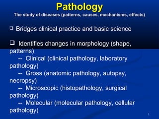
  • 1. 1 PathologyPathology The study of diseases (patterns, causes, mechanisms, effects)The study of diseases (patterns, causes, mechanisms, effects)  Bridges clinical practice and basic science  Identifies changes in morphology (shape, patterns) -- Clinical (clinical pathology, laboratory pathology) -- Gross (anatomic pathology, autopsy, necropsy) -- Microscopic (histopathology, surgical pathology) -- Molecular (molecular pathology, cellular pathology)
  • 2. 2  Renders an histopathologic diagnosis  May require immunohistochemistry, molecular markers  Studies causes (etiology, pathoetiology, etiopathology)  -- Genetics  -- Environment (including iatrogenic)  -- Idiopathic (“idiot pathologists”?!)  Studies mechanisms of disease development (pathogenesis)  Studies progression of disease (pathophysiology)
  • 3. 3 Causes of Cell InjuryCauses of Cell Injury  Physical agents -- Mechanical trauma -- Temperature -- Radiation -- Electric shock -- Changes in atmospheric pressure  Chemical agents  Infectious agents  Inflammation  Nutritional imbalances -- Starvation -- Obesity -- Vitamin deficiency
  • 4. 4 Causes of Cell InjuryCauses of Cell Injury  Hypoxia (oxygen deficiency) -- Pneumonia -- Anemia -- Carbon monoxide (CO) poisoning -- Ischemia (poor blood flow) -- Infarction (loss of blood supply)  Immunologic reactions -- Allergy -- Anaphylaxis -- Autoimmune diseases (self-allergy, loss of tolerance)  Genetic defects (DNA alterations) -- Congenital malformations -- Inborn errors of metabolism  Aging -- Degeneration -- Cellular senescence
  • 5. 5 Biochemical Mechanisms of InjuryBiochemical Mechanisms of Injury  ATP depletion via: a- mitochondrial oxidative phosphorylation b- anaerobic glycolysis  Oxygen deprivation (ischemia, infarction)  Oxygen damage -- partially reduced O2 >> free radicals  Loss of calcium homeostasis  Defects in cell membrane permeability  Mitochondrial damage The most vulnerable intracellular systems:The most vulnerable intracellular systems:  Cell membrane integrity  mitochondrial aerobic respiration  Protein synthesis  Gene integrity
  • 6. Free Radical-Induced Cellular InjuryFree Radical-Induced Cellular Injury Highly reactive, unstable species, interact with proteins, lipid,Highly reactive, unstable species, interact with proteins, lipid, carbohydrates causing cellular injury.carbohydrates causing cellular injury. Generation of free radicalsGeneration of free radicals 1- Absorption of radiant energy (ultraviolet light & x-rays): H2O1- Absorption of radiant energy (ultraviolet light & x-rays): H2O OH* & H*OH* & H* 2- Enzymatic metabolism of exogenous chemicals or drugs: CCL42- Enzymatic metabolism of exogenous chemicals or drugs: CCL4 CCL3CCL3 3- Reduction-oxidation reaction during normal metabolic processes: O23- Reduction-oxidation reaction during normal metabolic processes: O2-- ,, H2O2, OH*H2O2, OH* 4- Transition metals (Iron & Copper), Fenton reaction, superoxide& iron4- Transition metals (Iron & Copper), Fenton reaction, superoxide& iron  maximal oxidative cellular damagemaximal oxidative cellular damage 5- Nitric oxide.5- Nitric oxide.
  • 7. 7 Free Radicals in InjuryFree Radicals in Injury Generation, Injury and Neutralization by AntioxidantsGeneration, Injury and Neutralization by Antioxidants GSSG = oxidized glutathione; NADPH = reduced nicotinamide adenine dinucleotide phosphate; NO = nitric oxide Major injuries:  Lipid peroxidation of membranes yield peroxides and begin an autocatalytic chain reaction  Single-strand DNA breaks (thymine)  Cross-linking of proteins (sulfhydryl-mediated) enhances degradation rate and loss of enzyme activity Antioxidants:  Superoxide dismutase (SOD)  Glutathione (GSH) peroxidase  Catalase (in peroxisomes)  Vitamins E, A, C and beta-carotene  Free ionized iron and copper
  • 8. Removal of Free RadicalsRemoval of Free Radicals •Decay: SuperoxideDecay: Superoxide O2 & H2O2O2 & H2O2 • Inactivation:Inactivation: • Antioxidants: lipid soluble vitamins, ascorbic acidAntioxidants: lipid soluble vitamins, ascorbic acid glutathionglutathion block initiation of FR, inhibition, terminationblock initiation of FR, inhibition, termination of radical damage.of radical damage. • Binding of storage or transport proteins.Binding of storage or transport proteins. • Enzymes acting as a free radical-Scavenging system:Enzymes acting as a free radical-Scavenging system: Catalase, Superoxide dismutases, GlutathioneCatalase, Superoxide dismutases, Glutathione peroxidases.peroxidases.
  • 9. Chemical InjuryChemical Injury Mechanisms of chemical injury:Mechanisms of chemical injury: I- DirectI- Direct:: binding to some critical molecular component: mercury of mercuricbinding to some critical molecular component: mercury of mercuric chloride+SH group of cell membranechloride+SH group of cell membrane Increase permeability and inhibition of ATP-Increase permeability and inhibition of ATP- ase dependent transportase dependent transport II- Indirect:II- Indirect: conversion to reactive toxic metabolitesconversion to reactive toxic metabolites cell injury by directcell injury by direct covalent binding to membrane ptns and lipids or formation by reactive free radicalscovalent binding to membrane ptns and lipids or formation by reactive free radicals (CCL4, actetaminphen.(CCL4, actetaminphen. CCL4:CCL4: dry cleaning, CCL3 in SER of liver, initiate lipid peroxidation and autocatalyticdry cleaning, CCL3 in SER of liver, initiate lipid peroxidation and autocatalytic reactionreaction Swelling and breakdown of ER, dissociation of ribosomes, decrease hepaticSwelling and breakdown of ER, dissociation of ribosomes, decrease hepatic ptn synthesis (e.g. apoprotein), reduced lipid transportptn synthesis (e.g. apoprotein), reduced lipid transportfatty change, progressivefatty change, progressive heptocyte swelling, plasma membrane damage, deathheptocyte swelling, plasma membrane damage, death AcetaminophenAcetaminophen: Analgesic, metabolized by liver, toxic metabolites inactivated by: Analgesic, metabolized by liver, toxic metabolites inactivated by GSH, large dosesGSH, large doses acc. of metabolites due to GSH depletionacc. of metabolites due to GSH depletion binding to ptns &binding to ptns & nucleic acids-nucleic acids- increase drug toxicity & massive liver damageincrease drug toxicity & massive liver damage
  • 10. 10 Ischemia/Reperfusion InjuryIschemia/Reperfusion Injury Significant in myocardial and cerebral infarctionsSignificant in myocardial and cerebral infarctions  Restoration of blood flow brings concentrated calcium when cells aren’t healthy enough to regulate it  Recruits inflammatory cells, which release many free radicals -- membrane damage -- mitochondrial permeability transition  Damaged mitochondria cannot reduce oxygen well -- free radicals are produced  Compromised antioxidant defense mechanisms Problem?Problem? Vasoconstrictors in local anesthetics produce ischemia and reperfusion each time they are used.
  • 11. 11 MitochondrialMitochondrial DysfunctionDysfunction with Injurywith Injury  Are targets of most injuries  Nonselective pores allow protons out >> prevents ATP generation  Cytochrome c (electron transport protein) leaks out >> activates apoptotic death Apoptosis = programmed cell death
  • 12. 12 Reversible IschemicReversible Ischemic InjuryInjury Sequence of EventsSequence of Events Accumulation of inorganic phosphates, lactic acid (hydrolysis of phosphate esters)  Ribosomes detach from rough endoplasmic reticulum  Polysomes dissociate into monosomes
  • 13. 13 Increased Cytosolic Calcium in Cell InjuryIncreased Cytosolic Calcium in Cell Injury  Usually 10,000x lower than extracellular calcium  ATP-dependent  Ischemic/toxins >> influx of calcium across membrane  Activates phospholipases >> membrane damage  Activates proteases >> structural and membrane proteins catabolized  Activates endonucleases >> chromosomes fragment  May be irreversible  May kill the cell
  • 14. 14 Membrane DamageMembrane Damage If severe enough, may stimulate external attackIf severe enough, may stimulate external attack
  • 15. 15 Cell Responses to InjuryCell Responses to Injury  The cell adapts (reversible damage)  The cell is injured and heals (reversible damage)  The cell is injured and remains injured (atypical adaptation; usually irreversible damage)  The cell dies (irreversible damage) These are tempered by:These are tempered by:  Genetic factors  Immune factors  Environmental factors  Strength of the outside attack  Duration of the outside attack
  • 16. 16 Adapted Cell + Stress Injury Normal cell Reversibly injured cell Irreversibly Injured cell Dead cell +Stress Apoptosis Necrosis - Stress - Stress Overview
  • 17. 17 Reversible vs irreversible cell injury Reversible injury * Decreased ATP levels * Ion imbalance * Swelling * Decreased pH * Fatty change (liver) Irreversible injury * Amorphous densities in mitochondria * Severe membrane damage * Lysosomal rupture Extensive DNA damage
  • 18. 18  Reversible cell injuryReversible cell injury ((degeneration)degeneration) casedcased by mild injury of short duration andby mild injury of short duration and includes:includes: 1 Cell or Cloudy Swelling. Then can proceed to 2- Hydropic or Vacular Degeneration
  • 19. 19 Cell Swelling or HydropicCell Swelling or Hydropic DegenerationDegeneration  Characterized by:-Characterized by:- Swelling of cells -Swelling of cells - Granulation of cytoplasm.Granulation of cytoplasm. ** Organs affectedOrgans affected:: parenchymatous organsparenchymatous organs ** Microscopic appearanceMicroscopic appearance:: • Cytoplasm is fine red and granularCytoplasm is fine red and granular • Vacuolation of cytoplasm.Vacuolation of cytoplasm.  Nuclei are normalNuclei are normal Fate:Fate: ReversibleReversible
  • 20. 20 Reversible Ischemic InjuryReversible Ischemic Injury Microscopic ChangesMicroscopic Changes Accumulation of inorganic phosphates, lactic acid (hydrolysis of phosphate esters) Hydropic DegenerationHydropic Degeneration Cellular swelling in alcohol liver damageCellular swelling in alcohol liver damage Summary:Summary:  Swollen mitochondria  Swollen endoplasmic reticulum  Swollen cell (poor Na+ pump)  Release of ribosomes from rough ER  Mitochondrial densities  Membrane blebs (poor structure)  Autophagic vacuoles accumulate  Nuclear clumping
  • 22. 22 Subcellular response to injurySubcellular response to injury  Lysosomes (heterophagy; autophagy)  Smooth ER (induction)  Mitochondria (D number, size and shape)  Cytoskeleton (D phagocytosis, locomotion)  Nucleus (karyolysis, karyorrhexis, pyknosis)  Membranes (cellular and subcellullar)
  • 23. 23 Subcellular Response to InjurySubcellular Response to Injury  Lyzosomal CatabolismLyzosomal Catabolism::  Primary lyzosomes are membrane- bound intracellularPrimary lyzosomes are membrane- bound intracellular organells containing a variety of hydrolytic enzymes.organells containing a variety of hydrolytic enzymes.  These fuse with vacuoles containing materials neededThese fuse with vacuoles containing materials needed for digestion.for digestion.  They form Secondary lyzosomes or phagolyzosomes.They form Secondary lyzosomes or phagolyzosomes.  Lyzosomes are involved in the breakdown of ingestedLyzosomes are involved in the breakdown of ingested materials in one of two ways:materials in one of two ways: HeterophagyHeterophagy oror AutophagyAutophagy
  • 24. 24 Autophagy v. HeterophagyAutophagy v. Heterophagy DegeneratingDegenerating mitochondria withmitochondria with amorphousamorphous materialmaterial Autophagy:Autophagy:  Intracellular organelles and cytosol are sequestered (autophagic vacuoles)  In area of smooth ER fuse with preexisting lyzosomes to form autophagolysosomes.  Especially common is cell differentiation and removal of damaged organells.  Lipids, especially, may not be digested  Undigested debris: exocytosis or residual bodies, i.e. lipofuscin pigment, carbon Heterophagy:Heterophagy:  Outside stuff undergoes: endocystosis or phagocytosis (large particles) or pinocytosis (Soluble macromolecules)  Endocysted vacuoles fuse with lysosomes  Ex: phagocyteso of bacteria by neutrophil and necrotic tissues by macrophages.
  • 25. 25 Autophagy and Cell AtrophyAutophagy and Cell Atrophy
  • 26. 26 Intracellular AccumulationsIntracellular Accumulations Reversible cellular adaptationReversible cellular adaptation  Lipids (TG and Cholesterol)Lipids (TG and Cholesterol)  ProteinsProteins  PigmentsPigments  Glycogen (genetic disease)Glycogen (genetic disease)
  • 27. 27 Intracellular Accumulations (ReversibleIntracellular Accumulations (Reversible Cellular Adaptaion)Cellular Adaptaion) Three mechanisms by which the cell can acquireThree mechanisms by which the cell can acquire intracellular accumulations:intracellular accumulations:  Too much of a normal endogenous substance -- usually decreased metabolism from cell damage, e.g. fatty liver  Genetic or acquired defect of metabolism (inborn error of metabolism) -- errors of metabolism, packaging, transport, secretions -- storage diseases, alpha1-antitrypsin deficiency (protein folding & transport)  Exogenous deposits accumulate in cell (can’t degrade or transport it) -- carbon (black lung disease) -- silica (silicosis)
  • 28. 28 Fatty Change (Liver)Fatty Change (Liver) Many ways to get to sublethal cell damageMany ways to get to sublethal cell damage -Fatty Change refersFatty Change refers to any abnormalto any abnormal accumulation ofaccumulation of triglycerides withintriglycerides within parenchymal cells.parenchymal cells. - It is reversibleIt is reversible process.process. -When mild: noWhen mild: no effect.effect. -Severe cases:Severe cases: impair the cellularimpair the cellular function transiently.function transiently. -Micro: ClearMicro: Clear cytoplasmiccytoplasmic vacuoles that needsvacuoles that needs specific stains.specific stains.
  • 29. 29 Intracellular AccumulationsIntracellular Accumulations . Fatty LiverFatty Liver Lipofuscin in Heart MuscleLipofuscin in Heart Muscle
  • 30. 30 Fatty ChangeFatty Change  Characterized byCharacterized by - Accumulation of excess neutral fat in the cells.Accumulation of excess neutral fat in the cells. ** Causes:Causes: ** Hypoxia * Toxins * Chemicals * In the liver may be due to:Hypoxia * Toxins * Chemicals * In the liver may be due to: a- Excess fat brought to liver b- Diseases of livera- Excess fat brought to liver b- Diseases of liver c- Deficiency of lipotropic factors.c- Deficiency of lipotropic factors. Gross pictureGross picture:: The affected organ is enlarged, soft,The affected organ is enlarged, soft, Microscopic picture:Cells are:Microscopic picture:Cells are:pale yellow, rounded borders,pale yellow, rounded borders, *Swollen * Cytoplasm show cut surface bulges and greesy.*Swollen * Cytoplasm show cut surface bulges and greesy. multiple tiny fat globulesmultiple tiny fat globules * Nucleus is compressed, flattened* Nucleus is compressed, flattened against cell membrane.against cell membrane.
  • 32. Cholesterol and Cholesterol EstersCholesterol and Cholesterol Esters •Synthesis of cell membranesSynthesis of cell membranes • Accumulations manifested by intracellular vacuolesAccumulations manifested by intracellular vacuoles • Atherosclerosis:Atherosclerosis: lipids accumulate in smooth muscle cellslipids accumulate in smooth muscle cells and in macrophages of the wall of arteries.and in macrophages of the wall of arteries. *Cells:*Cells: vacuolesvacuoles *Extracellular:*Extracellular: Rhomboid cleft-like cavitiesRhomboid cleft-like cavities •Hereditary hyperlipidemiaHereditary hyperlipidemia:: in macrophages and mesenchymal cells-in macrophages and mesenchymal cells- XanthomasXanthomas • Inflammation and necrosis:Inflammation and necrosis: phagocytosis of membrane lipids derivedphagocytosis of membrane lipids derived from injured cellsfrom injured cells Foamy macrophagesFoamy macrophages
  • 33. 33 Cholesterolosis. Cholesterol-laden macrophages (foam cells) from a focus of gallbladder cholesterolosis (arrow).
  • 34. Intracellular Protein Accumulation •Excessive synthesis or Absorption or Defect in cellular transportExcessive synthesis or Absorption or Defect in cellular transport • Rounded eosinophilic droplets or masses in the cytoplasmRounded eosinophilic droplets or masses in the cytoplasm •Excessive AbsorptionExcessive Absorption: Protracted proteinuria: Protracted proteinuria •Excessive synthesisExcessive synthesis:: Russell bodiesRussell bodies •Defective intracellular transport and secretion of critical proteins:Defective intracellular transport and secretion of critical proteins: Alpha-1-Alpha-1- antitrypsin deficiency & Cystic fibrosisantitrypsin deficiency & Cystic fibrosis • Toxicity of aggregated abnormally folded protein:Toxicity of aggregated abnormally folded protein: NeurodegenerativeNeurodegenerative disorders (Alzheimer dis; microtubule-associated ptn & neurofilament) &disorders (Alzheimer dis; microtubule-associated ptn & neurofilament) & amylodosisamylodosis • Resistance of degradationResistance of degradation: Mallory body (alcoholic hyalin;: Mallory body (alcoholic hyalin; prekeratinprekeratin intermediate filamentsintermediate filaments
  • 35. 35 Intracellular ProteinIntracellular Protein AccumulationAccumulation Kidney (top), liver (bottom) Much less common than lipid accumulations. Kidney: albumin in pinocytic vesicles -- vesicles fuse with lysosomes -- result: hyalin droplets in cells -- nephrotic syndrome; proteinuria -- is reversible Liver: alcohol hyalin (Mallory bodies) -- intermediate filaments aggregate -- chronic alcohol abuse Plasma cells: Russell bodies -- immunoglobulins in RER
  • 36. GlycogenGlycogen •Abnormalities in the metabolism of glucose or glycogenAbnormalities in the metabolism of glucose or glycogen • Diabetes Mellitus:Diabetes Mellitus: Glycogen accumulation in renal tubularGlycogen accumulation in renal tubular epithelium, cardiac myocytes, beta cells of pancreasepithelium, cardiac myocytes, beta cells of pancreas • Enzyme defects:Enzyme defects: synthesis or breakdown of glycogensynthesis or breakdown of glycogen Glycogen storage diseaseGlycogen storage disease
  • 37. Pigments (Exogenous or Endogenous)Pigments (Exogenous or Endogenous) •Exogenous pigments:Exogenous pigments: •AnthracosisAnthracosis: Acc. of carbon in the macrophages of: Acc. of carbon in the macrophages of lungs and Lymphlungs and Lymph nodes Heavy acc.nodes Heavy acc. emphysema or coal workers pneumoconiosis.emphysema or coal workers pneumoconiosis. most common exogenous pigment (anthracosis) -- smoking, coal mining, urban living -- alveolar macrophages take it o tracheobronchial lymph nodes -- may induce emphysema and coal miner’s pneumoconiosis • Tattooing:Tattooing: Macrophages, extracellularly (persist for life)Macrophages, extracellularly (persist for life) • Endogenous pigments:Endogenous pigments: •Lipofuscin, melanine, certain hemoglobin derivativesLipofuscin, melanine, certain hemoglobin derivatives •Lipofuscin (the wear-and tear pigment):Lipofuscin (the wear-and tear pigment): Intra- cytoplasmic yellowbrownIntra- cytoplasmic yellowbrown fine pigment (Brown atrophy) lipid& phospholipids & proteins (peroxidationfine pigment (Brown atrophy) lipid& phospholipids & proteins (peroxidation of polyunsaturated lipids of cellular membraneof polyunsaturated lipids of cellular membrane)) • MelanineMelanine:: Non-hemoglobin-derived brown- black pigment formed byNon-hemoglobin-derived brown- black pigment formed by melanocytes by the oxidation of tyrosine.melanocytes by the oxidation of tyrosine.
  • 38. 38 Lipofuscin granules in a cardiac myocyte as shown by A, light microscopy (deposits indicated by arrows), and B, electron microscopy (note the perinuclear, intralysosomal location).
  • 39. • HemosiderinHemosiderin:: A hemoglobin-derived, golden-yellow to brownA hemoglobin-derived, golden-yellow to brown granular pigment composed of aggregates of ferritin micellesgranular pigment composed of aggregates of ferritin micelles Localized or systemicLocalized or systemic Local HemosidrosisLocal Hemosidrosis •Gross hemorrhage or rupture of small vessels (congestion)Gross hemorrhage or rupture of small vessels (congestion) • Lysosomal enzymes in macrophages convert hemoglobin toLysosomal enzymes in macrophages convert hemoglobin to hemosiderinhemosiderin Systemic HemosiderosisSystemic Hemosiderosis •Increased absorption of dietary iron (primary hemochromatosis)Increased absorption of dietary iron (primary hemochromatosis) • Impaired utilization of iron (Thalassemia)Impaired utilization of iron (Thalassemia) •Hemolytic anemiasHemolytic anemias •Repeated transfusionRepeated transfusion (exogenous load of iron)(exogenous load of iron)
  • 40. 40 Hemosiderin granules in liver cells. A, H&E section showing golden-brown, finely granular pigment. B, Prussian blue reaction, specific for iron.
  • 41. Pathologic CalcificationPathologic Calcification •Abnormal deposition of calcium salts in soft tissues • Dystrophic calcification: Non-viable or dying tissues, normal calcium serum level. (Atherosclerosis, Damaged heart valves, areas of coagulative, liquifactive or caseous necrosis). • Metastatic calcification: Vital tissue, hypercalcemia • Gross picture: fine, white granules or clumps, gritty deposits. • Microscopic picture: intracellular or extracellular basophilic deposits, by time heterotropic bone formation.
  • 42. Pathogenesis of Dystrophic Pathological CalcificationPathogenesis of Dystrophic Pathological Calcification •InitiationInitiation:: Extracellularly or intracellularlyExtracellularly or intracellularly •Extracellular initiation: membrane- bound vesicles derived from dead orExtracellular initiation: membrane- bound vesicles derived from dead or dying cells that concentrate calcium by their affinity for acidicdying cells that concentrate calcium by their affinity for acidic phospholipids.phospholipids. •Phosphates accumulation by the action of membrane boundPhosphates accumulation by the action of membrane bound phosphatases.phosphatases. • The cycle of calcium and phosphate binding is repeated-The cycle of calcium and phosphate binding is repeated- depositsdeposits • Initiation of intracellular calcification occurs in mitochondria of dead orInitiation of intracellular calcification occurs in mitochondria of dead or dying tissue.dying tissue. •Propagation of crystal formation:Propagation of crystal formation: depends on conc. of calcium anddepends on conc. of calcium and phosphates, the presence of inhibitors, structural components of extra-phosphates, the presence of inhibitors, structural components of extra- cellular matrix (collagen) as well as other matrix protein (osteonectin andcellular matrix (collagen) as well as other matrix protein (osteonectin and others).others).
  • 43. 43 Calcific Valves in Aortic StenosisCalcific Valves in Aortic Stenosis Thick, fibrotic cusps; masses of dystrophic calcificationThick, fibrotic cusps; masses of dystrophic calcification
  • 44. 44 Chronic Calcific PulpitisChronic Calcific Pulpitis Chronic Ischemic Pulpitis?Chronic Ischemic Pulpitis? DystrophicDystrophic CalcificationCalcification
  • 45. Causes of Metastatic CalcificationCauses of Metastatic Calcification (Hypercalcemia)(Hypercalcemia) A: Excessive mobilization of calcium from bone:A: Excessive mobilization of calcium from bone: 1- Hyperparathyroidism (primary or secondary).1- Hyperparathyroidism (primary or secondary). 2- Bony destructive lesions such as myelomas and metastatic carcinomas.2- Bony destructive lesions such as myelomas and metastatic carcinomas. 3- Disuse atrophy of bones.3- Disuse atrophy of bones. B- Excessive absorption of calcium from the gut:B- Excessive absorption of calcium from the gut: 1- Hypervitaminosis D.1- Hypervitaminosis D. 2- Excessive oral calcium intake.2- Excessive oral calcium intake. 3- Hypercalcemia of infancy.3- Hypercalcemia of infancy. Metastatic calcification affects Kidney (basement membrane of tubules),Metastatic calcification affects Kidney (basement membrane of tubules), Alveolar wall of lungs, Stomach (fundal glands0, Blood vessels especiallyAlveolar wall of lungs, Stomach (fundal glands0, Blood vessels especially internal elastic lamina and cornea.internal elastic lamina and cornea. It is based on that sites have relatively alkaline pH which favorsIt is based on that sites have relatively alkaline pH which favors precipitation of calcium.precipitation of calcium.
  • 46. 46 Metastatic CalcificationMetastatic Calcification Calcification in the wall of a blood vessel Calcification in the gastric submucosa
  • 47. Cellular Adaptation to InjuryCellular Adaptation to Injury A state that lies between the normal, unstressed cell andA state that lies between the normal, unstressed cell and the injured over stressed cell.the injured over stressed cell. Mechanisms:Mechanisms: •Up- or down-regulation of specific cellular receptorsUp- or down-regulation of specific cellular receptors • Induction of new protein synthesis : heat shock proteinInduction of new protein synthesis : heat shock protein (protection)(protection) •Switch from one type of protein to anotherSwitch from one type of protein to another • marked over-production of one type of protein (collagen)marked over-production of one type of protein (collagen) Atrophy, Hypertrophy, Hyperplasia, MetaplasiaAtrophy, Hypertrophy, Hyperplasia, Metaplasia
  • 48. 48 Reversible Cell AdaptationReversible Cell Adaptation AdaptationAdaptation ResultResult AtrophyAtrophy Decrease in cell sizeDecrease in cell size HypertrophyHypertrophy Increased cell size of an organ due to increase in theIncreased cell size of an organ due to increase in the size of cells.size of cells. HyperplasiaHyperplasia Increased cell size of an organ due to increase in theIncreased cell size of an organ due to increase in the number of cells.number of cells. MetaplasiaMetaplasia Stable change from one cell type to another cell typeStable change from one cell type to another cell type DysplasiaDysplasia Abnormal cell or tissue growthAbnormal cell or tissue growth
  • 49. 49 Cellular adaptations to stress: 1. Hyperplasia (more cells) 2. Hypertrophy (bigger cells) 3. Atrophy (smaller cells) 4. Metaplasia (different type of cells)
  • 50. HypertrophyHypertrophy An increase in the number of organelles and the size ofAn increase in the number of organelles and the size of the cells with subsequent increase in the size of organ duethe cells with subsequent increase in the size of organ due to an increase in the functional demands.to an increase in the functional demands. Types:Types: •physiologicalphysiological:: Uterus in pregnancy, muscle in athletesUterus in pregnancy, muscle in athletes •PathologicalPathological:: •Adaptive:Adaptive: Hollow muscular organs above a chronic partialHollow muscular organs above a chronic partial obstruction (Heart, stomach, intestine, bladder)obstruction (Heart, stomach, intestine, bladder) •Compensatory:Compensatory: Paired organ (kidney, lung)Paired organ (kidney, lung)
  • 51. 51 Hypertrophy * Larger cells * Not due to swelling * Increased synthesis of structural components * Results in larger organ * May occur with hyperplasia
  • 52. 52 Mechanism of HypetrophyMechanism of Hypetrophy •Increased synthesis of structural proteins via: •Transcription factors (i. e. c-fos and c-jun) •Growth factors (TGF-β, IGF-1, FGF) •Vasoactive agents (endothelien-1, AII)
  • 53. 53 HypertrophyHypertrophy Physiologic hypertrophy ofPhysiologic hypertrophy of myometrium during pregnancy due tomyometrium during pregnancy due to estrogen stimulationestrogen stimulation Physiologic hypertrophy of skeletalPhysiologic hypertrophy of skeletal musclemuscle from exercisefrom exercise Normal muscleNormal muscle
  • 54. 54 HypertrophyHypertrophy Enlargement of cardiac muscle in response to valve diseaseEnlargement of cardiac muscle in response to valve disease Normal HeartNormal Heart Hypertrophied Left VentricleHypertrophied Left Ventricle Normal Heart MuscleNormal Heart Muscle Hypertrophied Heart MuscleHypertrophied Heart Muscle
  • 56. 56 HyperplasiaHyperplasia  It is an increase in the number of cells in anIt is an increase in the number of cells in an organ or tissue.organ or tissue.  Physiologic Types:Physiologic Types: 1- Hormonal as in breast due to puberty and1- Hormonal as in breast due to puberty and pregnancypregnancy 2- Compensatory occurs when a portion of the2- Compensatory occurs when a portion of the tissue is removed or diseased.tissue is removed or diseased.  PathologicPathologic:: • Compensatory hyperplasia: Bone marrow, liver cirrhosisCompensatory hyperplasia: Bone marrow, liver cirrhosis • Hormonal hyperplasia: thyroid, breast, endometriumHormonal hyperplasia: thyroid, breast, endometrium • Irritation hyperplasia: lymphoid tissue in infection and toxemiaIrritation hyperplasia: lymphoid tissue in infection and toxemia
  • 57. Mechanism of hyperplasiaMechanism of hyperplasia Cell proliferation via increased production of TRANSCRIPTION FACTORS due to: * Increased production of GF * Increased levels of GF receptors * Activation of intracellular signaling Results in larger organ
  • 58. 58 HyperplasiaHyperplasia Endometrial hyperplasia in response to estrogenEndometrial hyperplasia in response to estrogen Hyperplastic GlandsHyperplastic Glands
  • 59. 59 HyperplasiaHyperplasia Nodular hyperplasia of the prostate glandNodular hyperplasia of the prostate gland NormalNormal
  • 60. 60 Thyroid hyperplasiaThyroid hyperplasia Normal ThyroidNormal Thyroid Hyperplastic ThyroidHyperplastic Thyroid
  • 61. Metaplasia A reversible change in which one differentiated adult cellA reversible change in which one differentiated adult cell type is replaced by another (epithelial or mesenchymal)type is replaced by another (epithelial or mesenchymal) of the same type (during postnatal life)of the same type (during postnatal life) - Columnar to squamous epithelium (most common epithelial type of metaplasia) - Chronic irritation i.e. (in trachea and bronchi of smokers) - Vit A deficiency squamous metaplasia in respirastory epithelium - May be some loss of function and predispose to malignancy
  • 62. Mechanism of MetaplasiaMechanism of Metaplasia ReprogrammingReprogramming 1. of stem cells present in normal tissues1. of stem cells present in normal tissues 2. of undifferentiated mesenchymal cells2. of undifferentiated mesenchymal cells in connective tissuein connective tissue Mediated by signals from:Mediated by signals from: cytokines, GF or ECMcytokines, GF or ECM Leading to induction ofLeading to induction of specific transcriptionspecific transcription factorsfactors
  • 63. 63 MetaplasiaMetaplasia Original TissueOriginal Tissue StimulusStimulus Metaplastic TissueMetaplastic Tissue Ciliated columnarCiliated columnar epithelium of bronchialepithelium of bronchial treetree Cigarette smokeCigarette smoke Squamous epitheliumSquamous epithelium Transitional epitheliumTransitional epithelium of bladderof bladder Trauma of bladderTrauma of bladder calculuscalculus Squamous epitheliumSquamous epithelium Columnar epithelium ofColumnar epithelium of gland ductsgland ducts Trauma of calculusTrauma of calculus Squamous epitheliumSquamous epithelium Esophageal squamousEsophageal squamous epitheliumepithelium Gastric acidGastric acid Columnar epitheliumColumnar epithelium Fibrocollagenous tissueFibrocollagenous tissue Chronic traumaChronic trauma Bone (osseous) tissueBone (osseous) tissue Columnar glandularColumnar glandular epitheliumepithelium Vitamin A deficiencyVitamin A deficiency Squamous epitheliumSquamous epithelium
  • 64. 64 Epithelial Metaplasia, the normal respiratory epithelium at the right and the squamous epithelium at the left .
  • 65. 65 Photomicrograph of the junction of normal epithelium (1) with metaplastic transitional epithelium (2).
  • 66. 66 Photomicrograph of the trachea from a smoker. Note that the columnar ciliated epithelium has been replaced by squamous epithelium.
  • 67. 67 This biopsy of the lower esophagus in a patient with chronic gastroesophageal reflux disease (GERD) shows columnar metaplasia (Barrett's esophagus), and the goblet cells are typical of an intestinal type of epithelium. Squamous epithelium typical of the normal esophagus appears at the right.
  • 68. 68 AtrophyAtrophy IIt is the shrinkage in the size of the cell by loss of cellt is the shrinkage in the size of the cell by loss of cell substancesubstance ((Balance between synthesis and degradation)Balance between synthesis and degradation)  Decreased synthesis and Increased catabolism  Influenced by: -- insulin -- thyroid-stimulating hormone -- glucocorticoids  Two systems for regulation of protein degradation ☺☺ -- lysosomal proteases and other enzymes degrade endocytosed molecules -- ubiquitin-proteasome pathway, primarily for cytosolic and nuclear proteins (senescent organelles)  Often accompanied by autophagic vacuoles -- some debris resists digestion: membrane-bound residual bodies
  • 69. 69 Types/Causes of AtrophyTypes/Causes of Atrophy Physiologic v. PathologicPhysiologic v. Pathologic  Hormone-related atrophy -- Ablation of pituitary gland>>less ACTH>>adrenal cortex atrophy -- Endometrial atrophy during menopause -- Myometrial atrophy post partum -- Thymus atrophy during adolescence -- Parathyroid atrophy with increasing age -- Old age>>reduced gonadotrophins>>testicular atrophy  Disuse atrophy -- Leg in cast -- Long-term hospitalization  Ischemic atrophy -- Brain atrophy after stroke  Denervation atrophy -- Spinal cord injury  Nutritional atrophy -- Mucosal atrophy in pernicious anemia (vitamin B12 deficiency)
  • 70. 70 Mechanism of Atrophy Reduction in structural components Decreased number of mitochondria, myofilaments, ER via proteolysis (lysosomal proteases; ubiquitin-proteosome system) Increase in number of autophagic vacuoles Residual bodies (i.e. lipofuscin  brown atrophy) NB: diminished function but not dead
  • 71. 71 Muscle fiber atrophy. The number of cells is the same as before the atrophy occurred, but the size of some fibers is reduced. This is a response to injury by "downsizing" to conserve the cell. In this case, innervation of the small fibers in the center was lost. This is a trichrome stain.
  • 72. 72 AtrophyAtrophy Atrophic Adrenal Gland from Corticosteroid UseAtrophic Adrenal Gland from Corticosteroid Use NormalNormal AtrophiedAtrophied
  • 73. 73 Brain AtrophyBrain Atrophy An aging processAn aging process 82 y/o male82 y/o male 25 y/o male25 y/o male
  • 74. 74
  • 75. 75 DysplasiaDysplasia Dysplasia meansDysplasia means abnormal organization of cellsabnormal organization of cells  At the cellular level, morphologically, it is characterized byAt the cellular level, morphologically, it is characterized by variations in size and shape of the cell (pleomorphism),variations in size and shape of the cell (pleomorphism), disorderly arrangement within the epithelium (loss of polarity)disorderly arrangement within the epithelium (loss of polarity) and nuclear changes, consisting of enlargement, irregularand nuclear changes, consisting of enlargement, irregular borders, and hyperchromasia of individual cell nuclei andborders, and hyperchromasia of individual cell nuclei and increased number of mitotic figures.increased number of mitotic figures.  It is considered pre-malignant (often arises in previouslyIt is considered pre-malignant (often arises in previously metaplastic epithelium), and can progress to malignantmetaplastic epithelium), and can progress to malignant squamous cell carcinoma, unless treated.squamous cell carcinoma, unless treated.  Basement membrane is always intactBasement membrane is always intact  In early stages it is reversibleIn early stages it is reversible
  • 76. 76 HISTOLOGICAL CRITERIA FOR DYSPLASIA  Pleomorphism of both cells & nuclei.  Increased nuclear cytoplasmic ratio.  Hyperchromatic or vesicular nuclei with prominence of nucleoli  Increased and specially presence of atypical mitotic figures  Loss of polarity   INTACT BASEMENT MEMBRANE
  • 77. 77  MILD DYSPLASIA  (INTRAEPITHELIAL NEOPLASIA-GRADE I)  These changes are confined to lower 1/3 of the thickness of the epithelium.  MODERATE DYSPLASIA  (INTRAEPITHELIAL NEOPLASIA-GRADE II)  These changes are confined to lower 1/2 of the thickness of the epithelium.  SEVERE DYSPLASIA  (INTRAEPITHELIAL NEOPLASIA-GRADE III)  These changes are confined to lower 2/3 of the thickness of the epithelium.   CARCINOMA IN SITU  (INTRAEPITHELIAL NEOPLASIA-GRADE III)  These changes involve the entire thickness of the epithelium.
  • 78. 78
  • 79. 79 At high magnification, the normal cervical squamous epithelium at the left merges into the dysplastic squamous epithelium at the right in which the cells are more disorderly and have darker nuclei with more irregular outlines.
  • 81. 81 •Irreversible cell injury caused by severe injury of long duration and includes: 1- Necrosis 2- Apoptosis
  • 82. 82 Sublethal v. Lethal Cell DamageSublethal v. Lethal Cell Damage
  • 83. 83 Nuclear Events in NecrosisNuclear Events in Necrosis Pyknosis, Karyorrhexis, KaryolysisPyknosis, Karyorrhexis, Karyolysis 1- Karyopyknosis means shrinkage and increased basophilia of the nucleus. 2- Karyorrhexis means fragmentation of the nucleus. 3- Karyolysis means fading of the nucleus KaryolysisKaryolysis KaryorrhexisKaryorrhexis NormalNormal
  • 84. 84 Proteins Liberated into BloodProteins Liberated into Blood Following NecrosisFollowing Necrosis Released enzymes can help with diagnosisReleased enzymes can help with diagnosis Cell DamagedCell Damaged Enzyme ReleasedEnzyme Released Cardiac muscle Creatine kinase (MB isoform) Aspartate transaminase (AST) Lactate dehydrogenase (LDH-1) Hepatocyte Alanine transaminase (ALT) Striated muscle Creatine kinase (MM isoform) Exocrine pancreas Amylase CytoplasmicCytoplasmic Changes inChanges in NecrosisNecrosis --Dead cells showDead cells show increased acidophilia.increased acidophilia. -Cells may have glassyCells may have glassy appearance.appearance. - Cytoplasm becomeCytoplasm become vacuolated and appearvacuolated and appear moth- eaten.moth- eaten. - Calcification of the deadCalcification of the dead cells may occur.cells may occur.
  • 85. 85 Mechanisms of Irreversible Injury (NECROSIS)Mechanisms of Irreversible Injury (NECROSIS) Two main characteristics of irreversible damage:Two main characteristics of irreversible damage:  Inability to reverse mitochondrial dysfunction  Profound membrane disturbances Cell membrane damage is the single most important problem:Cell membrane damage is the single most important problem:  Loss of membrane phospholipids  Lipid breakdown products are catabolic -- also: detergent effect on membranes  Cytoskeletal abnormalities -- from activate proteases (>Ca++ in cell) -- detachment of cell membrane from cytoskeleton  Toxic oxygen radicals cause additional damage and recruit leukocytes ☺☺
  • 86. 86 Types of NecrosisTypes of Necrosis  Coagulative necrosis: denatured proteins -- most common type -- cell outlines remain Gangrenous necrosis (form of ischemic coagulative necrosis) -- wet and dry (superimposed liquefactive necrosis) -- gas necrosis (Clostridium perfringens)  Liquefactive necrosis: enzymatic digestion (proteolysis) -- focal bacterial and some fungal infection (attract neutrophils) -- brain death  Caseous necrosis: tuberculosis (“cheesy”) -- granulomatous inflammation (granulomas) -- loss of architecture centrally (structureless, amorphus granular debris).  Fat necrosis (liquefied fat, released fatty acids) -- acute pancreatitis -- trauma/ischemia to fatty tissue - calcific fat necrosis --Shadowy outlines of necrotic fat cells with basophilic Ca and inflam. Cells. ☺☺
  • 87. 87 Coagulative necrosisCoagulative necrosis  Preservation of structurePreservation of structure  FirmFirm  Protein denaturationProtein denaturation  Hypoxic tissue death (except brain)Hypoxic tissue death (except brain)
  • 88. 88 This is an example of coagulative necrosis. This is the typical pattern with ischemia and infarction (loss of blood supply and resultant tissue anoxia). Here, there is a wedge-shaped pale area of coagulative necrosis (infarction) in the renal cortex of the kidney.
  • 90. 90 Caseous necrosisCaseous necrosis  Subset of coagulative necrosisSubset of coagulative necrosis  TBTB  Cheesy, whiteCheesy, white  Surrounded by inflammatory cellsSurrounded by inflammatory cells (granulomatous reaction)(granulomatous reaction)  Complete destruction of tissueComplete destruction of tissue
  • 92. 92 Caseous NecrosisCaseous Necrosis Tuberculosis of Lung, LiverTuberculosis of Lung, Liver GranulomaGranuloma
  • 93. 93 Liquefactive necrosisLiquefactive necrosis  Enzymatic digestionEnzymatic digestion  Liquid, viscous massLiquid, viscous mass  May contain pusMay contain pus  Bacterial infections (via inflammation)Bacterial infections (via inflammation)  Hypoxic brain injuryHypoxic brain injury
  • 94. 94 MeningitisMeningitis Liquefactive Necrosis of Central Nervous SystemLiquefactive Necrosis of Central Nervous System Loss of cell outlinesLoss of cell outlines
  • 96. 96 The liver shows a small abscess here filled with many neutrophils. This abscess is an example of localized liquefactive necrosis
  • 97. 97 Fat necrosisFat necrosis  Not a specific patternNot a specific pattern  Focal areas of fat digestionFocal areas of fat digestion  Ususally via release of lipases from pancreasUsusally via release of lipases from pancreas  FFA combine with Ca to produce “soaps”FFA combine with Ca to produce “soaps”
  • 98. 98 This is fat necrosis of the pancreas. Cellular injury to the pancreatic acini leads to release of powerful enzymes which damage fat by the production of soaps, and these appear grossly as the soft, chalky white areas seen here on the cut surfaces.
  • 100. GangreneGangrene DefDef: Gangrene is massive tissue necrosis followed by putrefaction: Gangrene is massive tissue necrosis followed by putrefaction Causes:Causes: 1- Necrosis1- Necrosis:: sudden ischemia or bacterial toxinssudden ischemia or bacterial toxins 2- Putrefaction2- Putrefaction:: Saprophytic bacteria that breaks down the protein ofSaprophytic bacteria that breaks down the protein of necrotic tissuenecrotic tissue liberation of hydrogen sulphide (H2S)liberation of hydrogen sulphide (H2S) foul odourfoul odour,, H2S + iron (derived from hemoglobinH2S + iron (derived from hemoglobin  iron sulphideiron sulphide  staining ofstaining of gangrenous tissue black.gangrenous tissue black. Types of GangreneTypes of Gangrene 1- Dry gangrene1- Dry gangrene 2- Moist gangrene2- Moist gangrene 3- Infective gangrene3- Infective gangrene 4- Gas gangrene4- Gas gangrene
  • 101. Classification of gangreneClassification of gangrene According to the amount of blood and tissue fluids in theAccording to the amount of blood and tissue fluids in the part affected at the time of its deathpart affected at the time of its death I- Dry gangreneI- Dry gangrene II- Moist gangreneII- Moist gangrene I- Dry GangreneI- Dry Gangrene •Dry gangrene of limb results from occlusion of its artery by thrombus,Dry gangrene of limb results from occlusion of its artery by thrombus, embolus, thromboangitis obliterans (Buerger’s disease), Ergot poisoningembolus, thromboangitis obliterans (Buerger’s disease), Ergot poisoning and Raynaud’s diseaseand Raynaud’s disease (spastic occlusion(spastic occlusion), surgical ligature.), surgical ligature. • Main arterial supply is cut off + poor collateral circulation= gangreneMain arterial supply is cut off + poor collateral circulation= gangrene • Artery (occluded) + venous and lymphatic drainage (open)+ surfaceArtery (occluded) + venous and lymphatic drainage (open)+ surface evaporationevaporation dry gangrenedry gangrene • Commonest example of dry gangrene:Commonest example of dry gangrene: Senile gangrene of limbSenile gangrene of limb..
  • 102. SenileSenile GangreneGangrene Usually affects old malesUsually affects old males Predisposing factors:Predisposing factors: 1)1) Atherosclerosis: common in old age, predispose to arterial thrombosis & poor collateral circulationAtherosclerosis: common in old age, predispose to arterial thrombosis & poor collateral circulation 2)2) Weak heart actionWeak heart action low blood pressurelow blood pressure  vascular stasisvascular stasis 3)3) Low body resistance due to nutritional disturbance, nephritis, anemia, etc..Low body resistance due to nutritional disturbance, nephritis, anemia, etc.. Pathological featuresPathological features: the gangrenous process follows the following steps:: the gangrenous process follows the following steps: 1- Arterial occlusion: spontaneous or initiated by slight injury caused by tight shoes1- Arterial occlusion: spontaneous or initiated by slight injury caused by tight shoes 2- Massive necrosis distal to occlusion2- Massive necrosis distal to occlusion (pale, cold due to ischemia(pale, cold due to ischemia), sensations are lost. Later on, the), sensations are lost. Later on, the necrotic arenecrotic are stains red (blood escaped from necrotic blood vessels),stains red (blood escaped from necrotic blood vessels), Drainage and evaporation ofDrainage and evaporation of blood and tissue fluidblood and tissue fluid dryness of dead partdryness of dead part Shrunken & mummifiedShrunken & mummified 3- saprophytic bacteria (bacillus subtitis & diphteroids) invade necrotic tissue3- saprophytic bacteria (bacillus subtitis & diphteroids) invade necrotic tissue  putrefaction (bacteriaputrefaction (bacteria + dead tissue)+ dead tissue) H2S (bad odour) & iron sulphide (black color)H2S (bad odour) & iron sulphide (black color) 4- Gangrenous process advances slowly along limb (gangrenous part irritates living one4- Gangrenous process advances slowly along limb (gangrenous part irritates living one  inflammation of tissue with thrombosis of the vesselsinflammation of tissue with thrombosis of the vessels more tissue necrosis & extension ofmore tissue necrosis & extension of gangrene.gangrene. 5- At level of good blood supply5- At level of good blood supply gangrene stops. Toxic products act as an irritantgangrene stops. Toxic products act as an irritant  acuteacute inflammation in the neighboring healthy partinflammation in the neighboring healthy part narrow red line between healthy and gangrenousnarrow red line between healthy and gangrenous partpart  line of demarcationline of demarcation.. 6- from healthy side granulation tissue grow towards gangrenous part with formation of groove on the6- from healthy side granulation tissue grow towards gangrenous part with formation of groove on the surfacesurface (line of separation(line of separation)) deepening of the groovedeepening of the groove conical stumpconical stump
  • 103. II Moist Gangrene (Wet gangrene)II Moist Gangrene (Wet gangrene) - Caused by sudden arterial and venous occlusion, mainly in internal organs (intestine;Caused by sudden arterial and venous occlusion, mainly in internal organs (intestine; no evaporation of fluid).no evaporation of fluid). - Excess tissue fluidExcess tissue fluid rapid putrefactionrapid putrefaction rapid spread of gangrenerapid spread of gangrene (line of demarcation(line of demarcation is poor and line of separation is absent, severe toxemia)is poor and line of separation is absent, severe toxemia) 1- Moist gangrene of intestine1- Moist gangrene of intestine:: strangulated hernia, intussusception, volvulus (venousstrangulated hernia, intussusception, volvulus (venous occlusion occur first) and mesenteric arterial occlusion.occlusion occur first) and mesenteric arterial occlusion. - Affected loop: congestion & edema, dark red and swollenAffected loop: congestion & edema, dark red and swollen arterial occlusionarterial occlusion  necrosisnecrosis invasion by putrefactive bacteria (lumen)invasion by putrefactive bacteria (lumen) putrefaction (rapid)putrefaction (rapid) black colorblack color (iron sulphide)(iron sulphide) - Severe toxemia, acute intestinal obstruction & peritonitis.Severe toxemia, acute intestinal obstruction & peritonitis. 2- Moist gangrene of limb2- Moist gangrene of limb:: severe crushing injury (occlusion of artery and vein bysevere crushing injury (occlusion of artery and vein by thrombosis and hematoma), diabetic patients.thrombosis and hematoma), diabetic patients. 3- Diabetic gangrene3- Diabetic gangrene: more common in diabetic female after 45 years (diabetic: more common in diabetic female after 45 years (diabetic hyperlipaemiahyperlipaemia early atherosclerosisearly atherosclerosis  Arterial occlusion.Arterial occlusion. Pathology:Pathology: initiated by mild injury, starts on big toe or sole of foot, at first dryinitiated by mild injury, starts on big toe or sole of foot, at first dry moistmoist (tissue hyperglycemia, poor body resistant(tissue hyperglycemia, poor body resistant  multiplication of bacteriamultiplication of bacteria inflammation andinflammation and occlusion of vessels), rapid spread,occlusion of vessels), rapid spread, poor line of demarcation, severe toxemia, littlepoor line of demarcation, severe toxemia, little tendency to self limitation.tendency to self limitation.
  • 104. III Infective gangrene:III Infective gangrene: A subtype of moist gangrene (bacteria causeA subtype of moist gangrene (bacteria cause tissue destruction and putrefaction).tissue destruction and putrefaction). Pathological bacteria (necrosis) + saprophytic bacteria (putrefaction)Pathological bacteria (necrosis) + saprophytic bacteria (putrefaction) Types:Types: a)a) Lung gangreneLung gangrene b)b) Cancrum oris: Cheeks of debilitated children,Cancrum oris: Cheeks of debilitated children, Treponema vincenti and BacillusTreponema vincenti and Bacillus fusiformisfusiformis, severe toxemia, bronchopneumonia., severe toxemia, bronchopneumonia. c)c) Noma pudendi: subcutaneous tissue of inguinal region.Noma pudendi: subcutaneous tissue of inguinal region. d)d) Phagenda: gangrene on top of syphilitic chancer.Phagenda: gangrene on top of syphilitic chancer. e)e) Synergistic gangrene: wounds draining deep seated abscessesSynergistic gangrene: wounds draining deep seated abscesses f)f) Bed soresBed sores IV Gas GangreneIV Gas Gangrene: Moist gangrene of muscles in deep wounds contaminated by: Moist gangrene of muscles in deep wounds contaminated by manured soil containing anaerobic spores. Tissue destructionmanured soil containing anaerobic spores. Tissue destruction local ischemialocal ischemia  germination of spores.germination of spores. • Saccharolytic bacteris & proteolytic bacteriaSaccharolytic bacteris & proteolytic bacteria • Putrefaction with excess production of gases, highly fatal, severe toxemiaPutrefaction with excess production of gases, highly fatal, severe toxemia degeneration and necrosis of parenchymatous organs.degeneration and necrosis of parenchymatous organs.
  • 105. Complications of GangareneComplications of Gangarene 1-Toxemia1-Toxemia: circulation of bacterial toxins in the blood causing: circulation of bacterial toxins in the blood causing pathological and clinical manifestations (acute & chronic)pathological and clinical manifestations (acute & chronic) constitutional symptoms, degeneration of parenchymatousconstitutional symptoms, degeneration of parenchymatous organs, Amyloidosis in chronic forms, necrosis andorgans, Amyloidosis in chronic forms, necrosis and hemorrhage of adrenal cortex, anemia.hemorrhage of adrenal cortex, anemia. 2- Bacteremia:2- Bacteremia: Transient presence of small number of bacteriaTransient presence of small number of bacteria in the blood stream without toxic manifestations (toothin the blood stream without toxic manifestations (tooth extraction)extraction) Fate:Fate: 1- phagocytosis by RES (small number).1- phagocytosis by RES (small number). 2- localization2- localization  pathological lesions (carbuncle, acutepathological lesions (carbuncle, acute osteomyelitis, subacute bacterial endocarditis)osteomyelitis, subacute bacterial endocarditis)
  • 106. 3- Septicemia:3- Septicemia: the circulation and multiplication of large amount ofthe circulation and multiplication of large amount of virulent bacteria and their toxins in blood stream, highly fatalvirulent bacteria and their toxins in blood stream, highly fatal Causes: pyogenic bacteria as staph, strept, pneumococci,Causes: pyogenic bacteria as staph, strept, pneumococci, gonococci) & Bacilli (proteus, anthrax)gonococci) & Bacilli (proteus, anthrax) Septic wound, puerperal sepsis, acute osteomyelitis + low bodySeptic wound, puerperal sepsis, acute osteomyelitis + low body resistanceresistance 4- Pyemia4- Pyemia:: Circulation of septic emboli in the blood stream and theirCirculation of septic emboli in the blood stream and their arrest in different organs causing multiple abscess, high mortalityarrest in different organs causing multiple abscess, high mortality rate.rate. 5-5- SapremiaSapremia: Presence of toxic metabolites in blood stream derived: Presence of toxic metabolites in blood stream derived from action of saprophytic bacteria on necrotic tissue (gangrene).from action of saprophytic bacteria on necrotic tissue (gangrene).
  • 107. 107 Postmortem ChangesPostmortem Changes  Somatic death = death of the body as a whole  Rigor mortis = stiffening of muscles (proteins precipitate) -- begins in involuntary muscles -- voluntary muscles in 4-10 hours -- passes off in 3-4 days -- unreliable as indicator of time of death  Livor mortis = red discoloration from pooling of blood at low points  Algor mortis = cooling of the body -- occurs gradually and rather evenly  Autolysis = self-digestion of tissues (no inflammation)  Putrefaction = gas and green color, from saprophytes in body (GI)  Post mortem clot – differs from antemortem clot (thrombus) -- “currant jelly” clot = rapid formation -- “chicken fat” clot = slow formation
  • 108. Apoptosis (Falling away)Apoptosis (Falling away) Def.: A programmed cellular death occurs when a cell within anDef.: A programmed cellular death occurs when a cell within an organism dies through activation of an internal suicide program.organism dies through activation of an internal suicide program. •FunctionFunction:: Elimination of unwanted cells selectively with minimalElimination of unwanted cells selectively with minimal disturbance to surrounding cells and the hostdisturbance to surrounding cells and the host ExamplesExamples •Programmed destruction of cells during embryogenesis.Programmed destruction of cells during embryogenesis. •Hormone- dependent involution of tissues.Hormone- dependent involution of tissues. •Cell deletion in proliferating cell populations (duct obstruction, intestinalCell deletion in proliferating cell populations (duct obstruction, intestinal crypts)crypts) • •Cell death by cytotoxic T cells.Cell death by cytotoxic T cells. • Deletion of auto-reactive T cells in thymus.Deletion of auto-reactive T cells in thymus. • A variety of mild injurious stimuli (heat, radiation, etc)A variety of mild injurious stimuli (heat, radiation, etc) irreparable DNAirreparable DNA damagedamage trigger cell suicide pathways (p53).trigger cell suicide pathways (p53).
  • 109. Morphological Features of ApoptosisMorphological Features of Apoptosis -- Cell shrinkage.Cell shrinkage. - Chromatin condensation and fragmentation.- Chromatin condensation and fragmentation. - Cellular blebbing and fragmentation into apoptotic bodies.- Cellular blebbing and fragmentation into apoptotic bodies. - Phagocytosis of apoptotic bodies by adjacent healthy cells or- Phagocytosis of apoptotic bodies by adjacent healthy cells or macrophages.macrophages. •No Inflammation.No Inflammation. • Apoptosis and necrosis can occur together depending on theApoptosis and necrosis can occur together depending on the severity of stimuli.severity of stimuli. • Apoptosis is not easy to be demonstrated in histological sections.Apoptosis is not easy to be demonstrated in histological sections.
  • 110. Mechanisms of ApoptosisMechanisms of Apoptosis 1- Signaling1- Signaling: Stimuli generates signals: Stimuli generates signals ** transmitted across the plasma membrane to intracellulartransmitted across the plasma membrane to intracellular regulatory molecules (e.g.FAS)regulatory molecules (e.g.FAS) •addressed directly at targets present within the cellsaddressed directly at targets present within the cells
  • 111. 2- Control and Integration2- Control and Integration Death signalsDeath signals Execution programExecution program 1- Adaptor protein : connection1- Adaptor protein : connection ** Mitochondrial permeability transitions: pore in inner mitochondrialMitochondrial permeability transitions: pore in inner mitochondrial membrane.membrane. * Death signals* Death signals  cytochrome c release from outer membrane tocytochrome c release from outer membrane to cytoplasmcytoplasm apoptosis.apoptosis. 2- bcl-2 family members2- bcl-2 family members: Regulate mitochondrial function and: Regulate mitochondrial function and suppress apoptosis.suppress apoptosis. ** Direct action on mitochondria to prevent increasedDirect action on mitochondria to prevent increased permeabilitypermeability • Effects mediated by interaction with other protein.Effects mediated by interaction with other protein.
  • 112. 3- Execution Phase3- Execution Phase Signaling and regulatory mechanismsSignaling and regulatory mechanisms 1- final proteolytic cascade (1- final proteolytic cascade (caspase family)caspase family) Caspases (inactive) activeCaspases (inactive) active Initiators and executorsInitiators and executors Executive caspases are responsible for the morphologicalExecutive caspases are responsible for the morphological changes characteristic of apoptosis.changes characteristic of apoptosis. 2-2- Extensive protein cross- linking (cytoskeleton)Extensive protein cross- linking (cytoskeleton) fragmentation (apoptotic bodies).fragmentation (apoptotic bodies). 3- DNA breakdown by endonucleases.3- DNA breakdown by endonucleases.
  • 113. 4- Removal of Dead Cells4- Removal of Dead Cells • Recognition: Marker molecules on theRecognition: Marker molecules on the surface of apoptotic fragments.surface of apoptotic fragments. • PhagocytosisPhagocytosis
  • 114. 114 Mechanism of ApoptosisMechanism of Apoptosis Programmed Cell DeathProgrammed Cell Death
  • 115. 115 Necrosis v. ApoptosisNecrosis v. Apoptosis Usually: single cellUsually: single cell EndonucleasesEndonucleases activatedactivated Necrosis Apoptosis 1- hypoxia Physiologic or Pathologic 2- Cell swelling Single cell 3- Nuclear changes Condensation 4-Diffuse DNA breakdown Internucleosomal 5- ATP depletion Gene activation Membrane injury, Endonucleases, proteases 6- Inflammation No inflammation, Phagocytosis, apoptotic bodies
  • 117.
  • 119. 119 Cellular Aging (Senescence)Cellular Aging (Senescence) Progressive accumulation of sublethal injury; diminished capacity to respond to injuryProgressive accumulation of sublethal injury; diminished capacity to respond to injury With increasing age:With increasing age:  Less oxidative phosphorylationLess oxidative phosphorylation  Less protein synthesis (structure, enzymes, receptors)Less protein synthesis (structure, enzymes, receptors)  Less ability to respond to stressLess ability to respond to stress Cell appearance with age:Cell appearance with age:  Irregular nucleusIrregular nucleus  Pleomorphic vacuolated mitochondriaPleomorphic vacuolated mitochondria  Less endoplasmic reticulumLess endoplasmic reticulum  Distorted Golgi apparatusDistorted Golgi apparatus  More lipofuscin pigment and fatty vesiclesMore lipofuscin pigment and fatty vesicles  More abnormally folded proteinsMore abnormally folded proteins  More glycosylation products (can cross-link with proteins)More glycosylation products (can cross-link with proteins)
  • 120. 120 Cellular Aging (Senescence)Cellular Aging (Senescence) Why? How?Why? How?  Predetermined by genetic programmingPredetermined by genetic programming -- adult fibroblasts stop dividing after 50 divisions-- adult fibroblasts stop dividing after 50 divisions -- neonatal fibroblasts stop after 65 divisions-- neonatal fibroblasts stop after 65 divisions -- Progeria (premature aging) fibroblasts stop-- Progeria (premature aging) fibroblasts stop after 35 divisionsafter 35 divisions -- Worms: can increase lifespan 5X genetically-- Worms: can increase lifespan 5X genetically  Telomere shortening (nontranscribed DNA on ends)Telomere shortening (nontranscribed DNA on ends) ---- new chromosome is slightly shorter than originalnew chromosome is slightly shorter than original -- somatic cells: severe truncation >> senescence-- somatic cells: severe truncation >> senescence -- germ cells: telomerase restores telomere after each division-- germ cells: telomerase restores telomere after each division -- stem cells: telomerase restores telomere after each division-- stem cells: telomerase restores telomere after each division -- cancer cells: telomerase restores telomere after each division-- cancer cells: telomerase restores telomere after each division
  • 121. 121 Cellular AgingCellular Aging (Senescence)(Senescence) Why? How?Why? How?  Wear-and-tearWear-and-tear -- Less recognition and repair of DNA damage-- Less recognition and repair of DNA damage -- DNA errors accumulate-- DNA errors accumulate -- Premature aging:-- Premature aging: -- Progeria (Werner syndrome)-- Progeria (Werner syndrome) -- Cockayne syndrome-- Cockayne syndrome -- Ataxia telangiectasia-- Ataxia telangiectasia  Free radical damageFree radical damage ---- evidenced by more lipofuscin with ageevidenced by more lipofuscin with age -- DNA damage: 10,000 base modifications/cell/day-- DNA damage: 10,000 base modifications/cell/day -- more radicals >> shorter life (animal models)-- more radicals >> shorter life (animal models) -- more antioxidants >> longer life (animal models)-- more antioxidants >> longer life (animal models) -- fewer calories/day >> fewer radicals >> slows age-related-- fewer calories/day >> fewer radicals >> slows age-related changeschanges
  • 122. 122

Notes de l'éditeur

  1. Normal cell has relative narrow range of functions and structure Limited changes in metabolism = homeostasis (increased Glc and TG metabolism in active contracting muscle) Stress = demands in excess of normal homeostatic changes leads to adaptations If stress exceeds adaptive response of cell - injury In addition, a variety of agents can directly injure cells (ie CN, , Hg, pH, temp, etc)
  2. Hypertrophy
  3. Lft: normal thyroid gland: rings of epithelial cells surrounding colloid. Rt: hyperplasia/hypertrophy of Graves d; note epithelial projections into colloid.
  4. Squamous thought to be more durable cell type Squamous epithelium don’t  mucus Acid reflux  col to squamous metaplasia (Barrett esophagus) Connective tissue metaplasia = formation of bone, cart, or adipose tissue in tissues that normally don’t contain them
  5. Glucocorticoids, thyroid hormone and TNF stim proteosome-mediated protein degradation; insulin opposes
  6. Kidney: atrophy via renal artery stenosis. NB: decrease in cortex (most metabolically active cells)