Inclusion bodies
•Download as PPT, PDF•
67 likes•32,686 views

Structures with distinct size, shape, location and staining properties that can be demonstrated in virus infected cells under the light microscope.

Recommended
More Related Content
What's hot
What's hot (20)
Similar to Inclusion bodies
Similar to Inclusion bodies (20)
More from SURAMYA BABU
More from SURAMYA BABU (11)
Recently uploaded
Recently uploaded (20)
Inclusion bodies
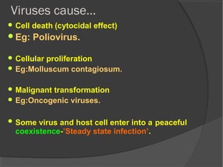
- 1. Viruses cause… Cell death (cytocidal effect) Eg: Poliovirus. Cellular proliferation Eg:Molluscum contagiosum. Malignant transformation Eg:Oncogenic viruses. Some virus and host cell enter into a peaceful coexistence-’Steady state infection’.
- 2. Seen in tissue culture Viral infection of cells leads to readily observable cellular changes called cytopathic effects Enteroviruses- crenation of cells and degeneration of cell sheet HEMA Measles- syncytium formation Adenovirus – bunch of grapes Herpes virus – discrete focal degeneration
- 3. Cytopathic effects Lead to observable cellularLead to observable cellular changes in tissue culture.changes in tissue culture.
- 4. Inclusion bodies….(SN,MCQs) Structures with distinct size, shape, location and staining properties that can be demonstrated in virus infected cells under the light microscope. CLASSIFICATION They may be situated at: Cytoplasm (intracytoplasmic)- pox virus Nucleus (intranuclear)- herpes Both - Measles
- 5. Appearance of Inclusion body…. • They are generally acidophilic. • Seen as pink structures when stained with giemsa or eosin methylene blue. • Some viruses form basophilic inclusions. Eg: Adenovirus.
- 6. General features… May be crystalline aggregates of virions. Made of virus antigens present at the site of virus synthesis. Represent degenerative changes produced by viral infection.
- 7. Intracytoplasmic inclusion bodies Negri bodies-Presumptive diagnosis of rabies. Guarineri bodies- small multiple inclusions. eg- Vaccinia(small pox) virus. PASCHEN Bodies Bollinger bodies- large inclusions. eg- Fowl pox virus. Molluscum bodies- very large 20-30µ. eg- Molluscum contagiosum.
- 8. Negribodies:Intracytoplasmic IB in the neurons, abundant in the cerebellum & hippocampusMCQ SQ MF
- 9. Negri body
- 10. Negri body in infected neuron.
- 11. Guarnieri bodies- small multiple inclusions. eg- Vaccinia(small pox) virus.
- 12. Guarnieri bodies
- 13. Molluscum bodies- very large 20-30µ. eg- Molluscum contagiosum.
- 14. Molluscum bodies
- 15. Intranuclear inclusion bodies Classified by Cowdry- 2types Cowdry type A inclusions: variable and granular Eg-herpesvirus, yellow fever virus. Cowdry type B inclusions: circumscribed and multiple Eg- Adenovirus, Poliovirus.