Ribozyme
•Download as DOCX, PDF•
5 likes•2,309 views

Ribozymes (ribonucleic acid enzymes) are RNA molecules that are capable of catalyzing specific biochemical reactions, similar to the action of protein enzymes.

Recommended
More Related Content
What's hot
What's hot (20)
Similar to Ribozyme
Similar to Ribozyme (20)
More from sworna kumari chithiraivelu
More from sworna kumari chithiraivelu (20)
Recently uploaded
Recently uploaded (20)
Ribozyme
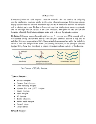
- 1. RIBOZYMES Ribozymes (ribonucleic acid enzymes) are RNA molecules that are capable of catalyzing specific biochemical reactions, similar to the action of protein enzymes. Ribozymes catalyze highly sequence-specific reactions determined by RNA-RNA interactions between the ribozyme and its substrate molecules. The key to the recognition of and binding to the substrate molecule, and the cleavage reaction, resides in the RNA molecule. Ribozyme can also catalyze the formation of peptide bond between adjacent amino acid by lowing the activation entropy. Definition: Ribozyme means ribonucleic acid enzyme. A ribozyme is an RNA molecule with a well defined tertiary structure that enables it to catalyze a chemical reaction. It may also be called an RNA enzyme or catalytic RNA. Many natural ribozymes catalyze either the hydrolysis of one of their own phosphodiester bonds (self-cleaving ribozymes), or the hydrolysis of bonds in other RNAs. Some have been found to catalyze the aminotransferase activity of the ribosome. Fig: Cleavage of RNA by ribozyme Types of ribozymes: RNase P ribozyme Hammer head ribozymes GIR1 branching ribozyme hepatitis delta virus (HDV) ribozyme hatchet ribozyme Pistol ribozyme VS ribozyme Twister ribozymes Twister sister ribozyme Group 1 introns Group2 introns RNase Pribozyme:
- 2. RNase P is a key enzyme in the biosynthesis of tRNAs.It is found in all cells and organelles that carry out tRNA synthesis. It is an RNA processing endonuclease that specically cleaves the tRNA precursors, releasing 5P-sequences and mature tRNAs . All known RNase P enzymes are ribonucleoproteins (RNPs) that contain an RNA subunit essential for catalysis. Fig.RNase P cleavage. a: Schematic representation of the cleavage reaction of the natural RNA substrate by the RNase P enzyme. In this reaction the 5P-end of the pre-tRNA is eliminated to generate mature tRNAs. The conserved triplet CCA at the 3P-end of the substrate RNA, needed for catalysis, is shown. b: Structure model of the minimal target RNA-EGS complex cleavable by bacterial RNase P, only a doublestranded domain carrying the CCA triplet is needed for cleavage.c: Target RNA-3/4EGS complex that can be recognized as substrate by eukaryotic RNase P. d: Representation of a minimized 3/4EGS-target complex. In all cases the arrow indicates the cleavage site. A thick line indicates the substrate RNA. Hepatitis delta virus (HDV) ribozyme: The hepatitis delta virus (HDV) ribozyme is a non-coding RNA found in the hepatitis delta virus that is necessary for viral replication and is the only known human virus that utilizes ribozyme activity to infect its host. The ribozyme acts to process the RNA transcripts to unit lengths in a self-cleavage reaction during replication of the hepatitis delta virus, which is thought to propagate by a double rolling circle mechanism. The hepatitis delta virus ribozyme is structurally and biochemically related to the Mammalian CPEB3 ribozyme.
- 3. Fig :secondary structure of HDV ribozyme Hammerhead ribozyme The hammerhead ribozyme is an RNA motif that catalyzes reversible cleavage and ligation reactions at a specific site within an RNA molecule. It is one of several catalytic RNAs (ribozymes) known to occur in nature. Structurally the hammerhead ribozyme is composed of three base paired helices, separated by short linkers of conserved sequences. These helices are called I, II and III. Hammerhead ribozymes can be classified into three types based on which helix the 5' and 3' ends are found in. If the 5' and 3' ends of the sequence contribute to stem I then it is a type I hammerhead ribozyme, to stem II is a type II and to stem III then it is a type III hammerhead ribozyme. Of the three possible topological types, type I can be found in the genomes of prokaryotes, eukaryotes and RNA plant pathogens, whereas type II have been only described in prokaryotes and type III are mostly found in plants, plant pathogens and prokaryotes Fig : Secondary structures and sequences of the minimal (A) and full-length (B) hammerhead ribozymes. Conserved and invariant nucleotides are shown explicitly. Watson-Crick base-paired helical stems are represented as ladder-like drawings. The arrow depicts the cleavage site, 3' to C17, on each construct.
- 4. Glucosamine-6-phosphate riboswitch ribozyme ( glmS ribozyme) The glucosamine-6-phosphate riboswitch ribozyme ( glmS ribozyme) is an RNA structure that resides in the 5' untranslated region (UTR) of the mRNA transcript of the glmS gene. This RNA regulates the glmS gene by responding to concentrations of a specific metabolite, glucosamine-6- phosphate (GlcN6P), in addition to catalyzing a self-cleaving chemical reaction upon activation. This cleavage leads to the degradation of the mRNA that contains the ribozyme, and lowers production of GlcN6P. The glmS gene encodes for an enzyme glutamine-fructose-6-phosphate amidotransferase, which catalyzes the formation of GlcN6P, a compound essential for cell wall biosynthesis, from fructose-6-phosphate and glutamine.[3] Thus, when GlcN6P levels are high, the glmS ribozyme is activated and the mRNA transcript is degraded but in the absence of GlcN6P the gene continues to be translated into glutamine-fructose-6-phosphate amidotransferase and GlcN6P is produced. GlcN6P is a cofactor for this cleavage reaction, as it directly participates as an acid-base catalyst. This RNA is the first riboswitch also found to be a self-cleaving ribozyme and, like many others, was discovered using a bioinformatics approach Group -I- like ribozymes (GIR1) branching ribozyme: The Lariat capping ribozyme (formerly called GIR1 branching ribozyme) is a 179 nucleotidet ribozyme with an apparent resemblance to a group I ribozyme. It is found within a complex type of group I introns also termed twin-ribozyme introns. Rather than splicing, it catalyses a branching reaction in which the 2'OH of an internal residue is involved in a nucleophilic attack at a nearby phosphodiester bond. As a result, the RNA is cleaved at an internal processing site (IPS), leaving a 3'OH and a downstream product with a tiny lariat at its 5' end. The lariat has the first and the third nucleotide joined by a 2',5' phosphodiester bond and is referred to as 'the lariat cap' because it caps an intron-encoded mRNA. The resulting lariat cap seems to contribute by increasing the half-life of the Homing Enodnuclease mRNA. Fig : secondary structure of Group -I- like ribozymes (GIR1) branching ribozyme hairpin ribozyme
- 5. The hairpin ribozyme is a small section of RNA that can act as a ribozyme. Like the hammerhead ribozyme it is found in RNA satellites of plant viruses. It was first identified in the minus strand of the tobacco ringspot virus (TRSV) satellite RNA where it catalyzes self- cleavage and joining (ligation) reactions to process the products of rolling circle virus replication into linear and circular satellite RNA molecules. The hairpin ribozyme is similar to the hammerhead ribozyme in that it does not require a metal ion for the reaction. Fig :Secondary structure of a minimal hairpin ribozyme with substrate RNA bound The hairpin ribozyme is an RNA motif that catalyzes RNA processing reactions essential for replication of the satellite RNA molecules in which it is embedded. These reactions are self- processing, i.e. a molecule rearranging its own structure. Both cleavage and end joining reactions are mediated by the ribozyme motif, leading to a mixture of interconvertible linear and circular satellite RNA molecules. These reactions are important for processing the large multimeric RNA molecules that are generated by rolling circle replication. At the end of the replication cycle, these large intermediates of satellite RNA replication are processed down to unit length molecules (circular or linear) before they can be packaged by viruses and carried to other cells for further rounds of replication. Group-I introns: Splicing of group I introns is processed by two sequential ester-transfer reactions.[3] The exogenous guanosine or guanosine nucleotide (exoG) first docks onto the active G-binding site located in P7, and its 3'-OH is aligned to attack the phosphodiester bond at the 5' splice site located in P1, resulting in a free 3'-OH group at the upstream exon and the exoG being attached to the 5' end of the intron. Then the terminal G (omega G) of the intron swaps the exoG and occupies the G-binding site to organize the second ester-transfer reaction: the 3'-OH group of the upstream exon in P1 is aligned to attack the 3' splice site in P10, leading to the ligation of the adjacent upstream and downstream exons and release of the catalytic intron.
- 6. Two-metal-ion mechanism seen in protein polymerases and phosphatases was proposed to be used by group I and group II introns to process the phosphoryl transfer reactions,[5]which was unambiguously proven by a recently resolved high-resolution structure of the Azoarcus group I intron Group II introns: Group II introns are a large class of self-catalytic ribozymes and mobile genetic elements found within the genes of all three domains of life. Ribozyme activity (e.g., self-splicing) can occur under high-salt conditions in vitro. However, assistance from proteins is required for in vivo splicing. In contrast to group I introns, intron excision occurs in the absence of GTP and involves the formation of a lariat, with an A-residue branchpoint strongly resembling that found in lariats formed during splicing of nuclear pre-mRNA. It is hypothesized that pre-mRNA splicing (see spliceosome) may have evolved from group II introns, due to the similar catalytic mechanism as well as the structural similarity of the Domain V substructure to the U6/U2 extended snRNA.
- 7. Hatchet ribozyme The hatchet ribozyme is an RNA structure that catalyzes its own cleavage at a specific site. In other words, it is a self-cleaving ribozyme. Hatchet ribozymes were discovered by a bioinformatics strategy as RNAs Associated with Genes Associated with Twister and Hammerhead ribozymes, or RAGATH.Subsequent biochemical analysis supports the conclusion of a ribozyme function, and determined further characteristics of the chemical reaction catalyzed by the ribozyme. Pistol ribozyme The pistol ribozyme is an RNA structure that catalyzes its own cleavage at a specific site. Pistol ribozymes cleave via internal phosphoester transfer. VS ribozyme The Varkud satellite (VS) ribozyme is an RNA enzyme that carries out the cleavage of a phosphodiester bond.Varkud satellite (VS) ribozyme is the largest known nucleolyic ribozyme
- 8. and found to be embedded in VS RNA. VS RNA is a long non-coding RNA exists as a satellite RNA and is found in mitochondria of Varkud-1C and few other strains of Neurospora. VS ribozyme contains features of both catalytic RNAs and group 1 introns. VS ribosyme has both cleavage and ligation activity and can perform both cleavage and ligation reactions efficiently in the absence of proteins. VS ribozyme undergo horizontal gene transfer with other Neuropora strains. VS ribozymes have nothing in common with other nucleolytic ribozymes. VS RNA has a unique primary, secondary, and tertiary structure. The secondary structure of the VS ribozyme consists of six helical domains (Figure). Stem loop I forms the substrate domain while stem-loop II-VI forms the catalytic domain. When these 2 domains are synthesized in vitro separately, they can perform the self-cleavage reaction by trans-acting. The substrate binds into a cleft which is made by two helices. The likely active site of the ribozyme is a very important nucleotide A756. The A730 loop and A756 nucleotide are critical to its function since they participate in the phosphoric transfer chemistry activity of the ribozyme Fig : secondary structure of VS rbozyme Twister ribozyme The twister ribozyme is a catalytic RNA structure capable of self-cleavage. The nucleolytic activity of this ribozyme has been demonstrated both in vivo and in vitro and has one of the fastest catalytic rates of naturally occurring ribozymes with similar function.The twister ribozyme is considered to be a member of the small self-cleaving ribozyme family which includes the hammerhead, hairpin, hepatitis delta virus (HDV), Varkud satellite (VS), and glmS ribozymes. Similar to other nucleolytic ribozymes, the twister ribozyme selectively cleaves phopshodiester bonds, through an SN2-related mechanism, into a 2',3'-cyclic phosphate and 5' hydroxyl product. Twister sister ribozyme:
- 9. The twister sister ribozyme (TS) is an RNA structure that catalyzes its own cleavage at a specific site. In other words, it is a self-cleaving ribozyme. The twister sister ribozyme was discovered by a bioinformatics strategy as an RNA Associated with Genes Associated with Twister and Hammerhead ribozymes, or RAGATH. The twister sister ribozyme has a possible structural similarity to twister ribozymes. Generally, nucleolytic ribozymes cleave a specific phosphodiester linkage by SN2 mechanism. The O2' acts as a nucleophile to attack the adjacent P, with O5’ as a leaving group. The catalytic products are a cyclic 2’,3’ phosphate and a 5’- hydroxyl.The catalytic activity of twister sister increases with pH and depends on divalent metal ion. The cleavage speed increases 10 fold with each increase in pH unit and reach a plateau near pH 7. Fig :Schematic and tertiary structure of the twister-sister ribozyme. a Schematic of the secondary fold of the dC62-containing four-way junctional twister-sister ribozyme. b Schematic of the tertiary fold based on the crystal structure of the dC62-containing four-way junctional twister-sister ribozyme. Artificial Ribozymes a. Since the discovery of ribozymes that exist in living organisms, there has been interest in the study of new synthetic ribozymes made in the laboratory. For example, artificially- produced self-cleaving RNAs those have good enzymatic activity have been produced. Tang and Breaker isolated self-cleaving RNAs by in vitro selection of RNAs originating from random-sequence RNAs. Some of the synthetic ribozymes that were produced had
- 10. novel structures, while some were similar to the naturally occurring hammerhead ribozyme. b. The techniques used to discover artificial ribozymes involve Darwinian evolution. This approach takes advantage of RNA's dual nature as both a catalyst and an informational polymer, making it easy for an investigator to produce vast populations of RNA catalysts using polymerase enzymes. The ribozymes are mutated by reverse transcribing them with reverse transcriptase into various cDNA and amplified with mutagenic PCR. The selection parameters in these experiments often differ. One approach for selecting a ligase ribozyme involves using biotin tags, which are covalently linked to the substrate. If a molecule possesses the desired ligase activity, a streptavidin matrix can be used to recover the active molecules. c. Lincoln and Joyce developed an RNA enzyme system capable of self replication in about an hour. By utilizing in vitro evolution of a candidate enzyme mixture, a pair of RNA enzymes emerged, in which each synthesizes the other from synthetic oligonucleotides, with no protein present. Applications of Ribozymes a. Catalytic RNAs (ribozymes) are capable of specifically cleaving RNA molecules, a property that enables them to act as potential antiviral and anti-cancer agents, as well as powerful tools for functional genomic studies. b. Recently, ribozymes have been used successfully to inhibit gene expression in a variety of biological systems in vitro and in vivo. c. Phase I clinical trials using ribozyme gene therapy to treat AIDS patients have been conducted. d. A type of synthetic ribozyme directed against HIV RNA called gene shears has been developed and has entered clinical testing for HIV infection References Weinberg Z, Kim PB, Chen TH, Li S, Harris KA, Lünse CE, Breaker RR (2015). "New classes of self-cleaving ribozymes revealed by comparative genomics analysis". Nat. Chem. Biol. 11 (8): 606–10. doi:10.1038/nchembio.1846. PMC 4509812. PMID 26167874. Li S, Lünse CE, Harris KA, Breaker RR (2015). "Biochemical analysis of hatchet self-cleaving ribozymes". RNA. 21 (11): 18451. doi:10.1261/rna.052522.115. PMC 4604424. PMID 26385510.
- 11. Harris KA, Lünse CE, Li S, Brewer KI, Breaker RR (2015). "Biochemical analysis of pistol self- cleaving ribozymes". RNA. 21 (11):1852–8 Saville BJ, Collins RA (1990). "A site-specific self-cleavage reaction performed by a novel RNA in Neurospora ribozymes". Cell. 61 (4): 685–696. Elena Puerta-Ferna.ndez, Cristina Romero-Lo.pez, Alicia Barroso-delJesus, Alfredo Berzal- Herranz Ribozymes: recent advances in the development of RNA tools. FEMS Microbiology Reviews 27 (2003) 75-97 Ferré-D'Amaré AR, Zhou K, Doudna JA (October 1998). "Crystal structure of a hepatitis delta virus ribozyme". Nature. 395 (6702): 567–74. Symons, RH (1997). "Plant pathogenic RNAs and RNA catalysis". Nucleic Acids Res. 25 (14): 2683–2689.