Short case...Acute postinfectious transverse myelitis
•
4 j'aime•411 vues

Short case....Acute postinfectious transverse myelitis

Recommandé
Contenu connexe
Similaire à Short case...Acute postinfectious transverse myelitis
Similaire à Short case...Acute postinfectious transverse myelitis (20)
Plus de Professor Yasser Metwally
Plus de Professor Yasser Metwally (20)
Dernier
Dernier (20)
Short case...Acute postinfectious transverse myelitis
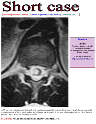
- 1. Short case publication... version 2.7| Edited by professor Yasser Metwally | November 2008 Short case Edited by Professor Yasser Metwally Professor of neurology Ain Shams university school of medicine Cairo, Egypt Visit my web site at: http://yassermetwally.com A 33 years old patient presented clinically with paraplegia associated with a mid-dorsal sensory level of acute onset and a regressive course. Clinical manifestations were bilateral and symmetrical. An antecedent upper respiratory infection was present 2 weeks before the neurological disease. DIAGNOSIS: ACUTE POSTINFECTIOUS TRANSVERSE MYELITIS
- 2. Figure 1. A centrally located multisegmental (3 to 8 spinal segments) MRI T2 hyperintensity that occupies more than two thirds of the cross-sectional area of the cord is characteristic of transverse myelitis. The MRI T2 hyperintensity commonly shows a slow regression with clinical improvement. The central spinal cord MRI T2 hyperintensity represents evenly distributed central cord edema. MRI T1 Hypointensity might be present in the same spinal segments that show T2 hyperintensity although to a lesser extent. The MRI T2 hyperintensity is central, bilateral, more or less symmetrical and multisegmental. Notice the MRI T2 central isointensity, or dot (within and in the core of the MRI T2 hyperintensity) which is believed to represent central gray matter squeezed by the uniform, evenly distributed edematous changes of the cord. (central dot sign). Figure 2. Postpontrast MRI T1 study. Notice contrast enhancement that is peripheral to and maximal at or near the segmental MRI T2 hyperintensity. In idiopathic transverse myelitis enhancement is peripheral to the centrally located area of high T2 signal intensity rather than in the very same area. The prevalence of cord enhancement is significantly higher in patients with cord expansion.
- 3. Figure 3. MRI T2 study showing a centrally located multisegmental (3 to 8 spinal segments) MRI T2 hyperintensity that occupies more than two thirds of the cross-sectional area of the cord is characteristic of transverse myelitis. The MRI T2 hyperintensity commonly shows a slow regression with clinical improvement. The central spinal cord MRI T2 hyperintensity represents evenly distributed central cord edema. Table 1. The MRI picture characteristic of idiopathic transverse myelitis A centrally located multisegmental (3 to 8 spinal segments) MRI T2 hyperintensity that occupies more than two thirds of the cross-sectional area of the cord is characteristic of transverse myelitis. The MRI T2 hyperintensity commonly shows a slow regression with clinical improvement. The central spinal cord MRI T2 hyperintensity represents evenly distributed central cord edema. MRI T1 Hypointensity might be present in the same spinal segments that show T2 hyperintensity although to a lesser extent. The MRI T2 hyperintensity is central, bilateral, more or less symmetrical and multisegmental. MRI T2 central isointensity, or dot (within and in the core of the MRI T2 hyperintensity) might be present and is believed to represent central gray matter squeezed by the uniform, evenly distributed edematous changes of the cord. (central dot sign). It might not be of any clinical significance. Contrast enhancement is commonly focal or peripheral and maximal at or near the segmental MRI T2 hyperintensity. In idiopathic transverse myelitis enhancement is peripheral to the centrally located area of high T2 signal intensity rather than in the very same area. The prevalence of cord enhancement is significantly higher in patients with cord expansion. Spinal cord expansion might or might not be present and when present is usually multisegmental and better appreciated on the sagittal MRI T1 images. Spinal cord expansion tapers smoothly to the normal cord, and is of lesser extent than the high T2 signal abnormality. Multiple sclerosis plaques (and subsequent T2 hyperintensity) are located peripherally, are less than 2 vertebral segments in length, and occupies less than half the cross-sectional area of the cord. In contrast to transverse myelitis, enhancement in MS occurs in the same location of high-signal-intensity lesions seen on T2-weighted images. References 1. Metwally, MYM: Textbook of neurimaging, A CD-ROM publication, (Metwally, MYM editor) WEB-CD agency for electronic publishing, version 9.4a October 2008
- 4. Addendum A new version of short case is uploaded in my web site every week (every Saturday and remains available till Friday.) To download the current version follow the link quot;http://pdf.yassermetwally.com/short.pdfquot;. You can download the long case version of this short case during the same week from: http://pdf.yassermetwally.com/case.pdf or visit web site: http://pdf.yassermetwally.com To download the software version of the publication (crow.exe) follow the link: http://neurology.yassermetwally.com/crow.zip At the end of each year, all the publications are compiled on a single CD-ROM, please contact the author to know more details. Screen resolution is better set at 1024*768 pixel screen area for optimum display For an archive of the previously reported cases go to www.yassermetwally.net, then under pages in the right panel, scroll down and click on the text entry quot;downloadable short cases in PDF formatquot; Also to view a list of the previously published case records follow the following link (http://wordpress.com/tag/case- record/) or click on it if it appears as a link in your PDF reader