SlideShare a Scribd company logo
1 of 5
Download to read offline
Short case publication... Version 2.15| Edited by professor Yasser Metwally | February 2009


                                                                                                   Short case

                                                                                                  Edited by
                                                                                          Professor Yasser Metwally
                                                                                            Professor of neurology
                                                                                        Ain Shams university school of medicine
                                                                                                   Cairo, Egypt

                                                                                             Visit my web site at:
                                                                                          http://yassermetwally.com


A 40 years old female patient presented clinically with manifestations of increased intracranial pressure, long tract
manifestations, ponto-medullary cranial nerve palsies. and cerebellar manifestations. Hearing was intact.

DIAGNOSIS: CEREBELLOPONTINE ANGLE MENINGIOMA
Figure 1. Postcontrast MRI T1 images showing a densely enhanced cerebellopontine angle meningioma, Notice the
wide base attachment, the enhanced meningeal tail, and the moderate compression of the brain stem. A CSF cleft is
also evident.


 Figure 2. Postcontrast MRI T1 images showing a densely enhanced cerebellopontine angle meningioma, Notice the
 wide base attachment, the enhanced meningeal tail, and the moderate compression of the brain stem. A CSF cleft
 is also evident.
Figure 3. Postcontrast MRI T1
       images showing a densely enhanced
       cerebellopontine angle meningioma,
       Notice the wide base attachment, the
       enhanced meningeal tail, and the
       moderate compression of the brain
       stem. A CSF cleft is also evident.


Figure 4. The meningioma (syncytial subtype)
is hyperintense on the MRI T2 images
Table 1. MRI appearance of the various types of meningiomas

Type                            Comment
Fibroblastic meningiomas        Fibroblastic meningiomas are composed of large, narrow spindle cells. The distinct
                                feature is the presence of abundant reticulum and collagen fibers between
                                individual cells. On MR imaging, fibroblastic meningiomas with cells embedded in
                                a dense collagenous matrix appear as low signal intensity in Tl-weighted and T2-
                                weighted pulse sequences.
Transitional meningiomas        Transitional meningiomas are characterized by whorl formations in which the
                                cells are wrapped together resembling onion skins. The whorls may degenerate
                                and calcify, becoming psammoma bodies. Marked calcifications can be seen in this
                                histologic type. MR imaging of transitional meningiomas thus also demonstrates
                                low signal intensity on Tl- weighted and T2-weighted images, with the
                                calcifications contributing to the low signal intensity.
Syncytial meningiomas           Syncytial (meningothelial, endotheliomatous) meningiomas contain polygonal
                                cells, poorly defined and arranged in lobules. Syncytial meningiomas composed of
                                sheets of contiguous cells with sparse interstitium might account for higher signal
                                intensity in T2-weighted images. Microcystic changes and nuclear vesicles can also
                                contribute to increased signal intensity.
Angioblastic meningiomas        Angioblastic meningiomas are highly cellular and vascular tumors with a spongy
                                appearance. Increased signal in T2-weighted pulse sequence of these tumors is due
                                to high cellularity with increase in water content of tumor.
Table 2. MRI characteristics of meningiomas

Pathological type   T2 MRI appearance

Fibroblastic        Hypointense on the T2 images because of the existence of dense collagen and fibrous tissue

Transitional        Hypointense on the T2 images because of the existence of densely calcified psammoma
                    bodies
Syncytial           Hyperintense on the T2 images because of the existence of high cell count, microcysts or
                    significant tissue oedema
Angioblastic        Same as the syncytial type. Blood vessels appear as signal void convoluted structures

Table 3. MRI characteristics of meningiomas


MRI feature          Description

Vascular rim         The peripheries of meningiomas are supplied by branches from the anterior or middle
                     cerebral arteries that encircle the tumour and form the characteristic vascular rim
Meningeal tail       The tail extends to a variable degree away from the meningioma site and probably
                     represents a meningeal reaction to the tumour
Hypointense cleft    Hypointense cleft between the tumour and the brain that probably represents blood
                     vessels or a CSF interface
The dural tail or quot;dural flairquot;

The dural tail is a curvilinear region of dural enhancement adjacent to the bulky hemispheric tumor. The finding
was originally thought to represent dural infiltration by tumor, and resection of all enhancing dura mater was
thought to be appropriate. However, later studies helped confirm that most of the linear dural enhancement,
especially when it was more than a centimeter away from the tumor bulk, was probably caused by a reactive
process. This reactive process includes both vasocongestion and accumulation of interstitial edema, both of which
increase the thickness of the dura mater. Because the dural capillaries are quot;nonneural,quot; they do not form a
blood-brain barrier, and, with accumulation of water within the dura mater, contrast material enhancement
occurs.


References

1. Metwally, MYM: Textbook of neurimaging, A CD-ROM publication, (Metwally, MYM editor) WEB-CD agency for
electronic publishing, version 10.1a January 2009


Addendum

   A new version of short case is uploaded in my web site every week (every Saturday and remains available till Friday.)
   To download the current version follow the link quot;http://pdf.yassermetwally.com/short.pdfquot;.
   You can download the long case version of this short case during the same week from: http://pdf.yassermetwally.com/case.pdf or visit
   web site: http://pdf.yassermetwally.com
   To download the software version of the publication (crow.exe) follow the link: http://neurology.yassermetwally.com/crow.zip
   At the end of each year, all the publications are compiled on a single CD-ROM, please contact the author to know
   more details.
   Screen resolution is better set at 1024*768 pixel screen area for optimum display
   For an archive of the previously reported cases go to www.yassermetwally.net, then under pages in the right panel,
   scroll down and click on the text entry quot;downloadable short cases in PDF formatquot;
   Also to view a list of the previously published case records follow the following link (http://wordpress.com/tag/case-
   record/) or click on it if it appears as a link in your PDF reader

More Related Content

What's hot

Presentation1, radiological imaging of cavernous sinus lesions.
Presentation1, radiological imaging of cavernous sinus lesions.Presentation1, radiological imaging of cavernous sinus lesions.
Presentation1, radiological imaging of cavernous sinus lesions.
Abdellah Nazeer
 
Presentation1, radiological imaging of trigeminal schwanoma.
Presentation1, radiological imaging of trigeminal schwanoma.Presentation1, radiological imaging of trigeminal schwanoma.
Presentation1, radiological imaging of trigeminal schwanoma.
Abdellah Nazeer
 
Presentation1.pptx, radiological imaging of spinal cord tumour.
Presentation1.pptx, radiological imaging of spinal cord tumour.Presentation1.pptx, radiological imaging of spinal cord tumour.
Presentation1.pptx, radiological imaging of spinal cord tumour.
Abdellah Nazeer
 
Presentation1, radiological imaging of intra cranial dermoid tumours.
Presentation1, radiological imaging of intra cranial dermoid tumours.Presentation1, radiological imaging of intra cranial dermoid tumours.
Presentation1, radiological imaging of intra cranial dermoid tumours.
Abdellah Nazeer
 

What's hot (17)

Spinal tumors drsant91
Spinal tumors drsant91Spinal tumors drsant91
Spinal tumors drsant91
 
Short case...Epidural empyema
Short case...Epidural empyemaShort case...Epidural empyema
Short case...Epidural empyema
 
Presentation1, radiological imaging of cavernous sinus lesions.
Presentation1, radiological imaging of cavernous sinus lesions.Presentation1, radiological imaging of cavernous sinus lesions.
Presentation1, radiological imaging of cavernous sinus lesions.
 
Knee cystic lesions
Knee cystic lesionsKnee cystic lesions
Knee cystic lesions
 
IntraCranial Aneurysm by Dr. Avinash
IntraCranial Aneurysm by Dr. AvinashIntraCranial Aneurysm by Dr. Avinash
IntraCranial Aneurysm by Dr. Avinash
 
Intra muscular non - traumatic lesions - ultrasonographic evaluation
Intra   muscular non - traumatic lesions - ultrasonographic evaluationIntra   muscular non - traumatic lesions - ultrasonographic evaluation
Intra muscular non - traumatic lesions - ultrasonographic evaluation
 
Presentation1, radiological imaging of trigeminal schwanoma.
Presentation1, radiological imaging of trigeminal schwanoma.Presentation1, radiological imaging of trigeminal schwanoma.
Presentation1, radiological imaging of trigeminal schwanoma.
 
Soft tissue tumors
Soft tissue tumorsSoft tissue tumors
Soft tissue tumors
 
MRI: Collapsed Vertebral Bodies
MRI: Collapsed Vertebral BodiesMRI: Collapsed Vertebral Bodies
MRI: Collapsed Vertebral Bodies
 
Case record...Idiopathic postinfectious transverse myelitis'
Case record...Idiopathic postinfectious transverse myelitis'Case record...Idiopathic postinfectious transverse myelitis'
Case record...Idiopathic postinfectious transverse myelitis'
 
Benign brain tumours
Benign brain tumoursBenign brain tumours
Benign brain tumours
 
Pseudosarcoma
Pseudosarcoma Pseudosarcoma
Pseudosarcoma
 
Cns tumours
Cns tumoursCns tumours
Cns tumours
 
Presentation1.pptx, radiological imaging of spinal cord tumour.
Presentation1.pptx, radiological imaging of spinal cord tumour.Presentation1.pptx, radiological imaging of spinal cord tumour.
Presentation1.pptx, radiological imaging of spinal cord tumour.
 
Presentation1, radiological imaging of intra cranial dermoid tumours.
Presentation1, radiological imaging of intra cranial dermoid tumours.Presentation1, radiological imaging of intra cranial dermoid tumours.
Presentation1, radiological imaging of intra cranial dermoid tumours.
 
Spot diagnosis for radiology second master degree
Spot diagnosis for radiology second master degreeSpot diagnosis for radiology second master degree
Spot diagnosis for radiology second master degree
 
PITUITARY TUMORS
PITUITARY TUMORSPITUITARY TUMORS
PITUITARY TUMORS
 

Similar to Short case...Cerebellopontine angle meningioma

Similar to Short case...Cerebellopontine angle meningioma (20)

Case record...Cerebellopontine angle meningioma
Case record...Cerebellopontine angle meningiomaCase record...Cerebellopontine angle meningioma
Case record...Cerebellopontine angle meningioma
 
Case record...Spinal meningioma
Case record...Spinal meningiomaCase record...Spinal meningioma
Case record...Spinal meningioma
 
Short case...Occipital lobe cavernoma
Short case...Occipital lobe cavernomaShort case...Occipital lobe cavernoma
Short case...Occipital lobe cavernoma
 
Case record...Parasagittal meningioma
Case record...Parasagittal meningiomaCase record...Parasagittal meningioma
Case record...Parasagittal meningioma
 
Short case...Spinal dural arteriovenous fistula
Short case...Spinal dural arteriovenous fistulaShort case...Spinal dural arteriovenous fistula
Short case...Spinal dural arteriovenous fistula
 
Short case...brain stem glioma
Short case...brain stem gliomaShort case...brain stem glioma
Short case...brain stem glioma
 
Short case...Multiple dural arteriovenous fistulas
Short case...Multiple dural arteriovenous fistulasShort case...Multiple dural arteriovenous fistulas
Short case...Multiple dural arteriovenous fistulas
 
Short case......Primary subependymal CNS lymphoma (MRI approach)
Short case......Primary subependymal CNS lymphoma (MRI approach)Short case......Primary subependymal CNS lymphoma (MRI approach)
Short case......Primary subependymal CNS lymphoma (MRI approach)
 
Short case...Acute postinfectious transverse myelitis
Short case...Acute postinfectious transverse myelitisShort case...Acute postinfectious transverse myelitis
Short case...Acute postinfectious transverse myelitis
 
Short case...Multiple craniocervical anomalies
Short case...Multiple craniocervical anomaliesShort case...Multiple craniocervical anomalies
Short case...Multiple craniocervical anomalies
 
Short case...tectal plate glioma
Short case...tectal plate gliomaShort case...tectal plate glioma
Short case...tectal plate glioma
 
Meninges and related tumors
Meninges and related tumorsMeninges and related tumors
Meninges and related tumors
 
Topic of the month: Radiological pathology of meningiomas
Topic of the month: Radiological pathology of meningiomasTopic of the month: Radiological pathology of meningiomas
Topic of the month: Radiological pathology of meningiomas
 
Short case...Neuro-Behcet
Short case...Neuro-BehcetShort case...Neuro-Behcet
Short case...Neuro-Behcet
 
Short case...Lymphomatous leptomeningitis
Short case...Lymphomatous leptomeningitisShort case...Lymphomatous leptomeningitis
Short case...Lymphomatous leptomeningitis
 
Short case...Spinal multiple sclerosis
Short case...Spinal multiple sclerosisShort case...Spinal multiple sclerosis
Short case...Spinal multiple sclerosis
 
Short case...Cervical vascular spondylotic myelopathy
Short case...Cervical vascular spondylotic myelopathyShort case...Cervical vascular spondylotic myelopathy
Short case...Cervical vascular spondylotic myelopathy
 
imaging of itraconal mass EOS 2015.pptx
imaging of itraconal mass EOS 2015.pptximaging of itraconal mass EOS 2015.pptx
imaging of itraconal mass EOS 2015.pptx
 
Short case...Multiple sclerosis
Short case...Multiple sclerosisShort case...Multiple sclerosis
Short case...Multiple sclerosis
 
Short case...Hypoparathyroidism
Short case...HypoparathyroidismShort case...Hypoparathyroidism
Short case...Hypoparathyroidism
 

More from Professor Yasser Metwally

More from Professor Yasser Metwally (20)

The Egyptian Zoo in Cairo 2015
The Egyptian Zoo in Cairo 2015The Egyptian Zoo in Cairo 2015
The Egyptian Zoo in Cairo 2015
 
End of the great nile river in Ras Elbar
End of the great nile river in Ras ElbarEnd of the great nile river in Ras Elbar
End of the great nile river in Ras Elbar
 
The Lion and The tiger in Egypt
The Lion and The tiger in EgyptThe Lion and The tiger in Egypt
The Lion and The tiger in Egypt
 
The monkeys in Egypt
The monkeys in EgyptThe monkeys in Egypt
The monkeys in Egypt
 
The Snake, the Scorpion, the turtle in Egypt
The Snake, the Scorpion, the turtle in EgyptThe Snake, the Scorpion, the turtle in Egypt
The Snake, the Scorpion, the turtle in Egypt
 
The Egyptian Parrot
The Egyptian ParrotThe Egyptian Parrot
The Egyptian Parrot
 
The Egyptian Deer
The Egyptian DeerThe Egyptian Deer
The Egyptian Deer
 
The Egyptian Pelican
The Egyptian PelicanThe Egyptian Pelican
The Egyptian Pelican
 
The Flamingo bird in Egypt
The Flamingo bird in EgyptThe Flamingo bird in Egypt
The Flamingo bird in Egypt
 
Egyptian Cats
Egyptian CatsEgyptian Cats
Egyptian Cats
 
Radiological pathology of epileptic disorders
Radiological pathology of epileptic disordersRadiological pathology of epileptic disorders
Radiological pathology of epileptic disorders
 
Radiological pathology of cerebrovascular disorders
Radiological pathology of cerebrovascular disordersRadiological pathology of cerebrovascular disorders
Radiological pathology of cerebrovascular disorders
 
Radiological pathology of spontaneous cerebral hemorrhage
Radiological pathology of spontaneous cerebral hemorrhageRadiological pathology of spontaneous cerebral hemorrhage
Radiological pathology of spontaneous cerebral hemorrhage
 
Radiological pathology of cerebral amyloid angiography
Radiological pathology of cerebral amyloid angiographyRadiological pathology of cerebral amyloid angiography
Radiological pathology of cerebral amyloid angiography
 
Radiological pathology of cerebral microbleeds
Radiological pathology of cerebral microbleedsRadiological pathology of cerebral microbleeds
Radiological pathology of cerebral microbleeds
 
The Egyptian Zoo in Cairo
The Egyptian Zoo in CairoThe Egyptian Zoo in Cairo
The Egyptian Zoo in Cairo
 
Progressive multifocal leukoencephalopathy
Progressive multifocal leukoencephalopathyProgressive multifocal leukoencephalopathy
Progressive multifocal leukoencephalopathy
 
Progressive multifocal leukoencephalopathy
Progressive multifocal leukoencephalopathyProgressive multifocal leukoencephalopathy
Progressive multifocal leukoencephalopathy
 
Issues in radiological pathology: Radiological pathology of watershed infarct...
Issues in radiological pathology: Radiological pathology of watershed infarct...Issues in radiological pathology: Radiological pathology of watershed infarct...
Issues in radiological pathology: Radiological pathology of watershed infarct...
 
Radiological pathology of cortical laminar necrosis
Radiological pathology of cortical laminar necrosisRadiological pathology of cortical laminar necrosis
Radiological pathology of cortical laminar necrosis
 

Recently uploaded

Jual Obat Aborsi Hongkong ( Asli No.1 ) 085657271886 Obat Penggugur Kandungan...
Jual Obat Aborsi Hongkong ( Asli No.1 ) 085657271886 Obat Penggugur Kandungan...Jual Obat Aborsi Hongkong ( Asli No.1 ) 085657271886 Obat Penggugur Kandungan...
Jual Obat Aborsi Hongkong ( Asli No.1 ) 085657271886 Obat Penggugur Kandungan...
ZurliaSoop
 
1029-Danh muc Sach Giao Khoa khoi 6.pdf
1029-Danh muc Sach Giao Khoa khoi  6.pdf1029-Danh muc Sach Giao Khoa khoi  6.pdf
1029-Danh muc Sach Giao Khoa khoi 6.pdf
QucHHunhnh
 

Recently uploaded (20)

Jual Obat Aborsi Hongkong ( Asli No.1 ) 085657271886 Obat Penggugur Kandungan...
Jual Obat Aborsi Hongkong ( Asli No.1 ) 085657271886 Obat Penggugur Kandungan...Jual Obat Aborsi Hongkong ( Asli No.1 ) 085657271886 Obat Penggugur Kandungan...
Jual Obat Aborsi Hongkong ( Asli No.1 ) 085657271886 Obat Penggugur Kandungan...
 
This PowerPoint helps students to consider the concept of infinity.
This PowerPoint helps students to consider the concept of infinity.This PowerPoint helps students to consider the concept of infinity.
This PowerPoint helps students to consider the concept of infinity.
 
microwave assisted reaction. General introduction
microwave assisted reaction. General introductionmicrowave assisted reaction. General introduction
microwave assisted reaction. General introduction
 
General Principles of Intellectual Property: Concepts of Intellectual Proper...
General Principles of Intellectual Property: Concepts of Intellectual  Proper...General Principles of Intellectual Property: Concepts of Intellectual  Proper...
General Principles of Intellectual Property: Concepts of Intellectual Proper...
 
Spatium Project Simulation student brief
Spatium Project Simulation student briefSpatium Project Simulation student brief
Spatium Project Simulation student brief
 
Unit-IV; Professional Sales Representative (PSR).pptx
Unit-IV; Professional Sales Representative (PSR).pptxUnit-IV; Professional Sales Representative (PSR).pptx
Unit-IV; Professional Sales Representative (PSR).pptx
 
Sociology 101 Demonstration of Learning Exhibit
Sociology 101 Demonstration of Learning ExhibitSociology 101 Demonstration of Learning Exhibit
Sociology 101 Demonstration of Learning Exhibit
 
Unit-IV- Pharma. Marketing Channels.pptx
Unit-IV- Pharma. Marketing Channels.pptxUnit-IV- Pharma. Marketing Channels.pptx
Unit-IV- Pharma. Marketing Channels.pptx
 
Kodo Millet PPT made by Ghanshyam bairwa college of Agriculture kumher bhara...
Kodo Millet  PPT made by Ghanshyam bairwa college of Agriculture kumher bhara...Kodo Millet  PPT made by Ghanshyam bairwa college of Agriculture kumher bhara...
Kodo Millet PPT made by Ghanshyam bairwa college of Agriculture kumher bhara...
 
2024-NATIONAL-LEARNING-CAMP-AND-OTHER.pptx
2024-NATIONAL-LEARNING-CAMP-AND-OTHER.pptx2024-NATIONAL-LEARNING-CAMP-AND-OTHER.pptx
2024-NATIONAL-LEARNING-CAMP-AND-OTHER.pptx
 
Python Notes for mca i year students osmania university.docx
Python Notes for mca i year students osmania university.docxPython Notes for mca i year students osmania university.docx
Python Notes for mca i year students osmania university.docx
 
SKILL OF INTRODUCING THE LESSON MICRO SKILLS.pptx
SKILL OF INTRODUCING THE LESSON MICRO SKILLS.pptxSKILL OF INTRODUCING THE LESSON MICRO SKILLS.pptx
SKILL OF INTRODUCING THE LESSON MICRO SKILLS.pptx
 
ComPTIA Overview | Comptia Security+ Book SY0-701
ComPTIA Overview | Comptia Security+ Book SY0-701ComPTIA Overview | Comptia Security+ Book SY0-701
ComPTIA Overview | Comptia Security+ Book SY0-701
 
Unit-V; Pricing (Pharma Marketing Management).pptx
Unit-V; Pricing (Pharma Marketing Management).pptxUnit-V; Pricing (Pharma Marketing Management).pptx
Unit-V; Pricing (Pharma Marketing Management).pptx
 
How to Create and Manage Wizard in Odoo 17
How to Create and Manage Wizard in Odoo 17How to Create and Manage Wizard in Odoo 17
How to Create and Manage Wizard in Odoo 17
 
Grant Readiness 101 TechSoup and Remy Consulting
Grant Readiness 101 TechSoup and Remy ConsultingGrant Readiness 101 TechSoup and Remy Consulting
Grant Readiness 101 TechSoup and Remy Consulting
 
SOC 101 Demonstration of Learning Presentation
SOC 101 Demonstration of Learning PresentationSOC 101 Demonstration of Learning Presentation
SOC 101 Demonstration of Learning Presentation
 
On National Teacher Day, meet the 2024-25 Kenan Fellows
On National Teacher Day, meet the 2024-25 Kenan FellowsOn National Teacher Day, meet the 2024-25 Kenan Fellows
On National Teacher Day, meet the 2024-25 Kenan Fellows
 
1029-Danh muc Sach Giao Khoa khoi 6.pdf
1029-Danh muc Sach Giao Khoa khoi  6.pdf1029-Danh muc Sach Giao Khoa khoi  6.pdf
1029-Danh muc Sach Giao Khoa khoi 6.pdf
 
Basic Civil Engineering first year Notes- Chapter 4 Building.pptx
Basic Civil Engineering first year Notes- Chapter 4 Building.pptxBasic Civil Engineering first year Notes- Chapter 4 Building.pptx
Basic Civil Engineering first year Notes- Chapter 4 Building.pptx
 

Short case...Cerebellopontine angle meningioma

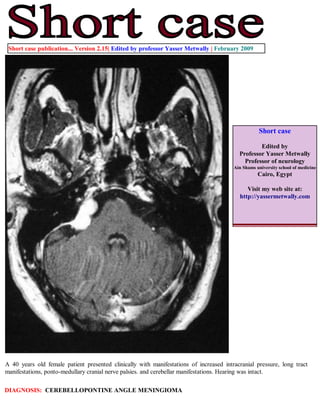
  • 1. Short case publication... Version 2.15| Edited by professor Yasser Metwally | February 2009 Short case Edited by Professor Yasser Metwally Professor of neurology Ain Shams university school of medicine Cairo, Egypt Visit my web site at: http://yassermetwally.com A 40 years old female patient presented clinically with manifestations of increased intracranial pressure, long tract manifestations, ponto-medullary cranial nerve palsies. and cerebellar manifestations. Hearing was intact. DIAGNOSIS: CEREBELLOPONTINE ANGLE MENINGIOMA
  • 2. Figure 1. Postcontrast MRI T1 images showing a densely enhanced cerebellopontine angle meningioma, Notice the wide base attachment, the enhanced meningeal tail, and the moderate compression of the brain stem. A CSF cleft is also evident. Figure 2. Postcontrast MRI T1 images showing a densely enhanced cerebellopontine angle meningioma, Notice the wide base attachment, the enhanced meningeal tail, and the moderate compression of the brain stem. A CSF cleft is also evident.
  • 3. Figure 3. Postcontrast MRI T1 images showing a densely enhanced cerebellopontine angle meningioma, Notice the wide base attachment, the enhanced meningeal tail, and the moderate compression of the brain stem. A CSF cleft is also evident. Figure 4. The meningioma (syncytial subtype) is hyperintense on the MRI T2 images
  • 4. Table 1. MRI appearance of the various types of meningiomas Type Comment Fibroblastic meningiomas Fibroblastic meningiomas are composed of large, narrow spindle cells. The distinct feature is the presence of abundant reticulum and collagen fibers between individual cells. On MR imaging, fibroblastic meningiomas with cells embedded in a dense collagenous matrix appear as low signal intensity in Tl-weighted and T2- weighted pulse sequences. Transitional meningiomas Transitional meningiomas are characterized by whorl formations in which the cells are wrapped together resembling onion skins. The whorls may degenerate and calcify, becoming psammoma bodies. Marked calcifications can be seen in this histologic type. MR imaging of transitional meningiomas thus also demonstrates low signal intensity on Tl- weighted and T2-weighted images, with the calcifications contributing to the low signal intensity. Syncytial meningiomas Syncytial (meningothelial, endotheliomatous) meningiomas contain polygonal cells, poorly defined and arranged in lobules. Syncytial meningiomas composed of sheets of contiguous cells with sparse interstitium might account for higher signal intensity in T2-weighted images. Microcystic changes and nuclear vesicles can also contribute to increased signal intensity. Angioblastic meningiomas Angioblastic meningiomas are highly cellular and vascular tumors with a spongy appearance. Increased signal in T2-weighted pulse sequence of these tumors is due to high cellularity with increase in water content of tumor. Table 2. MRI characteristics of meningiomas Pathological type T2 MRI appearance Fibroblastic Hypointense on the T2 images because of the existence of dense collagen and fibrous tissue Transitional Hypointense on the T2 images because of the existence of densely calcified psammoma bodies Syncytial Hyperintense on the T2 images because of the existence of high cell count, microcysts or significant tissue oedema Angioblastic Same as the syncytial type. Blood vessels appear as signal void convoluted structures Table 3. MRI characteristics of meningiomas MRI feature Description Vascular rim The peripheries of meningiomas are supplied by branches from the anterior or middle cerebral arteries that encircle the tumour and form the characteristic vascular rim Meningeal tail The tail extends to a variable degree away from the meningioma site and probably represents a meningeal reaction to the tumour Hypointense cleft Hypointense cleft between the tumour and the brain that probably represents blood vessels or a CSF interface
  • 5. The dural tail or quot;dural flairquot; The dural tail is a curvilinear region of dural enhancement adjacent to the bulky hemispheric tumor. The finding was originally thought to represent dural infiltration by tumor, and resection of all enhancing dura mater was thought to be appropriate. However, later studies helped confirm that most of the linear dural enhancement, especially when it was more than a centimeter away from the tumor bulk, was probably caused by a reactive process. This reactive process includes both vasocongestion and accumulation of interstitial edema, both of which increase the thickness of the dura mater. Because the dural capillaries are quot;nonneural,quot; they do not form a blood-brain barrier, and, with accumulation of water within the dura mater, contrast material enhancement occurs. References 1. Metwally, MYM: Textbook of neurimaging, A CD-ROM publication, (Metwally, MYM editor) WEB-CD agency for electronic publishing, version 10.1a January 2009 Addendum A new version of short case is uploaded in my web site every week (every Saturday and remains available till Friday.) To download the current version follow the link quot;http://pdf.yassermetwally.com/short.pdfquot;. You can download the long case version of this short case during the same week from: http://pdf.yassermetwally.com/case.pdf or visit web site: http://pdf.yassermetwally.com To download the software version of the publication (crow.exe) follow the link: http://neurology.yassermetwally.com/crow.zip At the end of each year, all the publications are compiled on a single CD-ROM, please contact the author to know more details. Screen resolution is better set at 1024*768 pixel screen area for optimum display For an archive of the previously reported cases go to www.yassermetwally.net, then under pages in the right panel, scroll down and click on the text entry quot;downloadable short cases in PDF formatquot; Also to view a list of the previously published case records follow the following link (http://wordpress.com/tag/case- record/) or click on it if it appears as a link in your PDF reader