Short case...Epidural empyema
•
0 likes•569 views

Short case...Epidural empyema

Recommended
More Related Content
What's hot
What's hot (20)
Similar to Short case...Epidural empyema
Similar to Short case...Epidural empyema (20)
More from Professor Yasser Metwally
More from Professor Yasser Metwally (20)
Recently uploaded
Recently uploaded (20)
Short case...Epidural empyema
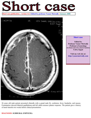
- 1. Short case publication... version 1.6 | Edited by professor Yasser Metwally | January 2008 Short case Edited by Professor Yasser Metwally Professor of neurology Ain Shams university school of medicine Cairo, Egypt Visit my web site at: http://yassermetwally.com 26 years old male patient presented clinically with a grand male fit, confusion, fever, headache, and nausea. Examination showed bilateral papilledema and left sided extensor planter response. The patient gave a history of nasal sinusitis two weeks before clinical presentation. DIAGNOSIS: SUBDURAL EMPYEMA
- 2. Figure 1. MRI T1 pre and post contrast scans showing two hypointense, oval, elongated, lentiform-like cystic lesions in the medial frontal (near the frontopolar region) and medial occipital regions. The lateral borders of the lesions are crescent shaped and the medial borders are flattened. The signal intensity of the intracystic fluid is higher than that of the CSF. The lesions are extraaxial in location, orientated along the falx cerebri and interhemispheric fissure and their capsules are densely enhanced after contrast injection. The pachymeninges are thickened and showed dense contrast enhancement. Notice poor or nonvisualization of the cortical sulci, and medial displacement of the white matter digitations (quot;white matter bucklingquot;) mostly due to diffuse brain edema. Some degree of venous congestion is probably present. Figure 2. A case of subdural empyema (abscess). Postcontrast MRI T1 images showing bilateral oval and elongated extra-axial cystic lesions with densely enhanced capsules orientated along the falx cerebri, tentorial cerebelli and the interhemispheric fissure with medial fattened borders and lateral crescent-shaped borders. The pachymeninges are thickened and showed dense contrast enhancement. The nasal sinuses on the left side show thickened mucosa. Notice poor or nonvisualization of the cortical sulci, and medial displacement of the white matter digitations (quot;white matter bucklingquot;), mostly due to diffuse brain edema. Some degree of venous congestion is probably present.
- 3. Figure 3. A case of subdural empyema (abscess). Postcontrast MRI T1 images showing bilateral oval and elongated extra-axial cystic lesions with densely enhanced capsules orientated along the falx cerebri, tentorial cerebelli, with medial fattened borders and lateral crescent-shaped borders. Notice the medial frontopolar abscess with enhanced capsule, another abscess could be appreciated along the medial aspect of the tentorial ridge on the left side. The pachymeninges are thickened with dense contrast enhancement. The nasal sinuses show thickened mucosa. Notice poor or nonvisualization of the cortical sulci, and medial displacement of the white matter digitations (quot;white matter bucklingquot;), mostly due to diffuse brain edema. Some degree of venous congestion is probably present. Figure 4. MRI T2, and FLAIR studies showing the frontopolar extra-axial abscess surround by edema. The abscess has a hypointense capsule. The mechanism of the capsule hypointensity on the T2 weighted scan is not certain. Proposals in this respect include: 1-Relative lack of water in the fibrous capsule. 2-The presence of blood products in the abscess capsule[deoxyhaemoglobin induces T2 hypointensity,while methemoglobin induces T1 hyperintensity]. 3- The presence of paramagnetic free radicals within the phagocytosing macrophages which are heterogeneously distributed in the periphery of the abscess, paramagnetic free radicals induce T2 hypointensity. The signal intensity of the intracystic fluid is different from that of the CSF on the FLAIR image (C). Notice the parenchyma edema that surrounds the frontal extra-axial abscess. The subdural abscess along the tentorial ridge could also be seen as a hyperintense crescent- shaped lesion.
- 4. Figure 5. The extra-axial subdural abscesses have a hypointense capsule on the T2 and FLAIR images. The capsule is densely enhanced on the postcontrast T1 scans. Notice the parenchyma edema that surrounds the frontal extra-axial abscess. Box 1. Extra-axial subdural abscesses Extraaxial empyemas usually develop 1-2 weeks following sinusitis or mastoiditis by retrograde thrombophlebitis of the transdiploic veins. Subdural empyema (ie, abscess) is an intracranial focal collection of purulent material located between the dura mater and the arachnoid mater. About 95% of subdural empyemas are located within the cranium; most involve the frontal lobe, and 5% involve the spinal neuraxis. The subdural extra-axial abscesses are multiple, scattered along the falx and tentorial cerebelli, the interhemispheric fissure and extra-axial in location. The medial borders of the subdural abscesses are flattened and the lateral borders are crescent shaped. The medial growth of these abscesses are limited by the rigid dura resulting in flattening of the medial borders of the subdural abscesses along the the falx and tentorial cerebelli. SE is a primarily intracranial infection located between the dura mater and the arachnoid mater. It has a tendency to spread rapidly through the subdural space until limited by specific boundaries (eg, falx cerebri, tentorium cerebelli, base of the brain, foramen magnum). The subdural space has no septations except in areas where arachnoid granulations are attached to the dura mater. SE is usually unilateral. Pachymeningitis is invariable present with thickening and enhancement of the pachymeninges. The extraaxial subdural abscesses have a hypointense capsule on the T2 and flair images. The capsule is densely enhanced on the postcontrast T1 scans. The signal intensity of the fluid inside the abscess is different from that of the CSF.
- 5. Addendum A new version of this software is uploaded in my web site every week (every Saturday and remains available till Friday.) To download the current version follow the link quot;http://pdf.yassermetwally.com/short.pdfquot;. You can download the long case version of this short case during the same week from: http://pdf.yassermetwally.com/case.pdf or visit web site: http://pdf.yassermetwally.com To download the software version of the publication (crow.exe) follow the link: http://neurology.yassermetwally.com/ crow.zip At the end of each year, all the publications are compiled on a single CD-ROM, please contact the author to know more details. Screen resolution is better set at 1024*768 pixel screen area for optimum display References 1. Metwally, MYM: Textbook of neurimaging, A CD-ROM publication, (Metwally, MYM editor) WEB-CD agency for electronic publishing, version 9.1a January 2008