SlideShare a Scribd company logo
1 of 4
Download to read offline
Short case publication... Version 3.21 | Edited by professor Yasser Metwally | May 2010


                                                                                                           Short case

                                                                                                          Edited by
                                                                                                  Professor Yasser Metwally
                                                                                                    Professor of neurology
                                                                                                Ain Shams university school of medicine
                                                                                                           Cairo, Egypt

                                                                                                     Visit my web site at:
                                                                                                  http://yassermetwally.com


A 22 years old male patient presented clinically with bilateral cerebellar ataxia, bilateral pyramidal manifestations, lost tendon
jerk reflexes, peripheral neuropathy with peroneal muscular atrophy, enlarged peripheral nerves, kyphoscoliosis, and pes
cavus. Peripheral nerve biopsy revealed diffuse demyelinating neuropathy with onion bulb formation. The condition started
at the age of 12 and is gradually progressive. The clinical diagnosis of Friedreich ataxia was made.


DIAGNOSIS: FRIEDREICH'S ATAXIA
Figure 1. MRI of the brain showing normal findings


Figure 2. MRI T2 (A,B) and MRI T1 (C) showing marked atrophy of the uppermost part of the cervical spinal
cord
Figure 3. MRI T1 (A) and MRI T2 (B)
                                                                        showing marked atrophy of the
                                                                        uppermost part of the cervical spinal cord


Figure 4. MRI T2 images showings cervical cord atrophy, thinning with reduced anteroposterior diameter. Notice
the hyperintense line in posterior portion of cord. The thinned spinal cord is seen lying on the posterior wall of
spinal canal with increased signal intensity in its posterior and lateral compartments. The anterior subarachnoid
space is enlarged. The intramedullary signal changes reflect loss of myelinated fibers and gliosis.


References

1. Metwally, MYM: Textbook of neurimaging, A CD-ROM publication, (Metwally, MYM editor) WEB-CD agency for
electronic publishing, version 11.2a April 2010
Addendum

  A new version of short case is uploaded in my web site every week (every Saturday and remains available till Friday.)
  To download the current version follow the link "http://pdf.yassermetwally.com/short.pdf".
  You can download the long case version of this short case during the same week from: http://pdf.yassermetwally.com/case.pdf or visit web
  site: http://pdf.yassermetwally.com
  To download the software version of the publication (crow.exe) follow the link: http://neurology.yassermetwally.com/crow.zip
  At the end of each year, all the publications are compiled on a single CD-ROM, please contact the author to know more
  details.
  Also to view a list of the previously published case records follow the following link:
   (http://wordpress.com/tag/case-record/) or click on it if it appears as a link in your PDF reader
  To inspect the patient's full radiological study, click on the attachment icon (the paper clip icon in the left pane) of the acrobat
  reader then double click on the attached file
  Click here to download the long case version of this short case in PDF format

More Related Content

What's hot

Presentation2, radiological imaging of neurodegenerative and dementai disease...
Presentation2, radiological imaging of neurodegenerative and dementai disease...Presentation2, radiological imaging of neurodegenerative and dementai disease...
Presentation2, radiological imaging of neurodegenerative and dementai disease...
Abdellah Nazeer
 
Presentation1.pptx. radiological imaging of epilepsy.
Presentation1.pptx. radiological imaging of epilepsy.Presentation1.pptx. radiological imaging of epilepsy.
Presentation1.pptx. radiological imaging of epilepsy.
Abdellah Nazeer
 
Presentation1.pptx, radiological imaging of congenital anomalies of the spine...
Presentation1.pptx, radiological imaging of congenital anomalies of the spine...Presentation1.pptx, radiological imaging of congenital anomalies of the spine...
Presentation1.pptx, radiological imaging of congenital anomalies of the spine...
Abdellah Nazeer
 

What's hot (20)

Neuroimaging of alzheimer's
Neuroimaging of alzheimer'sNeuroimaging of alzheimer's
Neuroimaging of alzheimer's
 
Presentation2, radiological imaging of neurodegenerative and dementai disease...
Presentation2, radiological imaging of neurodegenerative and dementai disease...Presentation2, radiological imaging of neurodegenerative and dementai disease...
Presentation2, radiological imaging of neurodegenerative and dementai disease...
 
Tethered Cord Syndrome
Tethered Cord SyndromeTethered Cord Syndrome
Tethered Cord Syndrome
 
Presentation1.pptx. radiological imaging of epilepsy.
Presentation1.pptx. radiological imaging of epilepsy.Presentation1.pptx. radiological imaging of epilepsy.
Presentation1.pptx. radiological imaging of epilepsy.
 
SPINAL INTRAMEDULLARY TUMORS - COMPARISON OF MRI, INTRAOPERATIVE CHARACTERISTICS
SPINAL INTRAMEDULLARY TUMORS - COMPARISON OF MRI, INTRAOPERATIVE CHARACTERISTICSSPINAL INTRAMEDULLARY TUMORS - COMPARISON OF MRI, INTRAOPERATIVE CHARACTERISTICS
SPINAL INTRAMEDULLARY TUMORS - COMPARISON OF MRI, INTRAOPERATIVE CHARACTERISTICS
 
Presentation1.pptx, radiological imaging of congenital anomalies of the spine...
Presentation1.pptx, radiological imaging of congenital anomalies of the spine...Presentation1.pptx, radiological imaging of congenital anomalies of the spine...
Presentation1.pptx, radiological imaging of congenital anomalies of the spine...
 
Spina bifida
Spina bifidaSpina bifida
Spina bifida
 
Case record...Intramedullary ependymoma
Case record...Intramedullary ependymomaCase record...Intramedullary ependymoma
Case record...Intramedullary ependymoma
 
power point presentation on nerve injury introduction by clinical approach
power point presentation on nerve injury introduction by clinical approachpower point presentation on nerve injury introduction by clinical approach
power point presentation on nerve injury introduction by clinical approach
 
Facial nerve extratemporal
Facial nerve extratemporalFacial nerve extratemporal
Facial nerve extratemporal
 
Case record...Wilson disease
Case record...Wilson diseaseCase record...Wilson disease
Case record...Wilson disease
 
case report
case reportcase report
case report
 
Imaging in MESIAL TEMPORAL EPILESPY
Imaging in MESIAL TEMPORAL EPILESPYImaging in MESIAL TEMPORAL EPILESPY
Imaging in MESIAL TEMPORAL EPILESPY
 
Bony tumors of spine
Bony tumors of spineBony tumors of spine
Bony tumors of spine
 
Case record... Spinal dysraphism
Case record... Spinal dysraphismCase record... Spinal dysraphism
Case record... Spinal dysraphism
 
CRANIOSPINAL IRRADIATION-PART 1
CRANIOSPINAL IRRADIATION-PART 1CRANIOSPINAL IRRADIATION-PART 1
CRANIOSPINAL IRRADIATION-PART 1
 
Case record...Spinal multiple sclerosis
Case record...Spinal multiple sclerosisCase record...Spinal multiple sclerosis
Case record...Spinal multiple sclerosis
 
Central cord syndrome
Central cord syndromeCentral cord syndrome
Central cord syndrome
 
Imaging in epilepsy1
Imaging in epilepsy1Imaging in epilepsy1
Imaging in epilepsy1
 
Congenital Anomalies Of Spine And Spinal Cord
Congenital Anomalies Of Spine And Spinal CordCongenital Anomalies Of Spine And Spinal Cord
Congenital Anomalies Of Spine And Spinal Cord
 

Similar to Short case...Friedreich ataxia

Similar to Short case...Friedreich ataxia (20)

Short case...Neurofibromatosis type 2
Short case...Neurofibromatosis type 2Short case...Neurofibromatosis type 2
Short case...Neurofibromatosis type 2
 
Short case...Neuromyelitis optica
Short case...Neuromyelitis opticaShort case...Neuromyelitis optica
Short case...Neuromyelitis optica
 
Short case...Neuro-Behcet
Short case...Neuro-BehcetShort case...Neuro-Behcet
Short case...Neuro-Behcet
 
Short case...Cerebral palsy
Short case...Cerebral palsyShort case...Cerebral palsy
Short case...Cerebral palsy
 
Short case...Neuromyelitis optica
Short case...Neuromyelitis opticaShort case...Neuromyelitis optica
Short case...Neuromyelitis optica
 
Short case...Paraneoplastic leukoencephalopathy
Short case...Paraneoplastic leukoencephalopathyShort case...Paraneoplastic leukoencephalopathy
Short case...Paraneoplastic leukoencephalopathy
 
Short case...Lumbar spondylosis
Short case...Lumbar spondylosisShort case...Lumbar spondylosis
Short case...Lumbar spondylosis
 
Short case...Spinal multiple sclerosis
Short case...Spinal multiple sclerosisShort case...Spinal multiple sclerosis
Short case...Spinal multiple sclerosis
 
Short case...Parasellar craniopharyngioma
Short case...Parasellar craniopharyngiomaShort case...Parasellar craniopharyngioma
Short case...Parasellar craniopharyngioma
 
Short case...Spinal dural arteriovenous fistula withn congestive myelopathy
Short case...Spinal dural arteriovenous fistula withn congestive myelopathyShort case...Spinal dural arteriovenous fistula withn congestive myelopathy
Short case...Spinal dural arteriovenous fistula withn congestive myelopathy
 
Short case...Myxopapillary ependymoma
Short case...Myxopapillary ependymomaShort case...Myxopapillary ependymoma
Short case...Myxopapillary ependymoma
 
Short case...Spinal metastasis
Short case...Spinal metastasisShort case...Spinal metastasis
Short case...Spinal metastasis
 
Short case...Parasagittal meningioma
Short case...Parasagittal meningiomaShort case...Parasagittal meningioma
Short case...Parasagittal meningioma
 
Short case...Acute postinfectious transverse myelitis
Short case...Acute postinfectious transverse myelitisShort case...Acute postinfectious transverse myelitis
Short case...Acute postinfectious transverse myelitis
 
Short case...Subependymal primary CNS lymphoma
Short case...Subependymal primary CNS lymphomaShort case...Subependymal primary CNS lymphoma
Short case...Subependymal primary CNS lymphoma
 
Short case... Arterial ectasia of the vertebrobasilar system
Short case... Arterial ectasia of the vertebrobasilar systemShort case... Arterial ectasia of the vertebrobasilar system
Short case... Arterial ectasia of the vertebrobasilar system
 
Short case...Thalamic glioma
Short case...Thalamic gliomaShort case...Thalamic glioma
Short case...Thalamic glioma
 
Short case...Epidural secondary CNS lymphoma
Short case...Epidural secondary CNS lymphomaShort case...Epidural secondary CNS lymphoma
Short case...Epidural secondary CNS lymphoma
 
Short case...Spinal dural arteriovenous fistula
Short case...Spinal dural arteriovenous fistulaShort case...Spinal dural arteriovenous fistula
Short case...Spinal dural arteriovenous fistula
 
Short case...tectal plate glioma
Short case...tectal plate gliomaShort case...tectal plate glioma
Short case...tectal plate glioma
 

More from Professor Yasser Metwally

More from Professor Yasser Metwally (20)

The Egyptian Zoo in Cairo 2015
The Egyptian Zoo in Cairo 2015The Egyptian Zoo in Cairo 2015
The Egyptian Zoo in Cairo 2015
 
End of the great nile river in Ras Elbar
End of the great nile river in Ras ElbarEnd of the great nile river in Ras Elbar
End of the great nile river in Ras Elbar
 
The Lion and The tiger in Egypt
The Lion and The tiger in EgyptThe Lion and The tiger in Egypt
The Lion and The tiger in Egypt
 
The monkeys in Egypt
The monkeys in EgyptThe monkeys in Egypt
The monkeys in Egypt
 
The Snake, the Scorpion, the turtle in Egypt
The Snake, the Scorpion, the turtle in EgyptThe Snake, the Scorpion, the turtle in Egypt
The Snake, the Scorpion, the turtle in Egypt
 
The Egyptian Parrot
The Egyptian ParrotThe Egyptian Parrot
The Egyptian Parrot
 
The Egyptian Deer
The Egyptian DeerThe Egyptian Deer
The Egyptian Deer
 
The Egyptian Pelican
The Egyptian PelicanThe Egyptian Pelican
The Egyptian Pelican
 
The Flamingo bird in Egypt
The Flamingo bird in EgyptThe Flamingo bird in Egypt
The Flamingo bird in Egypt
 
Egyptian Cats
Egyptian CatsEgyptian Cats
Egyptian Cats
 
Radiological pathology of epileptic disorders
Radiological pathology of epileptic disordersRadiological pathology of epileptic disorders
Radiological pathology of epileptic disorders
 
Radiological pathology of cerebrovascular disorders
Radiological pathology of cerebrovascular disordersRadiological pathology of cerebrovascular disorders
Radiological pathology of cerebrovascular disorders
 
Radiological pathology of spontaneous cerebral hemorrhage
Radiological pathology of spontaneous cerebral hemorrhageRadiological pathology of spontaneous cerebral hemorrhage
Radiological pathology of spontaneous cerebral hemorrhage
 
Radiological pathology of cerebral amyloid angiography
Radiological pathology of cerebral amyloid angiographyRadiological pathology of cerebral amyloid angiography
Radiological pathology of cerebral amyloid angiography
 
Radiological pathology of cerebral microbleeds
Radiological pathology of cerebral microbleedsRadiological pathology of cerebral microbleeds
Radiological pathology of cerebral microbleeds
 
The Egyptian Zoo in Cairo
The Egyptian Zoo in CairoThe Egyptian Zoo in Cairo
The Egyptian Zoo in Cairo
 
Progressive multifocal leukoencephalopathy
Progressive multifocal leukoencephalopathyProgressive multifocal leukoencephalopathy
Progressive multifocal leukoencephalopathy
 
Progressive multifocal leukoencephalopathy
Progressive multifocal leukoencephalopathyProgressive multifocal leukoencephalopathy
Progressive multifocal leukoencephalopathy
 
Issues in radiological pathology: Radiological pathology of watershed infarct...
Issues in radiological pathology: Radiological pathology of watershed infarct...Issues in radiological pathology: Radiological pathology of watershed infarct...
Issues in radiological pathology: Radiological pathology of watershed infarct...
 
Radiological pathology of cortical laminar necrosis
Radiological pathology of cortical laminar necrosisRadiological pathology of cortical laminar necrosis
Radiological pathology of cortical laminar necrosis
 

Recently uploaded

Cardiac Impulse: Rhythmical Excitation and Conduction in the Heart
Cardiac Impulse: Rhythmical Excitation and Conduction in the HeartCardiac Impulse: Rhythmical Excitation and Conduction in the Heart
Cardiac Impulse: Rhythmical Excitation and Conduction in the Heart
MedicoseAcademics
 

Recently uploaded (20)

BMK Glycidic Acid (sodium salt) CAS 5449-12-7 Pharmaceutical intermediates
BMK Glycidic Acid (sodium salt)  CAS 5449-12-7 Pharmaceutical intermediatesBMK Glycidic Acid (sodium salt)  CAS 5449-12-7 Pharmaceutical intermediates
BMK Glycidic Acid (sodium salt) CAS 5449-12-7 Pharmaceutical intermediates
 
รายการตํารับยาแผนไทยแห่งชาติ ฉบับ พ.ศ. 2564.pdf
รายการตํารับยาแผนไทยแห่งชาติ ฉบับ พ.ศ. 2564.pdfรายการตํารับยาแผนไทยแห่งชาติ ฉบับ พ.ศ. 2564.pdf
รายการตํารับยาแผนไทยแห่งชาติ ฉบับ พ.ศ. 2564.pdf
 
Impact of cancers therapies on the loss in cardiac function, myocardial fffic...
Impact of cancers therapies on the loss in cardiac function, myocardial fffic...Impact of cancers therapies on the loss in cardiac function, myocardial fffic...
Impact of cancers therapies on the loss in cardiac function, myocardial fffic...
 
Denture base resins materials and its mechanism of action
Denture base resins materials and its mechanism of actionDenture base resins materials and its mechanism of action
Denture base resins materials and its mechanism of action
 
Hemodialysis: Chapter 2, Extracorporeal Blood Circuit - Dr.Gawad
Hemodialysis: Chapter 2, Extracorporeal Blood Circuit - Dr.GawadHemodialysis: Chapter 2, Extracorporeal Blood Circuit - Dr.Gawad
Hemodialysis: Chapter 2, Extracorporeal Blood Circuit - Dr.Gawad
 
DECIPHERING COMMON ECG FINDINGS IN ED.pptx
DECIPHERING COMMON ECG FINDINGS IN ED.pptxDECIPHERING COMMON ECG FINDINGS IN ED.pptx
DECIPHERING COMMON ECG FINDINGS IN ED.pptx
 
End Feel -joint end feel - Normal and Abnormal end feel
End Feel -joint end feel - Normal and Abnormal end feelEnd Feel -joint end feel - Normal and Abnormal end feel
End Feel -joint end feel - Normal and Abnormal end feel
 
linearity concept of significance, standard deviation, chi square test, stude...
linearity concept of significance, standard deviation, chi square test, stude...linearity concept of significance, standard deviation, chi square test, stude...
linearity concept of significance, standard deviation, chi square test, stude...
 
MRI Artifacts and Their Remedies/Corrections.pptx
MRI Artifacts and Their Remedies/Corrections.pptxMRI Artifacts and Their Remedies/Corrections.pptx
MRI Artifacts and Their Remedies/Corrections.pptx
 
180-hour Power Capsules For Men In Ghana
180-hour Power Capsules For Men In Ghana180-hour Power Capsules For Men In Ghana
180-hour Power Capsules For Men In Ghana
 
Creating Accessible Public Health Communications
Creating Accessible Public Health CommunicationsCreating Accessible Public Health Communications
Creating Accessible Public Health Communications
 
Video capsule endoscopy (VCE ) in children
Video capsule endoscopy (VCE ) in childrenVideo capsule endoscopy (VCE ) in children
Video capsule endoscopy (VCE ) in children
 
TUBERCULINUM-2.BHMS.MATERIA MEDICA.HOMOEOPATHY
TUBERCULINUM-2.BHMS.MATERIA MEDICA.HOMOEOPATHYTUBERCULINUM-2.BHMS.MATERIA MEDICA.HOMOEOPATHY
TUBERCULINUM-2.BHMS.MATERIA MEDICA.HOMOEOPATHY
 
Effects of vaping e-cigarettes on arterial health
Effects of vaping e-cigarettes on arterial healthEffects of vaping e-cigarettes on arterial health
Effects of vaping e-cigarettes on arterial health
 
Cardiac Impulse: Rhythmical Excitation and Conduction in the Heart
Cardiac Impulse: Rhythmical Excitation and Conduction in the HeartCardiac Impulse: Rhythmical Excitation and Conduction in the Heart
Cardiac Impulse: Rhythmical Excitation and Conduction in the Heart
 
Muscle Energy Technique (MET) with variant and techniques.
Muscle Energy Technique (MET) with variant and techniques.Muscle Energy Technique (MET) with variant and techniques.
Muscle Energy Technique (MET) with variant and techniques.
 
CURRENT HEALTH PROBLEMS AND ITS SOLUTION BY AYURVEDA.pptx
CURRENT HEALTH PROBLEMS AND ITS SOLUTION BY AYURVEDA.pptxCURRENT HEALTH PROBLEMS AND ITS SOLUTION BY AYURVEDA.pptx
CURRENT HEALTH PROBLEMS AND ITS SOLUTION BY AYURVEDA.pptx
 
SURGICAL ANATOMY OF ORAL IMPLANTOLOGY.pptx
SURGICAL ANATOMY OF ORAL IMPLANTOLOGY.pptxSURGICAL ANATOMY OF ORAL IMPLANTOLOGY.pptx
SURGICAL ANATOMY OF ORAL IMPLANTOLOGY.pptx
 
In-service education (Nursing Mangement)
In-service education (Nursing Mangement)In-service education (Nursing Mangement)
In-service education (Nursing Mangement)
 
Is Rheumatoid Arthritis a Metabolic Disorder.pptx
Is Rheumatoid Arthritis a Metabolic Disorder.pptxIs Rheumatoid Arthritis a Metabolic Disorder.pptx
Is Rheumatoid Arthritis a Metabolic Disorder.pptx
 

Short case...Friedreich ataxia

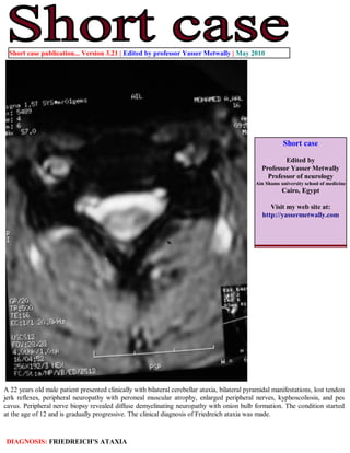
  • 1. Short case publication... Version 3.21 | Edited by professor Yasser Metwally | May 2010 Short case Edited by Professor Yasser Metwally Professor of neurology Ain Shams university school of medicine Cairo, Egypt Visit my web site at: http://yassermetwally.com A 22 years old male patient presented clinically with bilateral cerebellar ataxia, bilateral pyramidal manifestations, lost tendon jerk reflexes, peripheral neuropathy with peroneal muscular atrophy, enlarged peripheral nerves, kyphoscoliosis, and pes cavus. Peripheral nerve biopsy revealed diffuse demyelinating neuropathy with onion bulb formation. The condition started at the age of 12 and is gradually progressive. The clinical diagnosis of Friedreich ataxia was made. DIAGNOSIS: FRIEDREICH'S ATAXIA
  • 2. Figure 1. MRI of the brain showing normal findings Figure 2. MRI T2 (A,B) and MRI T1 (C) showing marked atrophy of the uppermost part of the cervical spinal cord
  • 3. Figure 3. MRI T1 (A) and MRI T2 (B) showing marked atrophy of the uppermost part of the cervical spinal cord Figure 4. MRI T2 images showings cervical cord atrophy, thinning with reduced anteroposterior diameter. Notice the hyperintense line in posterior portion of cord. The thinned spinal cord is seen lying on the posterior wall of spinal canal with increased signal intensity in its posterior and lateral compartments. The anterior subarachnoid space is enlarged. The intramedullary signal changes reflect loss of myelinated fibers and gliosis. References 1. Metwally, MYM: Textbook of neurimaging, A CD-ROM publication, (Metwally, MYM editor) WEB-CD agency for electronic publishing, version 11.2a April 2010
  • 4. Addendum A new version of short case is uploaded in my web site every week (every Saturday and remains available till Friday.) To download the current version follow the link "http://pdf.yassermetwally.com/short.pdf". You can download the long case version of this short case during the same week from: http://pdf.yassermetwally.com/case.pdf or visit web site: http://pdf.yassermetwally.com To download the software version of the publication (crow.exe) follow the link: http://neurology.yassermetwally.com/crow.zip At the end of each year, all the publications are compiled on a single CD-ROM, please contact the author to know more details. Also to view a list of the previously published case records follow the following link: (http://wordpress.com/tag/case-record/) or click on it if it appears as a link in your PDF reader To inspect the patient's full radiological study, click on the attachment icon (the paper clip icon in the left pane) of the acrobat reader then double click on the attached file Click here to download the long case version of this short case in PDF format