1. Resident Grand Rounds
Series Editor: Mark A. Perazella, MD
Rhabdomyolysis
Lauren A. Walter, MD
Michael H. Catenacci, MD
A 32-year-old man with no significant past medical history presented to the emergency department with complaints of weak-
ness, myalgias, and muscle stiffness. Physical examination was notable for a low-grade fever at 100.8°F, mild tachycardia at 110 bpm,
and moderate tenderness on palpation of the muscle groups of the calves, thighs, biceps, triceps, and shoulder girdle. Cardiac aus-
cultation revealed no murmurs, neurologic examination showed no focal deficits, and skin examination was normal. On further
questioning, the patient admitted to using crack cocaine for 2 days. Notable laboratory values were as follows: a white blood cell
count of 15,000 cells/µL (normal, 4500–11,000 cells/µL), creatine kinase level of 16,500 IU/L (normal, 40–150 IU/L), blood urea
nitrogen level of 29 mg/dL (normal, 8–23 mg/dL), creatinine level of 1.1 mg/dL (normal, 0.6–1.2 mg/dL), and potassium level of
4.0 mEq/L (normal, 40–150 mEq/L). Urine myoglobin was not detected, urinalysis was normal, and urine toxicologic screen
was positive for cocaine. Rapid influenza testing was negative. The patient was admitted to the medical floor with a diagnosis of
cocaine-induced rhabdomyolysis.
R
habdomyolysis is an uncommon disease cium levels are also maintained by active sequestration
process with profound sequelae if it is not into the sarcoplasmic reticulum and mitochondria.
identified and treated expediently. Approxi- Damage to the myocyte cell membrane may be
mately 26,000 cases of rhabdomyolysis are re- caused directly through trauma (eg, crush injury) or
ported annually in the United States.1 Rhabdomyolysis indirectly through lack of adequate energy in the form
accounts for an estimated 8% to 15% of cases of acute of ATP (eg, vigorous sustained exercise). Regardless of
renal failure and is associated with a mortality rate of the cause, damage to the sarcolemma leads to a loss of
5%.2,3 Morbidity and mortality are usually the result of ionic gradients, thus increasing intracellular calcium.4
hyperkalemia, metabolic acidosis, and acute renal fail- This influx of calcium increases the activity of intracel-
ure. Clinical presentation varies, ranging from a nearly lular proteolytic enzymes that degrade the muscle cell.
asymptomatic illness to a fulminant and life-threatening As the myocyte degenerates, intracellular compounds
disease process with multiorgan system failure. This are extruded into the extracellular fluid and plasma.
article reviews the pathophysiology, etiology, clinical fea- These compounds may include myoglobin, aldolase,
tures, and management of rhabdomyolysis. potassium, uric acid, lactate dehydrogenase, aspartate
transaminase, creatine kinase (CK), and phosphate.5,6
PATHOPHYSIOLOGY In excess, these substances may have toxic effects on
Rhabdomyolysis is a clinical syndrome caused by distant organ systems.
injury to striated muscle. Despite the numerous condi- During myocyte destruction, the level of free myo-
tions that can cause rhabdomyolysis, there is a single globin in the plasma increases, resulting in higher
common pathway involving injury to skeletal muscle, quantities of myoglobin that are filtered by the kid-
breakdown of the myocyte cell membrane, and release neys.7 Myoglobinemia and myoglobinuria have long
of intracellular contents into the extracellular fluid been associated with the development of acute kidney
and circulation. The normal cellular function of the injury in rhabdomyolysis.8 Myoglobin is directly toxic to
myocyte is maintained by ionic gradients generated by renal tubular cells, a process that is likely mediated by
adenosine triphosphate (ATP)–dependent pumps em-
bedded within the cell membrane. Sodium-potassium
pumps maintain a low intracellular sodium level, which Dr. Walter is a resident and Dr. Catenacci is an assistant professor and as-
favors efflux of calcium in exchange for sodium by a sistant residency program director; both are at the Department of Emergency
separate ion exchange channel. Low intracellular cal- Medicine, University of Alabama at Birmingham, Birmingham, AL.
www.turner-white.com Hospital Physician January 2008 25
2. Walter & Catenacci : Rhabdomyolysis : pp. 25–31
TAKE HOME POINTS
Direct toxicity
of myoglobin on
• The single common pathophysiologic pathway in renal tubular cells
rhabdomyolysis involves damage to the myocyte
cell membrane, extrusion of intracellular muscle
contents into the circulation, and toxic effects on
distant organ systems.
• The diagnosis of rhabdomyolysis is best achieved
through careful clinical suspicion in combination
with an elevated serum creatine kinase level. Acute
kidney injury
• Renal injury may be averted through aggressive
intravascular volume replacement, maintenance of
high urinary flows, urinary alkalinization, and man-
nitol therapy.
• Indications for dialysis include refractory hyperka-
Cast formation, Hypovolemia
lemia, refractory acidosis, and volume overload.
decreasing and decreased
• Early diagnosis, combined with aggressive treat- tubular flow renal perfusion
ment of complications, may decrease morbidity
and mortality.
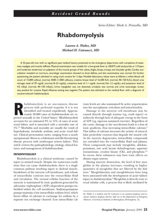
Figure. Etiology of acute kidney injury in patients with rhabdo-
myolysis.
free radicals.9,10 In the presence of acidosis and hypo-
volemia, myoglobin reacts with Tamm-Horsfall protein as DIC, is paramount to ensure their early detection
and precipitates into casts, which may then obstruct (Table 1).
tubular flow. Hypovolemia and overall decreased renal
perfusion also can compound renal injury. The Figure ETIOLOGY
depicts the multifactorial nature of acute kidney injury In the United States, the most common causes
in rhabdomyolysis patients. Rhabdomyolysis-induced of rhabdomyolysis are muscle overexertion, muscle
acute kidney injury is principally caused by damage to compression, and the use of illicit drugs (eg, cocaine,
the renal parenchyma and is thus classified as acute amphetamines) or alcohol.12 However, a myriad of eti-
intrinsic renal failure (AIRF). This syndrome is associ- ologies has been reported as inciting factors. Traumatic
ated with a low specific gravity of urine, pigmented causes of rhabdomyolysis include blunt trauma, crush
casts, and a high fractional excretion of sodium. injury, and strenuous exercise. Nontraumatic etiologies
The release of intracellular electrolytes from dying can be broadly divided into toxicologic, infectious, and
myocytes may be life-threatening. Rapid release of metabolic causes. The classic case of crush injury lead-
intracellular potassium, especially in the setting of ing to skeletal muscle destruction and rhabdomyolysis
acute kidney injury and metabolic acidosis, may pre- is a familiar textbook presentation. However, recogni-
cipitate malignant cardiac dysrhythmias. Heart blocks, tion of rhabdomyolysis caused by toxins, infections (eg,
ventricular tachycardia, ventricular fibrillation, pulse- influenza, HIV), or hereditary metabolic myopathies
less electrical activity, and asystole may occur with little requires a higher degree of clinical suspicion.
warning. Intracellular phosphorus may rapidly precipi-
tate in muscle tissues with calcium, which is reflected Toxins
by the early development of hyperphosphatemia and As illustrated by the case presentation, cocaine is a
hypocalcemia. common cause of rhabdomyolysis, particularly in urban
The hematologic system and clotting cascade also patient populations. Cocaine-induced muscle injury
may be affected by rhabdomyolysis. Necrosis of muscle, may occur through multiple mechanisms: vasospasm
in combination with release of tissue thromboplastin, with muscular ischemia, seizures, hyperpyrexia, coma
may lead to disseminated intravascular coagulation with muscle compression, and direct myofibrillar dam-
(DIC) and hemorrhagic complications.11 Aggressively age.13 In a series of patients with cocaine-induced rhab-
monitoring for complications of rhabdomyolysis, such domyolysis, 13 of 39 patients developed acute kidney
26 Hospital Physician January 2008 www.turner-white.com
3. Walter & Catenacci : Rhabdomyolysis : pp. 25–31
injury, the patients’ mean CK level was 12,187 IU/L Table 1. Complications Associated with Rhabdomyolysis
(range, 1756–85,000 IU/L), and 6 patients died.14 Acute renal failure
Numerous medications have been implicated in
Disseminated intravascular coagulation
cases of rhabdomyolysis, including zidovudine, colchi-
Electrolyte and metabolic derangements
cine, isoniazid, opiates, benzodiazepines, corticosteroids,
Hypoalbuminemia
statins, and fibric acid derivatives. Statin drugs inhibit the
3-hydroxy-3-methylglutaryl coenzyme A reductase and Hypocalcemia (early)
are potent reducers of low-density lipoprotein cholester- Hypercalcemia (late)
ol. As these drugs are commonly prescribed, they merit Hyperkalemia
particular attention. In rare cases, statin drugs may cause Hypernatremia
myopathy and life-threatening rhabdomyolysis. The inci- Hyperphosphatemia
dence of life-threatening rhabdomyolysis appears to be Hyperuricemia
quite low with statin monotherapy (0.44 per 10,000 years Cardiac dysrhythmias
of patient use), with the exception of cerivastatin, which Compartment syndromes
was withdrawn from the market voluntarily by its manu- Shock
facturer in 2001. Increased risk of developing rhabdo-
Death
myolysis occurs in elderly persons, in diabetic patients,
and when a statin is combined with a fibric acid deriva-
tive (5.98 per 10,000 years of patient use).15 Statins block
production of farnesyl pyrophosphate, an intermediate CLINICAL FEATURES
in the synthesis of coenzyme Q10 (Co Q10). Co Q10 is The clinical presentation of rhabdomyolysis is di-
important in mitochondrial energy production. It has verse. Some patients present with an acute medical
been hypothesized that statin-induced Co Q10 defi- or traumatic condition with rhabdomyolysis as a clear
ciency is involved in the pathogenesis of statin myopa- complication. In other patients, rhabdomyolysis may
thy, and that supplemental Co Q10 may reduce risk in be found by laboratory testing alone, prompting a
certain patient populations.16 search for an inciting condition. Classically, patients
with rhabdomyolysis report myalgias, muscle weakness
Infections and swelling, and dark-colored urine. Nonspecific
The pathogenesis of rhabdomyolysis associated systemic symptoms, such as malaise, fever, abdominal
with infections (whether bacterial, viral, or fungal) pain, and nausea and vomiting, may also be seen.
is thought to be the result of direct cell invasion and Initial assessment of symptoms may prove difficult in
cellular degeneration by the pathogen.17 In adult patients with altered mental status, intoxication, elec-
patients, Legionella species are classically associated trolyte imbalance, or uremic encephalopathy.
with rhabdomyolysis. Other bacteria linked to rhab- Physical examination may show signs of dehydra-
domyolysis include Salmonella species, group A β- tion, such as dry mucous membranes, decreased skin
hemolytic streptococci, Francisella tularensis, and Esche- turgor, and delayed capillary refill. The overlying skin
richia coli. Influenza A and B are the most common may be bruised or discolored if trauma has occurred.
viruses associated with rhabdomyolysis, while HIV re- With the development of a compartment syndrome,
mains an important consideration. Studies estimate that the affected area may demonstrate pain on passive
up to 25% of AIDS patients suffer from a myopathic range of motion, sensory deficits, motor deficits, or
disease that may be complicated by rhabdomyolysis.18 signs of vascular insufficiency (a late finding).
Genetic Disorders DIAGNOSIS
In cases where the etiology remains elusive, a genetic By definition, rhabdomyolysis is the breakdown of
disorder should be considered. Genetic disorders should skeletal muscle cells with the subsequent release of
be suspected particularly in pediatric patients with recur- intracellular contents. Assaying for elevated levels of
rent rhabdomyolysis after minimal to moderate exertion these intracellular contents establishes the diagnosis.
or following a viral infection. Any genetic disorder associ-
ated with decreased energy production may cause rhab- Serum Creatine Kinase
domyolysis, which can include disorders of carbohydrate Serum CK level is the most sensitive laboratory test
metabolism, fatty acid oxidation, nucleoside metabolism, for detecting rhabdomyolysis.19 As muscle cells degrade
myopathies, and mitochondrial defects. and release CK into the plasma, the degree of CK
www.turner-white.com Hospital Physician January 2008 27
4. Walter & Catenacci : Rhabdomyolysis : pp. 25–31
elevation correlates directly with the degree of muscle idly cleared via plasma metabolism and urinary excre-
necrosis. Serum CK levels begin to rise 2 to 12 hours tion. This rapid clearance results in normal levels of
after muscle injury, peak at 1 to 3 days, and usually de- myoglobin within 1 to 6 hours following onset of mus-
cline within 3 to 5 days after muscle injury ceases.20 CK cle injury. As such, serum and urine myoglobin levels
levels that remain persistently elevated are indicative of may be only transiently abnormal in some cases of
continued muscle injury, a compartment syndrome, or rhabdomyolysis and therefore should not be relied
decreased renal clearance due to acute kidney injury.2 upon for a definitive diagnosis.
Rhabdomyolysis cannot be defined by a specific
CK level. Most authorities would agree that a 5-fold or Other Diagnostic Studies
greater increase in serum CK is consistent with the di- Other useful laboratory tests include measurement
agnosis, although levels 40 times greater than normal of serum electrolytes. Early in the course of illness,
may often be seen.21 Early rhabdomyolysis should be hyperkalemia, hyperphosphatemia, and hypocalcemia
suspected in at-risk patients with only a 2- to 3-fold in- are seen frequently. Assessment of serum potassium
crease in serum CK. Serial CK levels should be trended levels is essential for averting malignant cardiac dys-
for progression in these patients. rhythmias in rhabdomyolysis-induced acute kidney
Clinical context is also important to consider when injury. Measurement of urine electrolytes and creati-
evaluating CK levels, as higher levels do not always in- nine allows for computation of fractional excretion of
dicate a higher risk for complications. Young healthy sodium, which may help differentiate AIRF caused
athletes may have elevated CK levels as a normal con- by rhabdomyolysis (> 1%) from prerenal azotemia
sequence of muscle damage during vigorous physical (< 1%). If concomitant DIC is present, thrombocyto-
exertion. These patients would not be expected to ex- penia, hypofibrinogenemia, prolonged bleeding times,
perience complications or progress to acute renal fail- and an elevated d-dimer level may be seen.
ure. However, elderly debilitated patients with lower Finally, directed laboratory testing aimed at un-
elevations in total CK levels could progress to renal covering the precipitating cause of rhabdomyolysis is
failure and therefore would represent a greater clinical important. Diagnostic evaluations may include toxi-
concern. Such distinctions are important to make in cologic testing, bacteriologic cultures, viral assays, and
terms of treatment decisions, expected complications, radiographic imaging. Genetic analysis, nerve testing,
and prognosis. Contrast the case of an elderly woman muscle biopsy, and the forearm ischemic test may be
with chronic kidney disease and a total CK level of indicated in patients who are suspected of having an
5000 IU/L with that of a young healthy marathon run- underlying genetic abnormality. The forearm ischemic
ner with a total CK level of 7000 IU/L after a race. This test is performed as follows. Baseline urine myoglobin,
elderly patient requires aggressive management, includ- venous lactate, CK, and ammonia levels are obtained at
ing rehydration, bicarbonate infusion, and admission, rest prior to testing. A sphygmomanometer cuff is then
whereas the marathon runner may only require oral placed on the arm and inflated to 200 mm Hg to in-
rehydration in the emergency department followed by a duce ischemia. The patient is instructed to repetitively
repeat CK level several hours later. grasp an object firmly in the hand for 2 to 3 minutes.
The blood pressure cuff is then released and removed
Serum and Urine Myoglobin from the arm, and laboratory testing is repeated at 0, 5,
Serum and urine myoglobin levels appear to be less 10, and 20 minutes. Elevation of lactate and ammonia
sensitive tests for establishing the diagnosis of rhabdo- to levels below what is normally expected during anaer-
myolysis. Myoglobin is a skeletal muscle protein involved obic metabolism is evidence of a pathway disturbance,
in oxidative metabolism. Necrotic muscle cells release and an enzyme deficiency is suggested.23
myoglobin, which is then excreted in the urine when
the plasma concentration exceeds 1.5 mg/dL. Myoglo- MANAGEMENT
binuria causes the typical reddish-brown urine discolor- Prevention of Complications
ation seen with rhabdomyolysis, clinically appreciable If patients present in extremis, attention should be
when urine myoglobin exceeds 100 mg/dL.22 As myo- given to basic airway, breathing, and circulatory mea-
globin is a heme-containing compound, myoglobinuria sures (Table 2). On stabilization, prevention of the early
will result in a positive urine dipstick for blood despite and late complications of rhabdomyolysis becomes para-
the absence of red blood cells on microscopic analysis. mount in all patients. Management strategies should
Following muscle necrosis, myoglobinemia occurs be tailored to clinical context, which considers risk of
before CK elevation does and subsequently is rap- progression and complications. Variables such as the
28 Hospital Physician January 2008 www.turner-white.com
5. Walter & Catenacci : Rhabdomyolysis : pp. 25–31
inciting factor, patient age, patient comorbidities, and the Table 2. Managing Rhabdomyolysis
presence of preexisting renal disease should be assessed Prehospital care
when deciding upon the aggressiveness of therapy.
If rhabdomyolysis is suspected, establish peripheral access and begin
Volume resuscitation with isotonic crystalloid is the IV rehydration with normal saline
primary therapy for preventing rhabdomyolysis-induced
Initial hospital stabilization/treatment
renal injury. Increasing intravascular volume increases
glomerular filtration rate (GFR), dilutes myoglobin and Supportive care: ABC measures; treat associated life-threatening
injuries
other nephrotoxins extruded during muscle injury, and
Confirm/establish diagnosis with history, physical examination, labora-
improves overall oxygen delivery to ischemic tissue. In-
tory studies (eg, creatine kinase, creatinine, electrolytes, etc)
fusions of 10 to 15 mL/kg/hr of normal saline should
Rehydrate aggressively with normal saline at 10–15 mL/kg/hr to
be used initially, followed by hypotonic saline after achieve urinary output of 2 mL/kg/hr; switch to hypotonic saline
initial resuscitation is completed.24 Fluids should be after resuscitation is complete
titrated to an ideal urinary output of 2 mL/kg/hr.25,26 Continue rehydration for first 24–72 hr in moderate to severe cases
Infusion should continue until adequate resuscitation or until patient is hemodynamically stable
has occurred and clinical and chemical evidence of In moderate to severe cases with risk of progression to acute renal
myoglobinuria has disappeared (usually by the third failure, preexisting renal disease, or evidence of metabolic acidosis
day of hospitalization). Patients may require impressive and dehydration, consider urinary alkalinization. The goal urine pH
amounts of fluid resuscitation to maintain adequate uri- of ≥ 6.5 is achieved by adding 3 ampules of sodium bicarbonate to
1 L of 5% dextrose in water; the solution is infused at an initial rate
nary output, as considerable fluid may be sequestered of 100 mL/hr
in injured muscles. For optimal outcomes, vigorous
In the nonoliguric patient, consider mannitol 1g/kg IV over 30 min,
intravenous fluid rehydration should be started in the followed by 5 g/hr IV, for a total of 120 g/day; use mannitol to
prehospital setting in crush injury patients at risk for de- assist diuresis only in patients who have received adequate volume
veloping rhabdomyolysis.27 In patients with significant replacement
comorbidities such as heart failure, central venous pres- Monitor for and treat hyperkalemia aggressively
sure monitoring may be required to optimally assess the Monitor urinary output and renal function closely
patient’s volume status. Monitor for coagulopathy, compartment syndromes, and sepsis in
Additional measures are indicated to prevent acute severe cases
kidney injury in patients at moderate to high risk of Consider hemodialysis in conjunction with a nephrologist for:
renal injury. Predictors for the development of acute Fulminant renal failure with uremic encephalopathy
kidney injury include preexisting renal disease, a peak Uremic pericardial effusion with tamponade physiology
CK level in excess of 6000 IU/L, dehydration (hema- Refractory hyperkalemia, volume overload, or metabolic acidosis
tocrit > 50%, serum sodium level >150 mEq/L, ortho-
Attempt to identify the inciting factor and stop further muscle
stasis, pulmonary wedge pressure < 5 mm Hg, urinary damage and disease progression
fractional excretion of sodium < 1%), sepsis, hyperka-
Disposition
lemia or hyperphosphatemia on admission, and the
presence of hypoalbuminemia.2 Two such preventive In mild to moderate cases with stable electrolytes that are respond-
ing to rehydration, admit to a general medicine ward
measures are urinary alkalinization with sodium bicar-
In patients with electrolyte abnormalities or underlying cardiac or
bonate and the use of the osmotic diuretic mannitol.
renal disease, admit to a monitored bed
Dehydration and metabolic acidosis favor precipi-
In severe cases, including those with fulminant renal failure with
tation of myoglobin in renal tubules, enhancing and sequelae (pulmonary edema, symptomatic hyperkalemia, oliguria/
exacerbating its nephrotoxic effects. Urinary alkaliniza- anuria), persistent hypotension, or DIC, admit to intensive care unit
tion is thought to enhance renal myoglobin clearance
by increasing its solubility. Although large randomized ABC = airway, breathing, circulation; DIC = disseminated intravascular
coagulation; IV = intravenous.
trials are lacking, urinary alkalinization is recommend-
ed in patients with moderate to high risk of renal fail-
ure, preexisting renal disease, evidence of metabolic cause hypocalcemia and hypokalemia. Therefore, se-
acidosis, or significant dehydration. The goal urine pH rial measurements of both serum electrolytes and uri-
of 6.5 or higher can be obtained by adding 1 ampule nary pH should be performed.
of sodium bicarbonate (44 mEq) to 1 L of 50% normal Mannitol is an osmotic diuretic commonly used to
saline or 2 to 3 ampules (88–132 mEq) to 1 L of 5% expand intravascular volume, promote renal vasodila-
dextrose in water. This solution is then administered tion, and increase GFR in rhabdomyolysis patients.
at a rate of 100 mL/hour. Of note, alkalinization can Mannitol increases urine flow, which may help prevent
www.turner-white.com Hospital Physician January 2008 29
6. Walter & Catenacci : Rhabdomyolysis : pp. 25–31
obstruction from myoglobin-containing casts.28 Man- ney injury may develop in 30% to 40% of patients with
nitol also may draw fluid from the interstitial space, rhabdomyolysis. Indications for emergent hemodialysis
thus decreasing muscle edema in a concomitant com- include hyperkalemia with evidence of cardiac instabil-
partment syndrome.29 Mannitol is administered intra- ity, refractory metabolic acidosis, volume overload with
venously either as 1 g/kg over 30 minutes or as 25 g pulmonary edema, uremic pericardial effusion with
initially followed by 5 g/hr for a total of 120 g/day.30 tamponade, and progressive renal failure with uremic
Mannitol should be given only after adequate volume encephalopathy. Early consultation with a nephrologist
resuscitation has occurred and should be avoided in is recommended.
cases of oliguria. Loop diuretics (eg, furosemide) have Early in rhabdomyolysis, hyperphosphatemia and
been used to enhance urinary output in some oliguric hypocalcemia are seen as myocyte-released phosphate
rhabdomyolysis patients.31 However, they may acidi- precipitates with calcium in injured muscle. Early
fy the urine and worsen myoglobin-induced toxicity. treatment should be limited, as late hypercalcemia
Therefore, loop diuretics should be avoided in patients and hypophosphatemia will develop in most patients.
who have not been adequately hydrated. Late hypercalcemia, more common with concomitant
As mentioned previously, monitoring and treat- renal failure in advanced disease, may require volume
ment of electrolyte derangements in rhabdomyolysis expansion and diuretic therapy.
patients is critical. Hyperkalemia is a life-threatening A compartment syndrome may cause or complicate
complication of rhabdomyolysis, causing cardiac insta- rhabdomyolysis. Compartment syndrome occurs when
bility and dysrhythmias. Conventional treatment for the circulation to tissues within a closed space is com-
hyperkalemia (eg, calcium salts, sodium bicarbonate, promised by increased pressure within that space.33
glucose, insulin, albuterol, sodium polystyrene) should This syndrome may develop either early or late in the
be employed. In the presence of profound hyper- clinical course of rhabdomyolysis, particularly in a
phosphatemia caused by muscle necrosis, calcium salts traumatized limb due to crush injury. If compartment
may be less effective, as the administered calcium may syndrome is suspected clinically and intracompartmen-
combine with the extracellular phosphate rapidly.32 tal pressures exceed 35 mm Hg, emergent fasciotomy
should be considered.34
Admission As mentioned previously, DIC may be a life-
All patients with rhabdomyolysis should be admitted threatening complication seen in rhabdomyolysis pa-
for intravenous hydration, serial laboratory evaluation, tients. DIC in this setting is usually worse during the
and management of potential complications. Clinical third through fifth days after admission.11 Serial labo-
context should determine level of admission. An un- ratory measurements of coagulation times, platelet
monitored bed may be appropriate for healthy patients counts, and fibrinogen levels may be necessary. Life-
who respond to rehydration and have stable electrolyte threatening hemorrhage can occur, and it should be
levels. A monitored bed may be most appropriate for treated with fresh frozen plasma.
the first 24 to 48 hours, particularly in elderly patients,
severely injured patients, and patients who have car- SUMMARY
diac or renal comorbidities, as these patients tend to The various etiologies and clinical presentations of
develop hyperkalemia and cardiac dysrhythmias. Place- rhabdomyolysis are diverse. With nontraumatic causes
ment in the intensive care unit is appropriate for pa- of rhabdomyolysis, the physician must maintain a high
tients who develop severe complications, such as acute clinical suspicion in patients with predisposing factors.
kidney injury requiring dialysis, cardiac instability due History and physical examination may be suggestive,
to hyperkalemia, shock, and DIC. but laboratory confirmation of elevated CK levels is
In the appropriate setting, an otherwise healthy young essential in making the diagnosis. Management rests
patient, typically an athlete with a minimally elevated CK on the prevention and early identification of compli-
level, may be considered for outpatient management. cations. Acute kidney injury may be averted through
These patients should be able to orally rehydrate, dem- early and aggressive rehydration, alkalinization of the
onstrate a falling CK level on serial testing, and have urine with sodium bicarbonate, and judicious use of
stable electrolyte levels. Close primary care follow-up and mannitol. Life-threatening hyperkalemia, compart-
detailed discharge instructions are important. ment syndromes, and DIC should be anticipated and
treated immediately. With prompt recognition and
Management of Complications aggressive treatment, the morbidity and mortality of
Despite instituting preventive measures, acute kid- rhabdomyolysis may be diminished. HP
30 Hospital Physician January 2008 www.turner-white.com
7. Walter & Catenacci : Rhabdomyolysis : pp. 25–31
16. Marcoff L, Thompson PD. The role of coenzyme Q10 in stain-associated my-
Corresponding author: Michael H. Catenacci, MD, Department of opathy: a systematic review. J Am Coll Cardiol 2007;49:2231–7.
Emergency Medicine, JTN 266, University of Alabama at Birmingham, 17. Huerta-Aldin AL, Varon J, Marik PE. Bench-to-bedside review: Rhabdomyolysis
—an overview for clinicians. Crit Care 2005;9:158–69.
619 19th Street South, Birmingham, AL 35249; mcaten@uab.edu. 18. Authier FJ, Chariot P, Gherardi RK. Skeletal muscle involvement in human
immunodeficiency virus (HIV)-infected patients in the era of highly active
antiretroviral therapy (HAART). Muscle Nerve 2005;32: 247–60.
REFERENCES 19. Vanholder R, Sever MS, Erek E, Lameire N. Rhabdomyolysis. J Am Soc
1. Graves EJ, Gillum BS. Detailed diagnoses and procedures, National Hospital Nephrol 2000;11:1553–61.
Discharge Survey, 1995. Vital Health Stat 13 1997(130):1–146. 20. Tan W, Herzlich BC, Funaro, et al. Rhabdomyolysis and myoglobinuric acute
2. Ward MM. Factors predictive of acute renal failure in rhabdomyolysis. Arch renal failure associated with classic heat stroke. South Med J 1995;88:1065–8.
Intern Med 1988;148:1553–7. 21. Line RL, Rust GS. Acute exertional rhabdomyolysis. Am Fam Physician
3. Fernandez WG, Hung OL, Braen GR, Chiang WK. Epidemiology of rhab- 1995;52:502–6.
domyolysis and risk of developing renal failure in an urban hospital setting. 22. Loun B, Astles R, Copeland KR, Sedor FA. Adaptation of a quantitative im-
Acad Emerg Med 2000;7:575. munoassay for urine myoglobin. Predictor in detecting renal dysfunction.
4. Lopez JR, Rojas B, Gonzalez MA, Terzic A. Myoplasmic Ca2+ concentration Am J Clin Pathol 1996;105:479–86.
during exertional rhabdomyolysis. Lancet 1995;345:424–5. 23. Sever MS, Vanholder R, Lameire N. Management of crush-related injuries
5. Bontempo LD. Rhabdomyolysis. In: Marx JA, Hockberger RS, Walls RM, after disasters. N Engl J Med 2006;354:1052–63.
editors. Rosen’s emergency medicine: concepts and clinical practice. 6th ed. 24. Slater MS, Mullins RJ. Rhabdomyolysis and myoglobinuric renal failure in
Philadelphia: Mosby/Elsevier; 2006:1975–83. trauma and surgical patients: a review. J Am Coll Surg 1998;186:693–716.
6. Hamer R. When exercise goes awry: exertional rhabdomyolysis. South Med J 25. Curry SC, Chang D, Connor D. Drug- and toxin-induced rhabdomyolysis.
1997;90:548–51. Ann Emerg Med 1989;18:1068–84.
7. Lappalainen H, Tiula E, Uotila L, Manttari M. Elimination kinetics of myo- 26. Better OS. The crush syndrome revisited (1940–1990). Nephron 1990;55:
globin and creatine kinase in rhabdomyolysis: implications for follow-up. Crit 97–103.
Care Med 2002;30:2212–5. 27. Better OS, Rubinstein I, Winaver JM, Knochel JP. Mannitol therapy revisited
8. Zager RA. Rhabdomyolysis and myohemoglobinuric acute renal failure [edi- (1940–1997). Kidney Int 1997;52:886–94.
torial]. Kidney Int 1996;49:314–26. 28. Better OS, Zinman C, Reis DN, et al. Hypertonic mannitol ameliorates intra-
9. Holt S, Moore K. Pathogenesis of renal failure in rhabdomyolysis: the role of compartmental tamponade in model compartment syndrome in the dog.
myoglobin. Exp Nephrol 2000;8:72–6. Nephron 1991;58:344–6.
10. Malinoski DJ, Slater MS, Mullins RJ. Crush injury and rhabdomyolysis. Crit 29. Better OS. Rescue and salvage of casualties suffering from the crush syn-
Care Clin 2004;20:171–92. drome after mass disasters. Mil Med 1999;164:366–9.
11. Sauret JM, Marinides G, Wang GK. Rhabdomyolysis. Am Fam Physician 30. Knottenbelt JD. Traumatic rhabdomyolysis from severe beating—experience
2002;65:907–12. of volume diuresis in 200 patients. J Trauma 1994;37:214–9.
12. Fernandez WG, Hung O, Bruno GR, et al. Factors predictive of acute renal 31. Visweswaran P, Guntupalli J. Rhabdomyolysis. Crit Care Clin 1999;15:415–28.
failure and need for hemodialysis among ED patients with rhabdomyolysis. 32. Edlich RF. Compartment syndrome. In: Edlich RF. Current emergency
Am J Emerg Med 2005;23:1–7. therapy. Rockville (MD): Aspen Publishers; 1985.
13. Crowe AV, Howse M, Bell GM, Henry JA. Substance abuse and the kidney. 33. Owen CA, Mubarak SJ, Hargens AR, et al. Intramuscular pressures with limb
QJM 2000;93:147–52. compression clarification of the pathogenesis of the drug-induced muscle-
14. Roth D, Alarcon FJ, Fernandez JA, et al. Acute rhabdomyolysis associated with compartment syndrome. N Eng J Med 1979;300:1169–72.
cocaine intoxication. N Engl J Med 1988;319:673–7. 34. Sinkeler FP, Wevers RA, Joosten EM, et al. Improvement of screening in exer-
15. Graham DJ, Staffa JA, Shatin D, et al. Incidence of hospitalized rhabdomyoly- tional myalgia with a standardized ischemic forearm test. Muscle Nerve 1986;
sis in patients treated with lipid-lowering drugs. JAMA 2004;292:2585–90. 9:731–7.
Copyright 2008 by Turner White Communications Inc., Wayne, PA. All rights reserved.
CAll FOR SubMISSIONS:
RESIDENT GRAND ROUNDS SERIES
The editors of Hospital Physician are currently seeking clinical review articles for the Resident Grand Rounds
series. This series is designed to provide residents with concise clinical review articles focusing on the diagnosis
and management of acute, complex conditions frequently encountered in the care of inpatients. The format
consists of a brief case scenario and focused discussion of diagnosis and management. Length is approximate-
ly 3500 words.
Residents and fellows are encouraged to contribute to this series under the guidance of a faculty mentor,
who must be closely involved in the writing process. Authors interested in contributing are asked to contact
the Editor, Robert Litchkofski (rlitchkofski@turner-white.com), or the Series Editor, Mark A. Perazella, MD
(Mark.Perazella@Yale.edu) to obtain author guidelines and discuss the appropriateness of their topic.
www.turner-white.com Hospital Physician January 2008 31