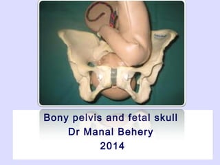
5. Fetal head
From an obstetrical point of view it’s the most
important part:
largest
least compressible part of the fetus.
most frequent presenting part
6.
7. Landmarks
Head is divided into 3areas
(1) the sinciput or brow portion;
(2) the vertex, or top of the head between the 2
fontanelles;
(3) Base large, ossified, firmly united, and
noncompressible
8. Sutures
Membrane-occupied spaces between the cranial
bones
1-Sagittal suture:
- lies btw the parietal bones
-extends in an AP direction btw the fontanelles
-divides the head into right and left sides
11. 4- frontal suture:
lies between the frontal bones
extends from the anterior fontanelle to the
glabella (the prominence between the
eyebrows).
12. Clinical importance of sutures
Position of fontanelle & sagittal suture can identify attitude and
position of vertex.
By plapating the sagittal suture during labour, degree of internal
rotation & molding of the head can be noticed.
In deep transverse arrest, this sagittal suture lies transversely at
the level of the ischial spines.
13.
14. Moulding…
Reshaping of the fetal skull:
Obliteration of the sutures.
Overlapping of the bones of the
vault:
One parietal bone overlaps the
other.
Both overlap the occipital
bone.
It accounts for diminution of
the biparietal diameter and
suboccipitobregmatic
diameters by 0.5-1 cm. 0r
even more.
15. A: Well flexed Head
B: Partially Flexed Head
C: Deflexed Head
D: Face Presentation
E: Brow presentation
16. The anterior fontanelle (bregma) :
diamond shaped area(2 × 3 cm) of
unossified membrane formed by
the junction of 4 suture.
17. The posterior fontanelle:
It is the triangular depressed area at the
junction of 3 suture:
closes at 6 to 8 weeks of life
Y- or T-shaped
23. Transverse Diameters of the Fetal
Skull
Biparietal Diameter 9.5 cm Between the 2 parietal
eminences
Bitemporal Diameter 8.5 cm.
Bimastoid Diameter 7.5 cm. Between the 2 mastoid
processes (Not reducible
nor destroyable even by
destructive procedures
Supra-subparietal 8.25 - 9 cm. Asynclitic head
28. Length Presentation
1-Suboccipito-bregmatic 9.5 cm. Flexed vertex
2-Suboccipito-frontal 10.5 cm. Partially deflexed vertex
3-Occipito-frontal 11.5 cm. Deflexed vertex
4-Mento-vertical 13.75-14 cm. Brow
5-Submento-bregmatic 9.5 cm. Face
6
29.
30. Engaging Diameters of Fetal Skull
Well Flexed Head Circle of 9.5 cm.
The engaging Diameter is the
Suboccipito-Bregmatic diameter
A deflexed Head An oval
The longer occipito-frontal
diameter Of 11.5 cm. Is exposed.
Greater Deflexion
of the Head
An oval
The longer mento vertical
diameter of 13.75-14 cm. is
exposed
Full Extension of
the Head
A circle of 9.5 cm.
The engaging dimeter is the
submento-vertical diameter
38. The Planes of the pelvis
Plane of the pelvic inlet.
Plane of the cavity: Plane of greatest Pelvic Dimensions
Plane of the mid pelvis (plane of obstetric outlet)
Plane of the Anatomical outlet
39. (Inlet (Pelvic brim)
passing with the
boundaries of
pelvic brim and
making an angle of
55o with the
horizon (angle of
pelvic inclination).
40. Pelvic inclination:
The plane of the
pelvic inlet makes
an angle of 55
degree with the
horizon in the
standing position"
41. The consequences of walking upright…
When a women stands erect:
The pelvic inlet makes an angle of about 55° with the horizon.
The pelvic outlet makes an angle of 15° with the horizon
If the angle made by the inlet is greater than 55° this may make
the descent of the fetal head in the pelvis difficult.
43. The True Conjugate = 11 cm
The Obstet. Conjugate = 10.5cm
The Diagonal Conjugate = 12 cm
44. (4) External conjugate:
From: The upper anterior
margin of the symphysis pubis.
To: The depression below the
tip of the 5th lumbar spine
(20 cm).
45. (5) Anatomical transverse diameter:
Between the farthest points on iliopectineal lines (= 13 cm).
It lies 4 cm infornt of the sacral promontory, 7 cm behind
the symphysis pubis.
(6) Obstetric
transverse
diameter:
It bisects the
true
conjugate
(12.5 cm)
ATD
OTD
TC
46. (7) The oblique
diameters: (12 cm)
The right extends from the
right joint to the left
eminence and vice versa.
(8) Sacrocotyloid
diameter: (9-9.5 cm)
The right diameter ends in
the right eminence & vice
versa.
48. bordered by:
the posterior midpoint of the pubis anteriorly
the upper part of the obturator foramina laterally
the junction of the 2nd and 3rd sacral vertebrae posteriorly.
The fetal head rotates to the anterior position in
this plane
2- pelvic cavity The plane of greatest
diameter:
49. The pelvic Outlet
(A)Anatomical outlet: Lozenge
-shaped, bounded by:
Lower border of symphysis
pubis.
Pubic arch.
Ischial tuberosities
Sacrotuberous & sacrospinous
ligaments.
Tip of the coccyx.
50. The Plane of the Outlet
Anterior Sagittal Plane
Posterior Sagittal Plane
51. Diameters of the pelvic outlet
(1)Anteroposterior
Anatomical anteroposterior
(11 cm)
b Obstetric anteroposterior
(13 cm)
52. (2) Transverse diameters
Bituberous (11 cm), between the inner aspects of
ischial tuberosities
Bispinous (10.5 cm), between the tips of ischial
spines.
Diameters of the pelvic outlet
53. Interspinous diam. = 10.5 cm.
Obstet. Ant. Post diam= 13 cm.
Anato. Ant. Post diam= 11 cm.
57. The Obstetric Pelvic Axis
This represents the
path that the
presenting part must
follow for delivery to
occur:
The upper part moves
downward
approximately in a
straight line till the level
of the ischial spine.
The trajectory then
changes to become a
curvilinear path directed
forward and downward
58. The Fetal Head Has Five Fifths…
0 : Head Not Palpable
1 : Sinciput felt – Occiput Not Felt
2 : Sinciput felt – Occiput Just Felt
3 : Sinciput easily felt – Occiput
Felt
4 : Sinciput High – Occiput easily
Felt
5 : Complete above pelvic brim
fifthabove
-5
0
+5
Pelvic Planes:
These are imaginary planes lie as follow:
(1) Plane of pelvic inlet:
passing with the boundaries of pelvic brim and making an angle of 55o with the horizon (angle of pelvic inclination).
(2) Plane of mid cavity ( plane of greatest pelvic dimensions):
- pass between the middle of the posterior surface of the symphysis pubis and the junction between 2nd and 3rd sacral vertebrae. Laterally, it passes to the centre of the acetabulum and the upper part of the greater sciatic notch.
- It is a round plane with diameter of 12.5 cm.
- Internal rotation of the head occurs when the biparietal diameter occupies this wide pelvic plane while the occiput is on the pelvic floor i.e. at the plane of the least pelvic dimensions.
(3) Plane of obstetric outlet (plane of least pelvic dimensions):
passes from the lower border of the symphysis pubis anteriorly, to the ischial spines laterally, to the tip of the sacrum posteriorly.
(4) Plane of anatomical outlet:
passes with the boundaries of anatomical outlet and consists of 2 triangular planes with one base which is the bituberous diameter.
a- Anterior sagittal plane: its apex at the lower border of the symphysis pubis.
b- Posterior sagittal plane: its apex at the tip of the coccyx.
Anterior sagittal diameter: 6-7 cm
from the lower border of the symphsis pubis to the centre of the bituberous diameter.
Posterior sagittal diameter: 7.5-10 cm
from the tip of the sacrum to the centre of the bituberous diameter
Effect of the inclination of the pelvis on the engagement of the fetal head
The ideal obstetric pelvis
outlet
Anatomical axis (curve of Carus):
- It is an imaginary line joining the centre points of the planes of the inlet, cavity and outlet.
- It is C shaped with the concavity directed forwards.
- It has no obstetric importance.