Igcse biology edexcel 2.68 2.76
•Télécharger en tant que PPTX, PDF•
7 j'aime•5,741 vues

Edexcell ppt Biology 2.68 - 2.76 Used in lessons to scaffold class teaching and as a revision resource for students

Recommandé
Contenu connexe
En vedette
En vedette (20)
Plus de Marc Rodriguez
Plus de Marc Rodriguez (14)
Dernier
Dernier (20)
Igcse biology edexcel 2.68 2.76
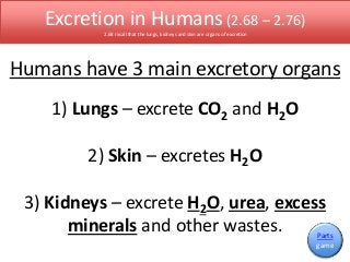
- 1. Excretion in Humans (2.68 – 2.76) 2.68 recall that the lungs, kidneys and skin are organs of excretion Humans have 3 main excretory organs 1) Lungs – excrete CO2 and H2O 2) Skin – excretes H2O 3) Kidneys – excrete H2O, urea, excess minerals and other wastes. Parts game
- 2. UREA 2.68 recall that the lungs, kidneys and skin are organs of excretion What’s urea? (not technically on syllabus) • All organisms produce ammonia as they metabolize nutrients (protein digestion/amino acids) • Ammonia is a nitrogenous waste that is toxic and must be removed from the body Solution: the liver turns the ammonia into urea, which is harmless. Therefore urea is a product of the metabolism of amino acids.
- 3. UREA 2.68 recall that the lungs, kidneys and skin are organs of excretion Many land animals and some bony fish (amphibians, mammals) dilute the toxic ammonia with water. This substance is called Urea & is filtered out by the kidneys. - The problem is they do lose water in the process - Requires Energy
- 4. Excretion & Osmoregulation 2.69 understand how the kidney carries out its roles of excretion and osmoregulation The Kidney: The functional unit of the kidney is the nephron. There are millions of nephrons in a single kidney. Nephrons have 2 jobs; Excretion - filtering the blood and reclaiming the “good bits” & removing waste Osmoregulation - balancing the water level of the body (water homeostasis)
- 5. Excretion System 2.70 describe the structure of the urinary system, including the kidneys, ureters, bladder and urethra
- 6. Excretion System 2.70 describe the structure of the urinary system, including the kidneys, ureters, bladder and urethra
- 7. Kidney Nephron and the Kidney 2.71 describe the structure of a nephron, to include Bowmancs capsule and glomerulus, convoluted tubules, loop of Henlé and collecting duct
- 8. Nephron and capilliaries 2.71 describe the structure of a nephron, to include Bowmancs capsule and glomerulus, convoluted tubules, loop of Henlé and collecting duct
- 9. How the Nephron works 2.71 describe the structure of a nephron, to include Bowmancs capsule and glomerulus, convoluted tubules, loop of Henlé and collecting duct 2.74 understand that selective reabsorption of glucose occurs at the proximal convoluted tubule How the nephron works: 1) Dirty blood enters the kidney via the afferent artery 2) The artery splits up into a ball of capillaries, called the glomerulus 3) The blood is under high pressure, so all small substances are forced out of the holes in the capillary walls. Only large proteins and cells stay behind. 4) The small substances (glucose, minerals, urea, water etc) move into the bowman’s capsule, which wraps around the glomerulus 5) The capsule leads into the PCT, which re-absorbs all glucose via active transport (i.e. it selectively removes the glucose from the nephron and returns it to the blood)
- 10. How the Nephron works 2.71 describe the structure of a nephron, to include Bowmancs capsule and glomerulus, convoluted tubules, loop of Henlé and collecting duct 6) The PCT leads to the Loop of Henlé, which re-absorbs the water by osmosis 7) The Loop leads to the DCT, which re-absorbs all minerals, amino acids and other “useful” substances by active transport 8) The remaining fluid (containing excess water, excess minerals and urea) passes into the collecting duct 9) The collecting ducts from other nephrons join and form the ureter, which leads to the bladder 10) The fluid is now called urine and is stored in the bladder for excretion 11) The bladder takes the urine to the outside world via the urethra This is the first role of the nephron (it’s role in excretion). Remember, the nephron has a second role in osmoregulation.
- 11. Excretion in Humans (2.68 – 2.76) 2.71 describe the structure of a nephron, to include Bowmancs capsule and glomerulus, convoluted tubules, loop of Henlé and collecting duct Kidney
- 12. Excretion in Humans (2.68 – 2.76) 2.72 describe ultrafiltration in the Bowmancs capsule and the composition of the glomerular filtrate The glomerulus filters blood and produces glomerular filtrate. This filtrate contains: water, glucose, salts and urea (amino acids). (Large molecules such as protein are too large to fit through the blood capillary walls.)
- 13. Excretion in Humans (2.68 – 2.76) 2.73 understand that water is reabsorbed into the blood from the collecting duct Teach Blood water levels are sensed by the hypothalamus in the brain. When water levels are too low, the hypothalamus tells the pituitary gland (also in the brain) to release the hormone Anti-Diuretic Hormone (ADH)
- 14. Excretion in Humans (2.68 – 2.76) 2.73 understand that water is reabsorbed into the blood from the collecting duct 2.75 describe the role of ADH in regulating the water content of the blood When blood water levels are too low; 1) Hypothalamus detects 2) Pituitary gland releases ADH into bloodstream 3) ADH travels all over the body 4) Only the cells in the collecting duct of the nephrons of the kidney have receptors for ADH, so only they respond to the hormone 5) The collecting duct becomes more permeable 6) Water is draw out of the collecting duct back into the blood 7) Water levels return to normal BBC
- 15. Review of Urine 2.76 understand that urine contains water, urea and salts. The waste, consisting of: • excess water • excess salts • urea is urine. This process can be summarized in three important steps: Revision 1) Ultra-Filtration - where lots of water, ions, urea and sugar are squeezed from the blood into the tubules. 2) Selective reabsorption – the useful substances (ions and sugars) are reabsorbed back into the blood from the tubules. The amount of water in the blood is regulated here to maintain it at a constant rate. This is known as ‘osmoregulation’. 3) Excretion of waste - urea and excess water and ions travel to the bladder as urine, to be released from the body.