This document summarizes key aspects of light microscopes and electron microscopes.
It explains that light microscopes have a resolution of 200nm, limiting their ability to distinguish objects closer than this. Electron microscopes use electron beams with shorter wavelengths, allowing resolution down to 0.2nm.
The document also distinguishes between magnification, which makes samples appear larger, and resolution, which is the ability to distinguish between close objects. While electron microscopes have much higher resolution than light microscopes, increasing magnification alone does not improve resolution.
1. Microscopes
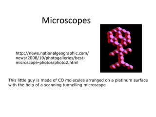
http://news.nationalgeographic.com/
news/2008/10/photogalleries/best-
microscope-photos/photo2.html
This little guy is made of CO molecules arranged on a platinum surface
with the help of a scanning tunnelling microscope
2. Learning Objectives:
explain and distinguish between resolution and
magnification with reference to light microscopy and
electron microscopy
3. Magnification vs. Resolution
Magnification is how Resolution is the
much bigger a sample ability to distinguish
appears to be under the between two points on
microscope than it is in an image - the amount
real life. of detail.
e.g. if two objects
Total Magnification = objective are less than
magnification x eyepiece magnification 200nm apart they
are seen as one
object.
Increasing the magnification does not increase
the resolution of the image!!
4.
5.
6.
7.
8. Invention of microscope
Light Microscope http://sciencevideos.wordpress.co
m/2008/01/26/cell-theory/
Resolution: 200nm
Therefore any images closer
together than 200nm will be seen as
1 object
Due to magnitude of the
wavelength of light
2 objects can only be seen if light
can pass between them
Human eye resolution = 100μm
Magnification: x4, x10,
x40
x100 (oil immersion)
11. Generates a beam of
electrons (0.004nm
wavelength)
Distinguishes between
objects 0.2nm apart.
How is this different from
a light microscope?
Uses magnets instead
Blood clot: platelets spin out a mesh
of lenses to focus the of fibrin.
beam onto the specimen Taken from a scanning electron
microscope
Image is projected
onto photographic
paper to make a grey
scale image
(Black & white
Electronmicrograph)
12. Transmission Scanning Electron
Electron Microscope
Microscope (TEM) (SEM)
Electron beam passes Electrons don't pass
through a thin sample through the specimen.
Electrons pass through They bounce off the
the denser parts less specimen.
easily – contrast 3D view
Image is 2D Magnification is x 100
Magnification is x 500 000
000
http://www.youtube.com/watch?
v=lrXMIghANbg
13. Electron microscope: advantages and
disadvantages
Advantages:
Resolution is 0.1nm
Detailed images or organelles
3D images-shows contours
(SEM)
Disadvantages:
Electron beams deflected by
air molecules - vacuum
needed.
Expensive
Skill & training needed
14. Coloured electron micrographs
Electron micrographs are always black, white &
grey when they are produced
Colours can be added afterwards using
computer software=false colour electron
micrographs