SlideShare une entreprise Scribd logo
1  sur  150
Interpretation of bone tumors at X-Ray.
Dr/ ABD ALLAH NAZEER. MD.
Osteochondroma:
Long bones++
The lesion is composed of cortical and medullary bone protruding from and
continuous with the underlying bone.
The areas of osseous continuity between parent bone and osteochondroma
may be broad, involving a large portion of the bone circumference (dimension
of the lesion base exceeding its length-sessile osteochondroma), or narrow,
with a bulbous tip (pedunculated osteochondroma).
Identifying the characteristic cortical and medullary continuity between lesion
and parent bone on radiographs is dependent on lesion type (sessile or
pedunculated), location, and image projection.
This relationship is usually well delineated in lesions of long bones, particularly
pedunculated osteochondromas, with standard radiographic projections
(although often only on one view) .
However, in osteochondromas of flat bones with complex anatomy (i.e., pelvis,
spine, scapula) and sessile lesions, the continuity and thus the diagnosis may
not be apparent on radiographs alone . Pedunculated lesions usually point
away from the nearest joint owing to the forces of the overlying tendons and
ligaments (although not typically attached to the osteochondroma.
X-ray appearance of different exostoses. (a) Solitary exostosis in a femur.
(b) Multiple exostoses in the knee joint, antero-posterior view. (c) Multiple
exostoses in the knee joint, lateral view. (d) Multiple exostoses in the
upper hip and trochanter (arrows indicate the exostoses sites).
Osteochondroma of scapula.
Osteoid osteoma.
Typical osteoid osteoma in the femur.
Notice the extensive cortical broadening.
Osteoblastoma in a child.
Osteoblastoma of the distal radius.
Osteoblastoma of the
right pubic ramus.
Unicameral bone cyst.
Unicameral bone cyst with fracture and fallen fragment sign.
Aneurysmal bone cyst - Proximal tibia.
Aneurysmal bone cyst - Distal tibia.
Aneurysmal bone cyst.
Giant cell tumour.
Giant cell tumor of the distal ulna.
Giant cell tumor of the distal radius.
Cortical fibrous defects and non-ossifying fibromas in children and young adults:
Cortical fibrous defect.
Non-ossifying fibroma.
Non-Ossifying Fibroma (Fibroxanthoma).
Fibrous Dysplasia
• Long lesion in a long bone
(often occurs in proximal femur).
• Expansion and bone deformity
• Lytic but becomes ground-glass
in appearance as the matrix
calcifies, and then becomes
sclerotic.
• Asymptomatic, but can fracture
• No periosteal reaction
• May be single or multiple
lesion in different locations.
Fibrous dysplasia (polyostotic form).
Fibrous dysplasia: various presentations with or without sclerotic margin,
with ground glass appearance, with calcifications or ossifications.
Fibrous dysplasia
Fibrous dysplasia with Coxa vara of proximal femur.
Enchondroma
• Most commonly seen in the phalanges
• Asymptomatic but commonly fractures
• Well-defined with narrow zone of transition
• Lobulated
• Can become slightly expansile
• Causes endosteal scalloping and cortical thinning
• Ollier's Disease = Multiple enchondromas
• Maffuci's Syndrome = Multiple enchondromas
with soft tissue hemangiomas
• Contain calcified chondroid matrix (irregular,
speckled) when located away from phalanges.
Enchondroma - Proximal humerus.
Plain X-Ray: Enchondroma of Proximal Phalanx.
A child with Ollier’s disease: a non-hereditary, sporadic,
skeletal disorder characterized by multiple enchondromas
Ollier’s disease
Mafucci’s syndrome. Plain radiograph of the left hand. Note multiple
enchondromas and soft-tissue hemangiomas with intralesional phleboliths.
Maffuci's syndrome.
Chondromyxoid fibroma - Proximal tibia.
Chondromyxoid fibroma.
Chondroblastoma.
Chondroblastoma.
Secondary synovial chondromatosis.
Synovial chondromatosis – Shoulder.
Chordoma:
• Uncommon malignant tumours: 40% of all sacral tumors
• Location
• 50% occur in sacrum,
• • • 40% of all sacral tumors
• Most common in 4th or 5th sacral segment
• 35% at skull base around clivus (Benign tumors but may locally
invade and compress)
• 15% in remainder of spine: Cervical spine most common
• Occurs between the ages of 30-70 with a 2:1 male: female ratio
• Imaging Findings
• Large presacral mass (>10cm) with displacement of the rectum
and/or Bladder
• Solid tumor with cystic areas in 50%
• Destroys multiple sacral and coccygeal segments
• Sequestered bone fragments are common
• Sclerotic rim in 50%
• May have amorphous calcifications, especially peripherally
• May cross the sacroiliac joint
Sacral chordoma.
Chordoma of skull base.
Brown tumor.
Brown tumor at the femur diaphysis.
Adamantinoma of the proximal tibia.
Adamantinoma Tibia.
Skull and femur hemangioma.
Computerized tomography demonstrates lesions with osseous spicules in keeping with hemangiomas
involved with left frontal bone, supraorbital wall, sphenoid sinus and ethmoid. The central portion of
this lesion revealed subtle striated trabeculations and retained a thin sclerotic rim of peripheral bone
condensation, and the boundary was clear. Both the inner and the outer tables of the skull were eroded
Hemangioma of the Long Bones
Intraosseous Hemangioma of the Nasal Septum.
Eosinophilic granuloma – Humerus.
Various presentations of eosinophilic granuloma.
Monostotic and polyostotic eosinophilic
granuloma in the skull.
Eosinophilic granuloma in the skull.
Ewing sarcoma.
Ewing sarcoma arising from the right iliac bone.
Ewing sarcoma of femur. Frontal radiograph and lateral radiographs of the femur demonstrate mottled,
osteolytic lesion (blue circle) with poorly marginated edges in the diaphysis of the bone. There is
sunburst periosteal reaction (red circle) and lamellated periosteal reaction (white arrows).
Ewing sarcoma
(a) X-ray anteroposterior and lateral views of proximal tibia and knee joint showing diaphyseal
osteosarcoma of tibia with sclerosis (arrow), cortical destruction on posteromedial side (arrow
heads) and new bone formation in the soft tissues (b) x-ray distal end of femur (anteroposterior
and lateral views) showing sclerosis/radio-opacity in sclerosing osteosarcoma.
X-ray of knee joint anteroposterior views showing surface osteosarcoma: (a)
parosteal (b) periosteal. See the under lying cortex is visibly intact in ‘a’ and lifting
of periosteum in ‘b’ (red arrow). However, both are on the surface of the bone
Telangiectatic type of osteosarcoma of the proximal tibia: (a) X-ray anteroposterior
and lateral views showing lysis and expansion (b) MRI showing fluid levels.
Osteosarcoma.
Parosteal Osteosarcoma.
Chondrosarcoma of the iliac bone and proximal humerus.
Chondrosarcoma of the distal femur and proximal humerus.
Chondrosarcoma.
Typical x-ray appearance of
osteolytic bone metastases.
Typical x-ray appearance of
osteoblastic bone metastases.
Anteroposterior radiograph of the lower legs demonstrates a permeative, lytic lesion in the metadiaphysis of
the left tibia. Note the aggressive periosteal reaction with Codman triangles and soft-tissue extension/swelling.
Soft-tissue involvement is easy to appreciate when the left leg is compared with the normal right leg.
Burkitt's lymphoma of the humerus.
62-year-old patient with lymphoma and low back pain. Anteroposterior (A) and profile (B) X-rays
showing a diffuse increase in bone density, with erosion on the left side of the L4 vertebral body (arrow).
The skull x-ray lateral and AP view reveals multiple oil droplet like punched out lytic defects throughout bony
calvarium. The appearance is highly suggestive of, but not completely diagnostic for, multiple myeloma.
Multiple myeloma.
Multiple Myeloma/Plasmacytoma.
Thank You.

Contenu connexe

Tendances

Avascular necrosis Radiology
Avascular necrosis RadiologyAvascular necrosis Radiology
Avascular necrosis Radiologyrajss007
 
MRI of Knee joint-- hossam massoud
MRI of Knee joint-- hossam massoudMRI of Knee joint-- hossam massoud
MRI of Knee joint-- hossam massoudHossam Massoud
 
Benign bone tumors
Benign bone tumorsBenign bone tumors
Benign bone tumorsmacshrestha
 
Systemic approach to bone tumor radiology
Systemic approach to bone tumor radiologySystemic approach to bone tumor radiology
Systemic approach to bone tumor radiologyRamin Sadeghi
 
Cervical Spine Trauma Imaging
Cervical Spine Trauma ImagingCervical Spine Trauma Imaging
Cervical Spine Trauma ImagingSCGH ED CME
 
Osteosarcoma Radiology Review
Osteosarcoma Radiology ReviewOsteosarcoma Radiology Review
Osteosarcoma Radiology ReviewRajesh Venunath
 
Avascular necrosis of scaphoid
Avascular necrosis of scaphoidAvascular necrosis of scaphoid
Avascular necrosis of scaphoidDr. Anshu Sharma
 
Imaging in rickets
Imaging in ricketsImaging in rickets
Imaging in ricketsVikram Patil
 
Presentation1.pptx, radiological imaging of benign bone tumour.
Presentation1.pptx, radiological imaging of benign bone tumour.Presentation1.pptx, radiological imaging of benign bone tumour.
Presentation1.pptx, radiological imaging of benign bone tumour.Abdellah Nazeer
 
An approach to malignant bone tumors
An approach to malignant bone tumors An approach to malignant bone tumors
An approach to malignant bone tumors Dr.Suhas Basavaiah
 
Bone tumor radiological approach
Bone tumor radiological approachBone tumor radiological approach
Bone tumor radiological approachSitanshu Barik
 
Presentation1, radiological imaging of degenerative and inflammatory disease ...
Presentation1, radiological imaging of degenerative and inflammatory disease ...Presentation1, radiological imaging of degenerative and inflammatory disease ...
Presentation1, radiological imaging of degenerative and inflammatory disease ...Abdellah Nazeer
 
Lytic lesions of bone
Lytic lesions of boneLytic lesions of bone
Lytic lesions of bonedevrajkandel1
 
Bone tumour , enchondroma , osteochondroma
Bone tumour , enchondroma , osteochondromaBone tumour , enchondroma , osteochondroma
Bone tumour , enchondroma , osteochondromaSagar Savsani
 
Benign Bone Tumors and Tumor Like Conditions
Benign Bone Tumors and Tumor Like Conditions Benign Bone Tumors and Tumor Like Conditions
Benign Bone Tumors and Tumor Like Conditions priyanka rana
 
Presentation1.pptx, radiological imaging of skeletal dysplasia
Presentation1.pptx, radiological imaging of skeletal dysplasiaPresentation1.pptx, radiological imaging of skeletal dysplasia
Presentation1.pptx, radiological imaging of skeletal dysplasiaAbdellah Nazeer
 
Interpretation of musculoskeletal x rays
Interpretation of musculoskeletal x  raysInterpretation of musculoskeletal x  rays
Interpretation of musculoskeletal x raysjatinder12345
 

Tendances (20)

Avascular necrosis Radiology
Avascular necrosis RadiologyAvascular necrosis Radiology
Avascular necrosis Radiology
 
MRI of Knee joint-- hossam massoud
MRI of Knee joint-- hossam massoudMRI of Knee joint-- hossam massoud
MRI of Knee joint-- hossam massoud
 
Benign bone tumors
Benign bone tumorsBenign bone tumors
Benign bone tumors
 
Systemic approach to bone tumor radiology
Systemic approach to bone tumor radiologySystemic approach to bone tumor radiology
Systemic approach to bone tumor radiology
 
Cervical Spine Trauma Imaging
Cervical Spine Trauma ImagingCervical Spine Trauma Imaging
Cervical Spine Trauma Imaging
 
Knee mri
Knee mriKnee mri
Knee mri
 
Osteosarcoma Radiology Review
Osteosarcoma Radiology ReviewOsteosarcoma Radiology Review
Osteosarcoma Radiology Review
 
Avascular necrosis of scaphoid
Avascular necrosis of scaphoidAvascular necrosis of scaphoid
Avascular necrosis of scaphoid
 
Imaging in rickets
Imaging in ricketsImaging in rickets
Imaging in rickets
 
Presentation1.pptx, radiological imaging of benign bone tumour.
Presentation1.pptx, radiological imaging of benign bone tumour.Presentation1.pptx, radiological imaging of benign bone tumour.
Presentation1.pptx, radiological imaging of benign bone tumour.
 
An approach to malignant bone tumors
An approach to malignant bone tumors An approach to malignant bone tumors
An approach to malignant bone tumors
 
Hip joint
Hip jointHip joint
Hip joint
 
Bone tumor radiological approach
Bone tumor radiological approachBone tumor radiological approach
Bone tumor radiological approach
 
Presentation1, radiological imaging of degenerative and inflammatory disease ...
Presentation1, radiological imaging of degenerative and inflammatory disease ...Presentation1, radiological imaging of degenerative and inflammatory disease ...
Presentation1, radiological imaging of degenerative and inflammatory disease ...
 
Lytic lesions of bone
Lytic lesions of boneLytic lesions of bone
Lytic lesions of bone
 
MRI of the shoulder
MRI of the shoulderMRI of the shoulder
MRI of the shoulder
 
Bone tumour , enchondroma , osteochondroma
Bone tumour , enchondroma , osteochondromaBone tumour , enchondroma , osteochondroma
Bone tumour , enchondroma , osteochondroma
 
Benign Bone Tumors and Tumor Like Conditions
Benign Bone Tumors and Tumor Like Conditions Benign Bone Tumors and Tumor Like Conditions
Benign Bone Tumors and Tumor Like Conditions
 
Presentation1.pptx, radiological imaging of skeletal dysplasia
Presentation1.pptx, radiological imaging of skeletal dysplasiaPresentation1.pptx, radiological imaging of skeletal dysplasia
Presentation1.pptx, radiological imaging of skeletal dysplasia
 
Interpretation of musculoskeletal x rays
Interpretation of musculoskeletal x  raysInterpretation of musculoskeletal x  rays
Interpretation of musculoskeletal x rays
 

En vedette

Tips for interpreting x ray in trauma
Tips for interpreting x ray in traumaTips for interpreting x ray in trauma
Tips for interpreting x ray in traumaChew Keng Sheng
 
Presentation2 Fractures
Presentation2 FracturesPresentation2 Fractures
Presentation2 FracturesZoe Palisoc
 
46 current evidence about femoral neck fracture treatment[1]
46 current evidence about femoral neck fracture treatment[1]46 current evidence about femoral neck fracture treatment[1]
46 current evidence about femoral neck fracture treatment[1]Vamsi Mohan
 
Benign bone tumors imaging
Benign bone tumors imagingBenign bone tumors imaging
Benign bone tumors imagingRiyaz Ahmed
 
L02 femoral neck fx
L02 femoral neck fxL02 femoral neck fx
L02 femoral neck fxClaudiu Cucu
 
X-RAY , SPECIAL ORTHOPEDIC BEDS
X-RAY , SPECIAL ORTHOPEDIC BEDSX-RAY , SPECIAL ORTHOPEDIC BEDS
X-RAY , SPECIAL ORTHOPEDIC BEDSRoxanneMae Birador
 
Imaging of Bone Tumors
Imaging of Bone TumorsImaging of Bone Tumors
Imaging of Bone TumorsSameer Peer
 
Tumor mega prosthesis
Tumor mega prosthesisTumor mega prosthesis
Tumor mega prosthesisSrinath Gupta
 
Musculoskeltal xray
Musculoskeltal xrayMusculoskeltal xray
Musculoskeltal xraykamn007
 
Presentation for skin traction
Presentation for skin tractionPresentation for skin traction
Presentation for skin tractionJayson Teruel
 
Cast & slab by dr. ahmed shedeed
Cast & slab by dr. ahmed shedeedCast & slab by dr. ahmed shedeed
Cast & slab by dr. ahmed shedeedAhmed-shedeed
 
Journal club presentation (chest tube) _.pptx dr shakil
Journal club presentation (chest tube) _.pptx dr shakilJournal club presentation (chest tube) _.pptx dr shakil
Journal club presentation (chest tube) _.pptx dr shakilShakil Ahmad
 
Principles of use of plaster of paris
Principles of use of plaster of parisPrinciples of use of plaster of paris
Principles of use of plaster of parisAsi-oqua Bassey
 
Introduction to musculoskeletal radiology
Introduction to musculoskeletal radiologyIntroduction to musculoskeletal radiology
Introduction to musculoskeletal radiologySubhanjan Das
 
Using Apps and Digital Tools for Health Campaigns
Using Apps and Digital Tools for Health CampaignsUsing Apps and Digital Tools for Health Campaigns
Using Apps and Digital Tools for Health CampaignsMike Robert
 

En vedette (20)

Approach to bone tumors
Approach to bone tumorsApproach to bone tumors
Approach to bone tumors
 
Tips for interpreting x ray in trauma
Tips for interpreting x ray in traumaTips for interpreting x ray in trauma
Tips for interpreting x ray in trauma
 
Bone Tumors Benign Ppt
Bone Tumors Benign PptBone Tumors Benign Ppt
Bone Tumors Benign Ppt
 
Presentation2 Fractures
Presentation2 FracturesPresentation2 Fractures
Presentation2 Fractures
 
46 current evidence about femoral neck fracture treatment[1]
46 current evidence about femoral neck fracture treatment[1]46 current evidence about femoral neck fracture treatment[1]
46 current evidence about femoral neck fracture treatment[1]
 
Benign bone tumors imaging
Benign bone tumors imagingBenign bone tumors imaging
Benign bone tumors imaging
 
L02 femoral neck fx
L02 femoral neck fxL02 femoral neck fx
L02 femoral neck fx
 
X-RAY , SPECIAL ORTHOPEDIC BEDS
X-RAY , SPECIAL ORTHOPEDIC BEDSX-RAY , SPECIAL ORTHOPEDIC BEDS
X-RAY , SPECIAL ORTHOPEDIC BEDS
 
Imaging of Bone Tumors
Imaging of Bone TumorsImaging of Bone Tumors
Imaging of Bone Tumors
 
Tumor mega prosthesis
Tumor mega prosthesisTumor mega prosthesis
Tumor mega prosthesis
 
Musculoskeltal xray
Musculoskeltal xrayMusculoskeltal xray
Musculoskeltal xray
 
Presentation for skin traction
Presentation for skin tractionPresentation for skin traction
Presentation for skin traction
 
Elbow
ElbowElbow
Elbow
 
Cast & slab by dr. ahmed shedeed
Cast & slab by dr. ahmed shedeedCast & slab by dr. ahmed shedeed
Cast & slab by dr. ahmed shedeed
 
Journal club presentation (chest tube) _.pptx dr shakil
Journal club presentation (chest tube) _.pptx dr shakilJournal club presentation (chest tube) _.pptx dr shakil
Journal club presentation (chest tube) _.pptx dr shakil
 
Cns trauma
Cns traumaCns trauma
Cns trauma
 
Principles of use of plaster of paris
Principles of use of plaster of parisPrinciples of use of plaster of paris
Principles of use of plaster of paris
 
Introduction to musculoskeletal radiology
Introduction to musculoskeletal radiologyIntroduction to musculoskeletal radiology
Introduction to musculoskeletal radiology
 
Femur fracture
Femur fractureFemur fracture
Femur fracture
 
Using Apps and Digital Tools for Health Campaigns
Using Apps and Digital Tools for Health CampaignsUsing Apps and Digital Tools for Health Campaigns
Using Apps and Digital Tools for Health Campaigns
 

Similaire à Presentation1.pptx, interpretation of x ray on bone tumour.

bone tumors-1.pptx
bone tumors-1.pptxbone tumors-1.pptx
bone tumors-1.pptxkattabharath
 
Benign bone tumours
Benign bone tumoursBenign bone tumours
Benign bone tumoursArif S
 
Musculo skeletal diseases
Musculo skeletal diseasesMusculo skeletal diseases
Musculo skeletal diseasesEneutron
 
Bone forming tumors rabins
Bone forming tumors rabinsBone forming tumors rabins
Bone forming tumors rabinsRobins Shah
 
Presentation1, radiological imaging of ostepopikilosis
Presentation1, radiological imaging of ostepopikilosisPresentation1, radiological imaging of ostepopikilosis
Presentation1, radiological imaging of ostepopikilosisAbdellah Nazeer
 
Orthopedic Aspects Of Metabolic Bone Disease By Xiu
Orthopedic Aspects Of Metabolic Bone Disease By XiuOrthopedic Aspects Of Metabolic Bone Disease By Xiu
Orthopedic Aspects Of Metabolic Bone Disease By XiuXiu Srithammasit
 
bonetumors-161023202240.pptx
bonetumors-161023202240.pptxbonetumors-161023202240.pptx
bonetumors-161023202240.pptxAmerManzoorPak
 
Classification of bone tumors
Classification of bone tumorsClassification of bone tumors
Classification of bone tumorsDpt Memon
 
bonetumors-161023202240 (1).pdf
bonetumors-161023202240 (1).pdfbonetumors-161023202240 (1).pdf
bonetumors-161023202240 (1).pdfShyamChadsania
 
bonetumors-161023202240.pdf
bonetumors-161023202240.pdfbonetumors-161023202240.pdf
bonetumors-161023202240.pdfShyamChadsania
 
Introduction skeletal radiology(11月20.)
Introduction   skeletal radiology(11月20.)Introduction   skeletal radiology(11月20.)
Introduction skeletal radiology(11月20.)ghalan
 
Osteochondroma
OsteochondromaOsteochondroma
Osteochondromavivhekpai
 

Similaire à Presentation1.pptx, interpretation of x ray on bone tumour. (20)

bone tumors-1.pptx
bone tumors-1.pptxbone tumors-1.pptx
bone tumors-1.pptx
 
Malignant bone tumours
Malignant bone tumoursMalignant bone tumours
Malignant bone tumours
 
Osteopetrosis
OsteopetrosisOsteopetrosis
Osteopetrosis
 
Osteopetrosis
OsteopetrosisOsteopetrosis
Osteopetrosis
 
Benign bone tumours
Benign bone tumoursBenign bone tumours
Benign bone tumours
 
Musculo skeletal diseases
Musculo skeletal diseasesMusculo skeletal diseases
Musculo skeletal diseases
 
Bone forming tumors rabins
Bone forming tumors rabinsBone forming tumors rabins
Bone forming tumors rabins
 
Xray bone-tumor
Xray bone-tumorXray bone-tumor
Xray bone-tumor
 
Presentation1, radiological imaging of ostepopikilosis
Presentation1, radiological imaging of ostepopikilosisPresentation1, radiological imaging of ostepopikilosis
Presentation1, radiological imaging of ostepopikilosis
 
Orthopedic Aspects Of Metabolic Bone Disease By Xiu
Orthopedic Aspects Of Metabolic Bone Disease By XiuOrthopedic Aspects Of Metabolic Bone Disease By Xiu
Orthopedic Aspects Of Metabolic Bone Disease By Xiu
 
bonetumors-161023202240.pptx
bonetumors-161023202240.pptxbonetumors-161023202240.pptx
bonetumors-161023202240.pptx
 
Classification of bone tumors
Classification of bone tumorsClassification of bone tumors
Classification of bone tumors
 
bonetumors-161023202240 (1).pdf
bonetumors-161023202240 (1).pdfbonetumors-161023202240 (1).pdf
bonetumors-161023202240 (1).pdf
 
bonetumors-161023202240.pdf
bonetumors-161023202240.pdfbonetumors-161023202240.pdf
bonetumors-161023202240.pdf
 
Bone tumors
Bone tumorsBone tumors
Bone tumors
 
Osteochondroma (dr. mahesh)
Osteochondroma (dr. mahesh)Osteochondroma (dr. mahesh)
Osteochondroma (dr. mahesh)
 
Introduction skeletal radiology(11月20.)
Introduction   skeletal radiology(11月20.)Introduction   skeletal radiology(11月20.)
Introduction skeletal radiology(11月20.)
 
Soft tissue s
Soft tissue sSoft tissue s
Soft tissue s
 
Radiology 5th year, 2nd lecture (Dr. Salah Mohammad Fatih)
Radiology 5th year, 2nd lecture (Dr. Salah Mohammad Fatih)Radiology 5th year, 2nd lecture (Dr. Salah Mohammad Fatih)
Radiology 5th year, 2nd lecture (Dr. Salah Mohammad Fatih)
 
Osteochondroma
OsteochondromaOsteochondroma
Osteochondroma
 

Plus de Abdellah Nazeer

Muculoskeletal Pediatic Imaging..pptx
Muculoskeletal Pediatic Imaging..pptxMuculoskeletal Pediatic Imaging..pptx
Muculoskeletal Pediatic Imaging..pptxAbdellah Nazeer
 
Presentation1, Ultrasound of the bowel loops and the lymph nodes..pptx
Presentation1, Ultrasound of the bowel loops and the lymph nodes..pptxPresentation1, Ultrasound of the bowel loops and the lymph nodes..pptx
Presentation1, Ultrasound of the bowel loops and the lymph nodes..pptxAbdellah Nazeer
 
Presentation1 Short cases MD..pptx
Presentation1 Short cases MD..pptxPresentation1 Short cases MD..pptx
Presentation1 Short cases MD..pptxAbdellah Nazeer
 
Presentation1, MD MCQ Cases..pptx
Presentation1, MD MCQ Cases..pptxPresentation1, MD MCQ Cases..pptx
Presentation1, MD MCQ Cases..pptxAbdellah Nazeer
 
Presentation1, Short Cases Quiz..pptx
Presentation1, Short Cases Quiz..pptxPresentation1, Short Cases Quiz..pptx
Presentation1, Short Cases Quiz..pptxAbdellah Nazeer
 
Presentation1, radiological imaging of lateral hindfoot impingement.
Presentation1, radiological imaging of lateral hindfoot impingement.Presentation1, radiological imaging of lateral hindfoot impingement.
Presentation1, radiological imaging of lateral hindfoot impingement.Abdellah Nazeer
 
Presentation2, radiological anatomy of the liver and spleen.
Presentation2, radiological anatomy of the liver and spleen.Presentation2, radiological anatomy of the liver and spleen.
Presentation2, radiological anatomy of the liver and spleen.Abdellah Nazeer
 
Presentation1, artifacts and pitfalls of the wrist and elbow joints.
Presentation1, artifacts and pitfalls of the wrist and elbow joints.Presentation1, artifacts and pitfalls of the wrist and elbow joints.
Presentation1, artifacts and pitfalls of the wrist and elbow joints.Abdellah Nazeer
 
Presentation1, artifact and pitfalls of the knee, hip and ankle joints.
Presentation1, artifact and pitfalls of the knee, hip and ankle joints.Presentation1, artifact and pitfalls of the knee, hip and ankle joints.
Presentation1, artifact and pitfalls of the knee, hip and ankle joints.Abdellah Nazeer
 
Presentation1, radiological imaging of artifact and pitfalls in shoulder join...
Presentation1, radiological imaging of artifact and pitfalls in shoulder join...Presentation1, radiological imaging of artifact and pitfalls in shoulder join...
Presentation1, radiological imaging of artifact and pitfalls in shoulder join...Abdellah Nazeer
 
Presentation1, radiological imaging of internal abdominal hernia.
Presentation1, radiological imaging of internal abdominal hernia.Presentation1, radiological imaging of internal abdominal hernia.
Presentation1, radiological imaging of internal abdominal hernia.Abdellah Nazeer
 
Presentation11, radiological imaging of ovarian torsion.
Presentation11, radiological imaging of ovarian torsion.Presentation11, radiological imaging of ovarian torsion.
Presentation11, radiological imaging of ovarian torsion.Abdellah Nazeer
 
Presentation1, musculoskeletal anatomy.
Presentation1, musculoskeletal anatomy.Presentation1, musculoskeletal anatomy.
Presentation1, musculoskeletal anatomy.Abdellah Nazeer
 
Presentation1, new mri techniques in the diagnosis and monitoring of multiple...
Presentation1, new mri techniques in the diagnosis and monitoring of multiple...Presentation1, new mri techniques in the diagnosis and monitoring of multiple...
Presentation1, new mri techniques in the diagnosis and monitoring of multiple...Abdellah Nazeer
 
Presentation1, radiological application of diffusion weighted mri in neck mas...
Presentation1, radiological application of diffusion weighted mri in neck mas...Presentation1, radiological application of diffusion weighted mri in neck mas...
Presentation1, radiological application of diffusion weighted mri in neck mas...Abdellah Nazeer
 
Presentation1, radiological application of diffusion weighted images in breas...
Presentation1, radiological application of diffusion weighted images in breas...Presentation1, radiological application of diffusion weighted images in breas...
Presentation1, radiological application of diffusion weighted images in breas...Abdellah Nazeer
 
Presentation1, radiological application of diffusion weighted images in abdom...
Presentation1, radiological application of diffusion weighted images in abdom...Presentation1, radiological application of diffusion weighted images in abdom...
Presentation1, radiological application of diffusion weighted images in abdom...Abdellah Nazeer
 
Presentation1, radiological application of diffusion weighted imges in neuror...
Presentation1, radiological application of diffusion weighted imges in neuror...Presentation1, radiological application of diffusion weighted imges in neuror...
Presentation1, radiological application of diffusion weighted imges in neuror...Abdellah Nazeer
 
Presentation1, mr physics.
Presentation1, mr physics.Presentation1, mr physics.
Presentation1, mr physics.Abdellah Nazeer
 
Presentation1. ct physics.
Presentation1. ct physics.Presentation1. ct physics.
Presentation1. ct physics.Abdellah Nazeer
 

Plus de Abdellah Nazeer (20)

Muculoskeletal Pediatic Imaging..pptx
Muculoskeletal Pediatic Imaging..pptxMuculoskeletal Pediatic Imaging..pptx
Muculoskeletal Pediatic Imaging..pptx
 
Presentation1, Ultrasound of the bowel loops and the lymph nodes..pptx
Presentation1, Ultrasound of the bowel loops and the lymph nodes..pptxPresentation1, Ultrasound of the bowel loops and the lymph nodes..pptx
Presentation1, Ultrasound of the bowel loops and the lymph nodes..pptx
 
Presentation1 Short cases MD..pptx
Presentation1 Short cases MD..pptxPresentation1 Short cases MD..pptx
Presentation1 Short cases MD..pptx
 
Presentation1, MD MCQ Cases..pptx
Presentation1, MD MCQ Cases..pptxPresentation1, MD MCQ Cases..pptx
Presentation1, MD MCQ Cases..pptx
 
Presentation1, Short Cases Quiz..pptx
Presentation1, Short Cases Quiz..pptxPresentation1, Short Cases Quiz..pptx
Presentation1, Short Cases Quiz..pptx
 
Presentation1, radiological imaging of lateral hindfoot impingement.
Presentation1, radiological imaging of lateral hindfoot impingement.Presentation1, radiological imaging of lateral hindfoot impingement.
Presentation1, radiological imaging of lateral hindfoot impingement.
 
Presentation2, radiological anatomy of the liver and spleen.
Presentation2, radiological anatomy of the liver and spleen.Presentation2, radiological anatomy of the liver and spleen.
Presentation2, radiological anatomy of the liver and spleen.
 
Presentation1, artifacts and pitfalls of the wrist and elbow joints.
Presentation1, artifacts and pitfalls of the wrist and elbow joints.Presentation1, artifacts and pitfalls of the wrist and elbow joints.
Presentation1, artifacts and pitfalls of the wrist and elbow joints.
 
Presentation1, artifact and pitfalls of the knee, hip and ankle joints.
Presentation1, artifact and pitfalls of the knee, hip and ankle joints.Presentation1, artifact and pitfalls of the knee, hip and ankle joints.
Presentation1, artifact and pitfalls of the knee, hip and ankle joints.
 
Presentation1, radiological imaging of artifact and pitfalls in shoulder join...
Presentation1, radiological imaging of artifact and pitfalls in shoulder join...Presentation1, radiological imaging of artifact and pitfalls in shoulder join...
Presentation1, radiological imaging of artifact and pitfalls in shoulder join...
 
Presentation1, radiological imaging of internal abdominal hernia.
Presentation1, radiological imaging of internal abdominal hernia.Presentation1, radiological imaging of internal abdominal hernia.
Presentation1, radiological imaging of internal abdominal hernia.
 
Presentation11, radiological imaging of ovarian torsion.
Presentation11, radiological imaging of ovarian torsion.Presentation11, radiological imaging of ovarian torsion.
Presentation11, radiological imaging of ovarian torsion.
 
Presentation1, musculoskeletal anatomy.
Presentation1, musculoskeletal anatomy.Presentation1, musculoskeletal anatomy.
Presentation1, musculoskeletal anatomy.
 
Presentation1, new mri techniques in the diagnosis and monitoring of multiple...
Presentation1, new mri techniques in the diagnosis and monitoring of multiple...Presentation1, new mri techniques in the diagnosis and monitoring of multiple...
Presentation1, new mri techniques in the diagnosis and monitoring of multiple...
 
Presentation1, radiological application of diffusion weighted mri in neck mas...
Presentation1, radiological application of diffusion weighted mri in neck mas...Presentation1, radiological application of diffusion weighted mri in neck mas...
Presentation1, radiological application of diffusion weighted mri in neck mas...
 
Presentation1, radiological application of diffusion weighted images in breas...
Presentation1, radiological application of diffusion weighted images in breas...Presentation1, radiological application of diffusion weighted images in breas...
Presentation1, radiological application of diffusion weighted images in breas...
 
Presentation1, radiological application of diffusion weighted images in abdom...
Presentation1, radiological application of diffusion weighted images in abdom...Presentation1, radiological application of diffusion weighted images in abdom...
Presentation1, radiological application of diffusion weighted images in abdom...
 
Presentation1, radiological application of diffusion weighted imges in neuror...
Presentation1, radiological application of diffusion weighted imges in neuror...Presentation1, radiological application of diffusion weighted imges in neuror...
Presentation1, radiological application of diffusion weighted imges in neuror...
 
Presentation1, mr physics.
Presentation1, mr physics.Presentation1, mr physics.
Presentation1, mr physics.
 
Presentation1. ct physics.
Presentation1. ct physics.Presentation1. ct physics.
Presentation1. ct physics.
 

Dernier

Cardiac Output, Venous Return, and Their Regulation
Cardiac Output, Venous Return, and Their RegulationCardiac Output, Venous Return, and Their Regulation
Cardiac Output, Venous Return, and Their RegulationMedicoseAcademics
 
Call Girls Bangalore - 450+ Call Girl Cash Payment 💯Call Us 🔝 6378878445 🔝 💃 ...
Call Girls Bangalore - 450+ Call Girl Cash Payment 💯Call Us 🔝 6378878445 🔝 💃 ...Call Girls Bangalore - 450+ Call Girl Cash Payment 💯Call Us 🔝 6378878445 🔝 💃 ...
Call Girls Bangalore - 450+ Call Girl Cash Payment 💯Call Us 🔝 6378878445 🔝 💃 ...gragneelam30
 
Call Girl In Indore 📞9235973566📞 Just📲 Call Inaaya Indore Call Girls Service ...
Call Girl In Indore 📞9235973566📞 Just📲 Call Inaaya Indore Call Girls Service ...Call Girl In Indore 📞9235973566📞 Just📲 Call Inaaya Indore Call Girls Service ...
Call Girl In Indore 📞9235973566📞 Just📲 Call Inaaya Indore Call Girls Service ...Sheetaleventcompany
 
Call Girls in Lucknow Just Call 👉👉8630512678 Top Class Call Girl Service Avai...
Call Girls in Lucknow Just Call 👉👉8630512678 Top Class Call Girl Service Avai...Call Girls in Lucknow Just Call 👉👉8630512678 Top Class Call Girl Service Avai...
Call Girls in Lucknow Just Call 👉👉8630512678 Top Class Call Girl Service Avai...soniyagrag336
 
Gastric Cancer: Сlinical Implementation of Artificial Intelligence, Synergeti...
Gastric Cancer: Сlinical Implementation of Artificial Intelligence, Synergeti...Gastric Cancer: Сlinical Implementation of Artificial Intelligence, Synergeti...
Gastric Cancer: Сlinical Implementation of Artificial Intelligence, Synergeti...Oleg Kshivets
 
tongue disease lecture Dr Assadawy legacy
tongue disease lecture Dr Assadawy legacytongue disease lecture Dr Assadawy legacy
tongue disease lecture Dr Assadawy legacyDrMohamed Assadawy
 
❤️Amritsar Escorts Service☎️9815674956☎️ Call Girl service in Amritsar☎️ Amri...
❤️Amritsar Escorts Service☎️9815674956☎️ Call Girl service in Amritsar☎️ Amri...❤️Amritsar Escorts Service☎️9815674956☎️ Call Girl service in Amritsar☎️ Amri...
❤️Amritsar Escorts Service☎️9815674956☎️ Call Girl service in Amritsar☎️ Amri...Sheetaleventcompany
 
Exclusive Call Girls Bangalore {7304373326} ❤️VVIP POOJA Call Girls in Bangal...
Exclusive Call Girls Bangalore {7304373326} ❤️VVIP POOJA Call Girls in Bangal...Exclusive Call Girls Bangalore {7304373326} ❤️VVIP POOJA Call Girls in Bangal...
Exclusive Call Girls Bangalore {7304373326} ❤️VVIP POOJA Call Girls in Bangal...Sheetaleventcompany
 
Bhawanipatna Call Girls 📞9332606886 Call Girls in Bhawanipatna Escorts servic...
Bhawanipatna Call Girls 📞9332606886 Call Girls in Bhawanipatna Escorts servic...Bhawanipatna Call Girls 📞9332606886 Call Girls in Bhawanipatna Escorts servic...
Bhawanipatna Call Girls 📞9332606886 Call Girls in Bhawanipatna Escorts servic...Dipal Arora
 
Race Course Road } Book Call Girls in Bangalore | Whatsapp No 6378878445 VIP ...
Race Course Road } Book Call Girls in Bangalore | Whatsapp No 6378878445 VIP ...Race Course Road } Book Call Girls in Bangalore | Whatsapp No 6378878445 VIP ...
Race Course Road } Book Call Girls in Bangalore | Whatsapp No 6378878445 VIP ...dishamehta3332
 
Dehradun Call Girls Service {8854095900} ❤️VVIP ROCKY Call Girl in Dehradun U...
Dehradun Call Girls Service {8854095900} ❤️VVIP ROCKY Call Girl in Dehradun U...Dehradun Call Girls Service {8854095900} ❤️VVIP ROCKY Call Girl in Dehradun U...
Dehradun Call Girls Service {8854095900} ❤️VVIP ROCKY Call Girl in Dehradun U...Sheetaleventcompany
 
💚Call Girls In Amritsar 💯Anvi 📲🔝8725944379🔝Amritsar Call Girl No💰Advance Cash...
💚Call Girls In Amritsar 💯Anvi 📲🔝8725944379🔝Amritsar Call Girl No💰Advance Cash...💚Call Girls In Amritsar 💯Anvi 📲🔝8725944379🔝Amritsar Call Girl No💰Advance Cash...
💚Call Girls In Amritsar 💯Anvi 📲🔝8725944379🔝Amritsar Call Girl No💰Advance Cash...Sheetaleventcompany
 
Chandigarh Call Girls Service ❤️🍑 9809698092 👄🫦Independent Escort Service Cha...
Chandigarh Call Girls Service ❤️🍑 9809698092 👄🫦Independent Escort Service Cha...Chandigarh Call Girls Service ❤️🍑 9809698092 👄🫦Independent Escort Service Cha...
Chandigarh Call Girls Service ❤️🍑 9809698092 👄🫦Independent Escort Service Cha...Sheetaleventcompany
 
Kolkata Call Girls Shobhabazar 💯Call Us 🔝 8005736733 🔝 💃 Top Class Call Gir...
Kolkata Call Girls Shobhabazar  💯Call Us 🔝 8005736733 🔝 💃  Top Class Call Gir...Kolkata Call Girls Shobhabazar  💯Call Us 🔝 8005736733 🔝 💃  Top Class Call Gir...
Kolkata Call Girls Shobhabazar 💯Call Us 🔝 8005736733 🔝 💃 Top Class Call Gir...Namrata Singh
 
Premium Call Girls Nagpur {9xx000xx09} ❤️VVIP POOJA Call Girls in Nagpur Maha...
Premium Call Girls Nagpur {9xx000xx09} ❤️VVIP POOJA Call Girls in Nagpur Maha...Premium Call Girls Nagpur {9xx000xx09} ❤️VVIP POOJA Call Girls in Nagpur Maha...
Premium Call Girls Nagpur {9xx000xx09} ❤️VVIP POOJA Call Girls in Nagpur Maha...Sheetaleventcompany
 
Low Cost Call Girls Bangalore {9179660964} ❤️VVIP NISHA Call Girls in Bangalo...
Low Cost Call Girls Bangalore {9179660964} ❤️VVIP NISHA Call Girls in Bangalo...Low Cost Call Girls Bangalore {9179660964} ❤️VVIP NISHA Call Girls in Bangalo...
Low Cost Call Girls Bangalore {9179660964} ❤️VVIP NISHA Call Girls in Bangalo...Sheetaleventcompany
 
👉 Chennai Sexy Aunty’s WhatsApp Number 👉📞 7427069034 👉📞 Just📲 Call Ruhi Colle...
👉 Chennai Sexy Aunty’s WhatsApp Number 👉📞 7427069034 👉📞 Just📲 Call Ruhi Colle...👉 Chennai Sexy Aunty’s WhatsApp Number 👉📞 7427069034 👉📞 Just📲 Call Ruhi Colle...
👉 Chennai Sexy Aunty’s WhatsApp Number 👉📞 7427069034 👉📞 Just📲 Call Ruhi Colle...rajnisinghkjn
 
Call Girls Rishikesh Just Call 9667172968 Top Class Call Girl Service Available
Call Girls Rishikesh Just Call 9667172968 Top Class Call Girl Service AvailableCall Girls Rishikesh Just Call 9667172968 Top Class Call Girl Service Available
Call Girls Rishikesh Just Call 9667172968 Top Class Call Girl Service Availableperfect solution
 
Cheap Rate Call Girls Bangalore {9179660964} ❤️VVIP BEBO Call Girls in Bangal...
Cheap Rate Call Girls Bangalore {9179660964} ❤️VVIP BEBO Call Girls in Bangal...Cheap Rate Call Girls Bangalore {9179660964} ❤️VVIP BEBO Call Girls in Bangal...
Cheap Rate Call Girls Bangalore {9179660964} ❤️VVIP BEBO Call Girls in Bangal...Sheetaleventcompany
 
Cara Menggugurkan Kandungan Dengan Cepat Selesai Dalam 24 Jam Secara Alami Bu...
Cara Menggugurkan Kandungan Dengan Cepat Selesai Dalam 24 Jam Secara Alami Bu...Cara Menggugurkan Kandungan Dengan Cepat Selesai Dalam 24 Jam Secara Alami Bu...
Cara Menggugurkan Kandungan Dengan Cepat Selesai Dalam 24 Jam Secara Alami Bu...Cara Menggugurkan Kandungan 087776558899
 

Dernier (20)

Cardiac Output, Venous Return, and Their Regulation
Cardiac Output, Venous Return, and Their RegulationCardiac Output, Venous Return, and Their Regulation
Cardiac Output, Venous Return, and Their Regulation
 
Call Girls Bangalore - 450+ Call Girl Cash Payment 💯Call Us 🔝 6378878445 🔝 💃 ...
Call Girls Bangalore - 450+ Call Girl Cash Payment 💯Call Us 🔝 6378878445 🔝 💃 ...Call Girls Bangalore - 450+ Call Girl Cash Payment 💯Call Us 🔝 6378878445 🔝 💃 ...
Call Girls Bangalore - 450+ Call Girl Cash Payment 💯Call Us 🔝 6378878445 🔝 💃 ...
 
Call Girl In Indore 📞9235973566📞 Just📲 Call Inaaya Indore Call Girls Service ...
Call Girl In Indore 📞9235973566📞 Just📲 Call Inaaya Indore Call Girls Service ...Call Girl In Indore 📞9235973566📞 Just📲 Call Inaaya Indore Call Girls Service ...
Call Girl In Indore 📞9235973566📞 Just📲 Call Inaaya Indore Call Girls Service ...
 
Call Girls in Lucknow Just Call 👉👉8630512678 Top Class Call Girl Service Avai...
Call Girls in Lucknow Just Call 👉👉8630512678 Top Class Call Girl Service Avai...Call Girls in Lucknow Just Call 👉👉8630512678 Top Class Call Girl Service Avai...
Call Girls in Lucknow Just Call 👉👉8630512678 Top Class Call Girl Service Avai...
 
Gastric Cancer: Сlinical Implementation of Artificial Intelligence, Synergeti...
Gastric Cancer: Сlinical Implementation of Artificial Intelligence, Synergeti...Gastric Cancer: Сlinical Implementation of Artificial Intelligence, Synergeti...
Gastric Cancer: Сlinical Implementation of Artificial Intelligence, Synergeti...
 
tongue disease lecture Dr Assadawy legacy
tongue disease lecture Dr Assadawy legacytongue disease lecture Dr Assadawy legacy
tongue disease lecture Dr Assadawy legacy
 
❤️Amritsar Escorts Service☎️9815674956☎️ Call Girl service in Amritsar☎️ Amri...
❤️Amritsar Escorts Service☎️9815674956☎️ Call Girl service in Amritsar☎️ Amri...❤️Amritsar Escorts Service☎️9815674956☎️ Call Girl service in Amritsar☎️ Amri...
❤️Amritsar Escorts Service☎️9815674956☎️ Call Girl service in Amritsar☎️ Amri...
 
Exclusive Call Girls Bangalore {7304373326} ❤️VVIP POOJA Call Girls in Bangal...
Exclusive Call Girls Bangalore {7304373326} ❤️VVIP POOJA Call Girls in Bangal...Exclusive Call Girls Bangalore {7304373326} ❤️VVIP POOJA Call Girls in Bangal...
Exclusive Call Girls Bangalore {7304373326} ❤️VVIP POOJA Call Girls in Bangal...
 
Bhawanipatna Call Girls 📞9332606886 Call Girls in Bhawanipatna Escorts servic...
Bhawanipatna Call Girls 📞9332606886 Call Girls in Bhawanipatna Escorts servic...Bhawanipatna Call Girls 📞9332606886 Call Girls in Bhawanipatna Escorts servic...
Bhawanipatna Call Girls 📞9332606886 Call Girls in Bhawanipatna Escorts servic...
 
Race Course Road } Book Call Girls in Bangalore | Whatsapp No 6378878445 VIP ...
Race Course Road } Book Call Girls in Bangalore | Whatsapp No 6378878445 VIP ...Race Course Road } Book Call Girls in Bangalore | Whatsapp No 6378878445 VIP ...
Race Course Road } Book Call Girls in Bangalore | Whatsapp No 6378878445 VIP ...
 
Dehradun Call Girls Service {8854095900} ❤️VVIP ROCKY Call Girl in Dehradun U...
Dehradun Call Girls Service {8854095900} ❤️VVIP ROCKY Call Girl in Dehradun U...Dehradun Call Girls Service {8854095900} ❤️VVIP ROCKY Call Girl in Dehradun U...
Dehradun Call Girls Service {8854095900} ❤️VVIP ROCKY Call Girl in Dehradun U...
 
💚Call Girls In Amritsar 💯Anvi 📲🔝8725944379🔝Amritsar Call Girl No💰Advance Cash...
💚Call Girls In Amritsar 💯Anvi 📲🔝8725944379🔝Amritsar Call Girl No💰Advance Cash...💚Call Girls In Amritsar 💯Anvi 📲🔝8725944379🔝Amritsar Call Girl No💰Advance Cash...
💚Call Girls In Amritsar 💯Anvi 📲🔝8725944379🔝Amritsar Call Girl No💰Advance Cash...
 
Chandigarh Call Girls Service ❤️🍑 9809698092 👄🫦Independent Escort Service Cha...
Chandigarh Call Girls Service ❤️🍑 9809698092 👄🫦Independent Escort Service Cha...Chandigarh Call Girls Service ❤️🍑 9809698092 👄🫦Independent Escort Service Cha...
Chandigarh Call Girls Service ❤️🍑 9809698092 👄🫦Independent Escort Service Cha...
 
Kolkata Call Girls Shobhabazar 💯Call Us 🔝 8005736733 🔝 💃 Top Class Call Gir...
Kolkata Call Girls Shobhabazar  💯Call Us 🔝 8005736733 🔝 💃  Top Class Call Gir...Kolkata Call Girls Shobhabazar  💯Call Us 🔝 8005736733 🔝 💃  Top Class Call Gir...
Kolkata Call Girls Shobhabazar 💯Call Us 🔝 8005736733 🔝 💃 Top Class Call Gir...
 
Premium Call Girls Nagpur {9xx000xx09} ❤️VVIP POOJA Call Girls in Nagpur Maha...
Premium Call Girls Nagpur {9xx000xx09} ❤️VVIP POOJA Call Girls in Nagpur Maha...Premium Call Girls Nagpur {9xx000xx09} ❤️VVIP POOJA Call Girls in Nagpur Maha...
Premium Call Girls Nagpur {9xx000xx09} ❤️VVIP POOJA Call Girls in Nagpur Maha...
 
Low Cost Call Girls Bangalore {9179660964} ❤️VVIP NISHA Call Girls in Bangalo...
Low Cost Call Girls Bangalore {9179660964} ❤️VVIP NISHA Call Girls in Bangalo...Low Cost Call Girls Bangalore {9179660964} ❤️VVIP NISHA Call Girls in Bangalo...
Low Cost Call Girls Bangalore {9179660964} ❤️VVIP NISHA Call Girls in Bangalo...
 
👉 Chennai Sexy Aunty’s WhatsApp Number 👉📞 7427069034 👉📞 Just📲 Call Ruhi Colle...
👉 Chennai Sexy Aunty’s WhatsApp Number 👉📞 7427069034 👉📞 Just📲 Call Ruhi Colle...👉 Chennai Sexy Aunty’s WhatsApp Number 👉📞 7427069034 👉📞 Just📲 Call Ruhi Colle...
👉 Chennai Sexy Aunty’s WhatsApp Number 👉📞 7427069034 👉📞 Just📲 Call Ruhi Colle...
 
Call Girls Rishikesh Just Call 9667172968 Top Class Call Girl Service Available
Call Girls Rishikesh Just Call 9667172968 Top Class Call Girl Service AvailableCall Girls Rishikesh Just Call 9667172968 Top Class Call Girl Service Available
Call Girls Rishikesh Just Call 9667172968 Top Class Call Girl Service Available
 
Cheap Rate Call Girls Bangalore {9179660964} ❤️VVIP BEBO Call Girls in Bangal...
Cheap Rate Call Girls Bangalore {9179660964} ❤️VVIP BEBO Call Girls in Bangal...Cheap Rate Call Girls Bangalore {9179660964} ❤️VVIP BEBO Call Girls in Bangal...
Cheap Rate Call Girls Bangalore {9179660964} ❤️VVIP BEBO Call Girls in Bangal...
 
Cara Menggugurkan Kandungan Dengan Cepat Selesai Dalam 24 Jam Secara Alami Bu...
Cara Menggugurkan Kandungan Dengan Cepat Selesai Dalam 24 Jam Secara Alami Bu...Cara Menggugurkan Kandungan Dengan Cepat Selesai Dalam 24 Jam Secara Alami Bu...
Cara Menggugurkan Kandungan Dengan Cepat Selesai Dalam 24 Jam Secara Alami Bu...
 

Presentation1.pptx, interpretation of x ray on bone tumour.

  • 1. Interpretation of bone tumors at X-Ray. Dr/ ABD ALLAH NAZEER. MD.
  • 2.
  • 3.
  • 4.
  • 5.
  • 6.
  • 7.
  • 8.
  • 9.
  • 10.
  • 11.
  • 12.
  • 13.
  • 14.
  • 15.
  • 16.
  • 17.
  • 18.
  • 19.
  • 20.
  • 21.
  • 22.
  • 23.
  • 24.
  • 25.
  • 26.
  • 27.
  • 28.
  • 29.
  • 30.
  • 31.
  • 32.
  • 33.
  • 34.
  • 35.
  • 36.
  • 37.
  • 38.
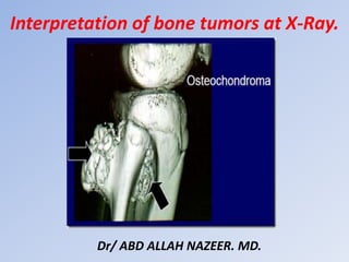
  • 39. Osteochondroma: Long bones++ The lesion is composed of cortical and medullary bone protruding from and continuous with the underlying bone. The areas of osseous continuity between parent bone and osteochondroma may be broad, involving a large portion of the bone circumference (dimension of the lesion base exceeding its length-sessile osteochondroma), or narrow, with a bulbous tip (pedunculated osteochondroma). Identifying the characteristic cortical and medullary continuity between lesion and parent bone on radiographs is dependent on lesion type (sessile or pedunculated), location, and image projection. This relationship is usually well delineated in lesions of long bones, particularly pedunculated osteochondromas, with standard radiographic projections (although often only on one view) . However, in osteochondromas of flat bones with complex anatomy (i.e., pelvis, spine, scapula) and sessile lesions, the continuity and thus the diagnosis may not be apparent on radiographs alone . Pedunculated lesions usually point away from the nearest joint owing to the forces of the overlying tendons and ligaments (although not typically attached to the osteochondroma.
  • 40.
  • 41.
  • 42. X-ray appearance of different exostoses. (a) Solitary exostosis in a femur. (b) Multiple exostoses in the knee joint, antero-posterior view. (c) Multiple exostoses in the knee joint, lateral view. (d) Multiple exostoses in the upper hip and trochanter (arrows indicate the exostoses sites).
  • 44.
  • 46. Typical osteoid osteoma in the femur. Notice the extensive cortical broadening.
  • 47.
  • 49. Osteoblastoma of the distal radius.
  • 51.
  • 54. Unicameral bone cyst with fracture and fallen fragment sign.
  • 55.
  • 56. Aneurysmal bone cyst - Proximal tibia.
  • 57. Aneurysmal bone cyst - Distal tibia.
  • 59.
  • 61. Giant cell tumor of the distal ulna.
  • 62. Giant cell tumor of the distal radius.
  • 63.
  • 64. Cortical fibrous defects and non-ossifying fibromas in children and young adults:
  • 66.
  • 69. Fibrous Dysplasia • Long lesion in a long bone (often occurs in proximal femur). • Expansion and bone deformity • Lytic but becomes ground-glass in appearance as the matrix calcifies, and then becomes sclerotic. • Asymptomatic, but can fracture • No periosteal reaction • May be single or multiple lesion in different locations.
  • 70.
  • 72. Fibrous dysplasia: various presentations with or without sclerotic margin, with ground glass appearance, with calcifications or ossifications.
  • 74. Fibrous dysplasia with Coxa vara of proximal femur.
  • 75. Enchondroma • Most commonly seen in the phalanges • Asymptomatic but commonly fractures • Well-defined with narrow zone of transition • Lobulated • Can become slightly expansile • Causes endosteal scalloping and cortical thinning • Ollier's Disease = Multiple enchondromas • Maffuci's Syndrome = Multiple enchondromas with soft tissue hemangiomas • Contain calcified chondroid matrix (irregular, speckled) when located away from phalanges.
  • 76.
  • 78. Plain X-Ray: Enchondroma of Proximal Phalanx.
  • 79. A child with Ollier’s disease: a non-hereditary, sporadic, skeletal disorder characterized by multiple enchondromas
  • 81. Mafucci’s syndrome. Plain radiograph of the left hand. Note multiple enchondromas and soft-tissue hemangiomas with intralesional phleboliths.
  • 83.
  • 84. Chondromyxoid fibroma - Proximal tibia.
  • 86.
  • 87.
  • 90.
  • 93. Chordoma: • Uncommon malignant tumours: 40% of all sacral tumors • Location • 50% occur in sacrum, • • • 40% of all sacral tumors • Most common in 4th or 5th sacral segment • 35% at skull base around clivus (Benign tumors but may locally invade and compress) • 15% in remainder of spine: Cervical spine most common • Occurs between the ages of 30-70 with a 2:1 male: female ratio • Imaging Findings • Large presacral mass (>10cm) with displacement of the rectum and/or Bladder • Solid tumor with cystic areas in 50% • Destroys multiple sacral and coccygeal segments • Sequestered bone fragments are common • Sclerotic rim in 50% • May have amorphous calcifications, especially peripherally • May cross the sacroiliac joint
  • 94.
  • 97.
  • 99. Brown tumor at the femur diaphysis.
  • 100.
  • 101. Adamantinoma of the proximal tibia.
  • 103.
  • 104.
  • 105. Skull and femur hemangioma.
  • 106. Computerized tomography demonstrates lesions with osseous spicules in keeping with hemangiomas involved with left frontal bone, supraorbital wall, sphenoid sinus and ethmoid. The central portion of this lesion revealed subtle striated trabeculations and retained a thin sclerotic rim of peripheral bone condensation, and the boundary was clear. Both the inner and the outer tables of the skull were eroded
  • 107. Hemangioma of the Long Bones
  • 108. Intraosseous Hemangioma of the Nasal Septum.
  • 109.
  • 111. Various presentations of eosinophilic granuloma.
  • 112. Monostotic and polyostotic eosinophilic granuloma in the skull. Eosinophilic granuloma in the skull.
  • 113.
  • 115. Ewing sarcoma arising from the right iliac bone.
  • 116. Ewing sarcoma of femur. Frontal radiograph and lateral radiographs of the femur demonstrate mottled, osteolytic lesion (blue circle) with poorly marginated edges in the diaphysis of the bone. There is sunburst periosteal reaction (red circle) and lamellated periosteal reaction (white arrows).
  • 118.
  • 119. (a) X-ray anteroposterior and lateral views of proximal tibia and knee joint showing diaphyseal osteosarcoma of tibia with sclerosis (arrow), cortical destruction on posteromedial side (arrow heads) and new bone formation in the soft tissues (b) x-ray distal end of femur (anteroposterior and lateral views) showing sclerosis/radio-opacity in sclerosing osteosarcoma.
  • 120. X-ray of knee joint anteroposterior views showing surface osteosarcoma: (a) parosteal (b) periosteal. See the under lying cortex is visibly intact in ‘a’ and lifting of periosteum in ‘b’ (red arrow). However, both are on the surface of the bone
  • 121. Telangiectatic type of osteosarcoma of the proximal tibia: (a) X-ray anteroposterior and lateral views showing lysis and expansion (b) MRI showing fluid levels.
  • 124.
  • 125. Chondrosarcoma of the iliac bone and proximal humerus.
  • 126. Chondrosarcoma of the distal femur and proximal humerus.
  • 127.
  • 129.
  • 130.
  • 131.
  • 132.
  • 133.
  • 134.
  • 135. Typical x-ray appearance of osteolytic bone metastases. Typical x-ray appearance of osteoblastic bone metastases.
  • 136.
  • 137.
  • 138.
  • 139.
  • 140. Anteroposterior radiograph of the lower legs demonstrates a permeative, lytic lesion in the metadiaphysis of the left tibia. Note the aggressive periosteal reaction with Codman triangles and soft-tissue extension/swelling. Soft-tissue involvement is easy to appreciate when the left leg is compared with the normal right leg.
  • 141. Burkitt's lymphoma of the humerus.
  • 142. 62-year-old patient with lymphoma and low back pain. Anteroposterior (A) and profile (B) X-rays showing a diffuse increase in bone density, with erosion on the left side of the L4 vertebral body (arrow).
  • 143.
  • 144. The skull x-ray lateral and AP view reveals multiple oil droplet like punched out lytic defects throughout bony calvarium. The appearance is highly suggestive of, but not completely diagnostic for, multiple myeloma.
  • 147.
  • 148.
  • 149.