
Recommandé
Contenu connexe
Tendances
Tendances (20)
Similaire à Blood Notes
Similaire à Blood Notes (20)
Plus de amyottmers
Dernier
Dernier (20)
Blood Notes
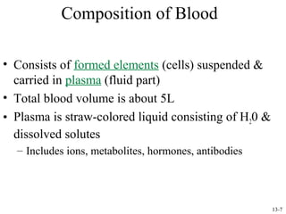
- 1. Composition of Blood • Consists of formed elements (cells) suspended & carried in plasma (fluid part) • Total blood volume is about 5L • Plasma is straw-colored liquid consisting of H20 & dissolved solutes – Includes ions, metabolites, hormones, antibodies 13-7
- 2. Physical Characteristics of Blood • Average volume of blood: – 5–6 L for males; 4–5 L for females (Normovolemia) – Hypovolemia - low blood volume – Hypervolemia - high blood volume • Viscosity (thickness) - 4 - 5 (where water = 1) • The pH of blood is 7.35–7.45; x = 7.4 • Salinity = 0.85% – Reflects the concentration of NaCl in the blood • Temperature is 38°C, slightly higher than “normal” body temperature • Blood accounts for approximately 8% of body weight
- 3. Plasma Proteins • Constitute 7-9% of plasma • Three types of plasma proteins: albumins, globulins, & fibrinogen – Albumin accounts for 60-80% • Creates colloid osmotic pressure that draws H 20 from interstitial fluid into capillaries to maintain blood volume & pressure • Globulins carry lipids – Gamma globulins are antibodies (immunoglobulins) • Fibrinogen serves as clotting factor – Converted to fibrin – Serum is fluid left when blood clots 13-8
- 4. Formed Elements • Are erythrocytes (RBCs) & leukocytes (WBCs) • RBCs are flattened biconcave discs – Shape provides increased surface area for diffusion – Lack nuclei & mitochondria – Each RBC contains 280 million hemoglobins 13-9
- 5. Leukocytes • Have nucleus, mitochondria, & amoeboid ability • Can squeeze through capillary walls (diapedesis) – Granular leukocytes help detoxify foreign substances & release heparin • Include eosinophils, basophils, & neutrophils 13-10
- 6. Leukocytes continued • Agranular leukocytes are phagocytic & produce antibodies • Include lymphocytes & monocytes 13-11
- 7. Platelets (thrombocytes) • Are smallest of formed elements, lack nucleus • Are fragments of megakaryocytes • Constitute most of mass of blood clots • Release serotonin to vasoconstrict & reduce blood flow to clot area • Secrete growth factors to maintain integrity of blood vessel wall • Survive 5-9 days 13-12
- 8. Components of Whole Blood Plasma (55% of whole blood) Buffy coat: leukocyctes and platelets (<1% of whole blood) 1 Withdraw blood 2 Centrifuge and place in tube • Hematocrit • Males: 47% ± 5% • Females: 42% ± 5% Erythrocytes (45% of whole blood) Formed elements
- 9. Hematopoiesis • Is formation of blood cells from stem cells in marrow (myeloid tissue) & lymphoid tissue • Erythropoiesis is formation of RBCs – Stimulated by erythropoietin (EPO) from kidney • Leukopoiesis is formation of WBCs – Stimulated by variety of cytokines • = autocrine regulators secreted by immune system 13-13
- 10. Life Cycle of Red Blood Cells
- 11. Erythropoiesis • 2.5 million RBCs are produced/sec • Lifespan of 120 days • Old RBCs removed from blood by phagocytic cells in liver, spleen, & bone marrow – Iron recycled back into hemoglobin production 13-14
- 12. Erythropoietin Mechanism Imb ala nce Start Normal blood oxygen levels Imb ala nce Increases O2-carrying ability of blood Stimulus: Hypoxia due to decreased RBC count, decreased availability of O2 to blood, or increased tissue demands for O2 Reduces O2 levels in blood Enhanced erythropoiesis increases RBC count Erythropoietin stimulates red bone marrow Kidney (and liver to a smaller extent) releases erythropoietin Figure 17.6
- 13. Dietary Requirements of Erythropoiesis • Erythropoiesis requires: – Proteins, lipids, and carbohydrates – Iron, vitamin B12, and folic acid • The body stores iron in Hb (65%), the liver, spleen, and bone marrow • Intracellular iron is stored in protein-iron complexes such as ferritin and hemosiderin • Circulating iron is loosely bound to the transport protein transferrin
- 14. RBC Antigens & Blood Typing • Antigens present on RBC surface specify blood type • Major antigen group is ABO system – – – – Type A blood has only A antigens Type B has only B antigens Type AB has both A & B antigens Type O has neither A or B antigens 13-15
- 15. Transfusion Reactions • People with Type A blood make antibodies to Type B RBCs, but not to Type A • Type B blood has antibodies to Type A RBCs but not to Type B • Type AB blood doesn’t have antibodies to A or B • Type O has antibodies to both Type A & B • If different blood types are mixed, antibodies will cause mixture to agglutinate 13-16
- 16. Transfusion Reactions continued • If blood types don't match, recipient’s antibodies agglutinate donor’s RBCs • Type O is “universal donor” because lacks A & B antigens • Insert fig. 13.6 – Recipient’s antibodies won’t agglutinate donor’s Type O RBCs • Type AB is “universal recipient” because doesn’t make anti-A or anti-B antibodies – Won’t agglutinate donor’s RBCs 13-17
- 17. Hemolytic Disease of the Newborn • May occur in an Rh- mom pregnanet with an Rh+ fetus • Hemolytic disease of the newborn – Rh+ antibodies of a sensitized Rh– mother cross the placenta and attack and destroy the RBCs of an Rh+ baby • Rh– mother becomes sensitized when Rh+ blood (from a previous pregnancy of an Rh+ baby or a Rh+ transfusion) causes her body to synthesis Rh+ antibodies • The drug RhoGAM can prevent the Rh– mother from becoming sensitized • Treatment of hemolytic disease of the newborn involves pre-birth transfusions and exchange transfusions after birth
- 18. Hemostasis • Is cessation of bleeding • Promoted by reactions initiated by vessel injury: – Vasoconstriction restricts blood flow to area – Platelet plug forms • Plug & surroundings are infiltrated by web of fibrin, forming clot 13-19
- 19. Role of Platelets • Platelets don't stick to intact endothelium because of presence of prostacyclin (PGI2--a prostaglandin) & NO QuickTime™ and a TIFF (LZW) decompressor are needed to see this picture. – Keep clots from forming & are vasodilators 13-20
- 20. Role of Platelets • Damage to endothelium allows platelets to bind to exposed collagen – von Willebrand factor increases bond by binding to both collagen & platelets – Platelets stick to collagen & release ADP, serotonin, & thromboxane A2 • = platelet release reaction 13-21
- 21. Role of Platelets continued • Serotonin & thromboxane A2 stimulate vasoconstriction, reducing blood flow to wound • ADP & thromboxane A2 cause other platelets to become sticky & attach & undergo platelet release reaction – This continues until platelet plug is formed 13-22
- 22. Role of Fibrin • Platelet plug becomes infiltrated by meshwork of fibrin • Clot now contains platelets, fibrin & trapped RBCs – Platelet plug undergoes plug contraction to form more compact plug 13-23
- 23. Conversion of Fibrinogen to Fibrin • Can occur via 2 pathways: – Intrinsic pathway clots damaged vessels & blood left in test tube • Initiated by exposure of blood to negatively charged surface of glass or blood vessel collagen – – This activates factor XII (a protease) which initiates a series of clotting factors Ca2+ & phospholipids convert prothrombin to thrombin » Thrombin converts fibrinogen to fibrin which polymerizes to form a mesh – Damage outside blood vessels releases tissue thromboplastin that triggers a clotting shortcut (= extrinsic pathway) 13-24
- 24. Fig 13.9 13-25
- 25. Dissolution of Clots • When damage is repaired, activated factor XII causes activation of kallikrein – Kallikrein converts plasminogen to plasmin • Plasmin digests fibrin, dissolving clot 13-26
- 26. Anticoagulants • Clotting can be prevented by Ca+2 chelators (e.g. sodium citrate or EDTA) – or heparin which activates antithrombin III (blocks thrombin) • Coumarin blocks clotting by inhibiting activation of Vit K – Vit K works indirectly by reducing Ca+2 availability 13-27
- 27. Prostaglandins (PGs) • Are produced in almost every organ • Belong to eicosanoid family -- all derived from arachidonic acid of plasma membrane 11-72
- 28. Prostaglandins (PGs) continued • Have wide variety of functions – Different PGs may exert antagonistic effects in tissues • Some promote smooth muscle contraction & some relaxation • Some promote clotting; some inhibit – Promotes inflammatory process of immune system – Plays role in ovulation – Inhibits gastric secretion in digestive system 11-73
- 29. Prostaglandins (PGs) continued • Cyclooxygenase (COX) 1 & 2 are involved in PG synthesis – Are targets of a number of inhibitory non-steroidal antiinflammatory drugs (NSAIDs) • Aspirin, indomethacin, ibuprofen inhibit both COX 1 & 2 thereby producing side effects • Celebrex & Vioxx only inhibit COX 2 & thus have few side effects 11-74
- 30. Erythrocyte Disorders • Polycythemia – Abnormal excess of erythrocytes • Increases viscosity, decreases flow rate of blood • Anemia – blood has abnormally low oxygen-carrying capacity – It is a symptom rather than a disease itself – Blood oxygen levels cannot support normal metabolism – Signs/symptoms include fatigue, paleness, shortness of breath, and chills
- 31. Anemia: Insufficient Erythrocytes • Hemorrhagic anemia – result of acute or chronic loss of blood • Hemolytic anemia – prematurely ruptured erythrocytes • Aplastic anemia – destruction or inhibition of red bone marrow
- 32. Anemia: Decreased Hemoglobin Content • Iron-deficiency anemia results from: – A secondary result of hemorrhagic anemia – Inadequate intake of iron-containing foods – Impaired iron absorption • Pernicious anemia results from: – Deficiency of vitamin B12 – Lack of intrinsic factor needed for absorption of B12 – Treatment is intramuscular injection of B12
- 33. Anemia: Abnormal Hemoglobin • Thalassemias – absent or faulty globin chain in hemoglobin – Erythrocytes are thin, delicate, and deficient in hemoglobin • Sickle-cell anemia – results from a defective gene – Codes for an abnormal hemoglobin called hemoglobin S (HbS) – This defect causes RBCs to become sickle-shaped in low oxygen situations
- 34. Polycythemia • Polycythemia – excess RBCs that increase blood viscosity • Three main polycythemias are: – Polycythemia vera – Secondary polycythemia – Blood doping
- 35. Leukocytes Disorders: Leukemias • Leukemia refers to cancerous conditions involving white blood cells • Leukemias are named according to the abnormal white blood cells involved – Myelocytic leukemia – involves myeloblasts – Lymphocytic leukemia – involves lymphocytes • Acute leukemia involves blast-type cells and primarily affects children • Chronic leukemia is more prevalent in older people
- 36. Leukemia • Immature white blood cells are found in the bloodstream in all leukemias • Bone marrow becomes totally occupied with cancerous leukocytes • Severe anemia ensues due to excess production of WBC’s • The white blood cells produced, though numerous, are not functional • Death is caused by internal hemorrhage and overwhelming infections • Treatments include irradiation, antileukemic drugs, and bone marrow transplants