This PowerPoint helps students to consider the concept of infinity.
Tuberculosis
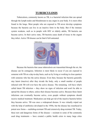
1. TUBERCULOSIS
Tuberculosis, commonly known as TB, is a bacterial infection that can spread
through the lymph nodes and bloodstream to any organ in your body. It is most often
found in the lungs. Most people who are exposed to TB never develop symptoms
because the bacteria can live in an inactive form in the body. But if the immune
system weakens, such as in people with HIV or elderly adults, TB bacteria can
become active. In their active state, TB bacteria cause death of tissue in the organs
they infect. Active TB disease can be fatal if left untreated.
Because the bacteria that cause tuberculosis are transmitted through the air, the
disease can be contagious. Infection is most likely to occur if you are exposed to
someone with TB on a day-to-day basis, such as by living or working in close quarters
with someone who has the active disease. Even then, because the bacteria generally
stay latent (inactive) after they invade the body, only a small number of people
infected with TB will ever have the active disease. The remaining will have what's
called latent TB infection -- they show no signs of infection and won't be able to
spread the disease to others, unless their disease becomes active. Because these latent
infections can eventually become active, even people without symptoms should
receive medical treatment. Medication can help get rid of the inactive bacteria before
they become active. TB was once a widespread disease. It was virtually wiped out
with the help of antibiotics developed in the 1950s, but the disease has resurfaced in
potent new forms -- multidrug-resistant TB and extensively drug-resistant TB. Today,
these new and dangerous forms of the disease -- resistant to some of the commonly
used drug treatments -- have created a public health crisis in many large cities
2. worldwide. If you have TB -- in its active or latent state -- you must seek medical
treatment.
Tuberculosis, MTB, or TB (short for tubercle bacillus), in the past also called
phthisis, phthisis pulmonalis, or consumption, is a widespread, and in many cases
fatal, infectious disease caused by various strains of mycobacteria, usually
Mycobacterium tuberculosis.[1] Tuberculosis typically attacks the lungs, but can also
affect other parts of the body. It is spread through the air when people who have an
active TB infection cough, sneeze, or otherwise transmit respiratory fluids through the
air. Most infections do not have symptoms, known as latent tuberculosis. About one in
ten latent infections eventually progresses to active disease which, if left untreated,
kills more than 50% of those so infected.
The classic symptoms of active TB infection are a chronic cough with blood-tinged
sputum, fever, night sweats, and weight loss (the latter giving rise to the
formerly common term for the disease, "consumption"). Infection of other organs
causes a wide range of symptoms. Diagnosis of active TB relies on radiology
(commonly chest X-rays), as well as microscopic examination and microbiological
culture of body fluids. Diagnosis of latent TB relies on the tuberculin skin test (TST)
and/or blood tests. Treatment is difficult and requires administration of multiple
antibiotics over a long period of time. Social contacts are also screened and treated if
necessary. Antibiotic resistance is a growing problem in multiple drug-resistant
tuberculosis (MDR-TB) infections. Prevention relies on screening programs and
vaccination with the bacillus Calmette-Guérin vaccine.
One-third of the world's population is thought to have been infected with M.
tuberculosis with new infections occurring in about 1% of the population each year. In
2007, an estimated 13.7 million chronic cases were active globally, while in 2010, an
estimated 8.8 million new cases and 1.5 million associated deaths occurred, mostly in
developing countries. The absolute number of tuberculosis cases has been decreasing
since 2006, and new cases have decreased since 2002. The rate of tuberculosis in
different areas varies across the globe; about 80% of the population in many Asian
and African countries tests positive in tuberculin tests, while only 5–10% of the
United States population tests positive.[1] More people in the developing world
3. contract tuberculosis because of a poor immune system, largely due to high rates of
HIV infection and the corresponding development of AIDS.
Signs and symptoms
The main symptoms of variants and stages of tuberculosis are given, with many
symptoms overlapping with other variants, while others are more (but not entirely)
specific for certain variants. Multiple variants may be present simultaneously.
Tuberculosis may infect any part of the body, but most commonly occurs in the lungs
(known as pulmonary tuberculosis). Extrapulmonary TB occurs when tuberculosis
develops outside of the lungs, although extrapulmonary TB may coexist with
pulmonary TB, as well. General signs and symptoms include fever, chills, night
sweats, loss of appetite, weight loss, and fatigue. Significant nail clubbing may also
occur.
Pulmonary
If a tuberculosis infection does become active, it most commonly involves the
lungs (in about 90% of cases). Symptoms may include chest pain and a prolonged
cough producing sputum. About 25% of people may not have any symptoms (i.e. they
remain "asymptomatic").Occasionally, people may cough up blood in small amounts,
and in very rare cases, the infection may erode into the pulmonary artery, resulting in
massive bleeding (Rasmussen's aneurysm). Tuberculosis may become a chronic
illness and cause extensive scarring in the upper lobes of the lungs. The upper lung
lobes are more frequently affected by tuberculosis than the lower ones. The reason for
this difference is not entirely clear. It may be due either to better air flow, or to poor
lymph drainage within the upper lungs.
Extrapulmonary
4. In 15–20% of active cases, the infection spreads outside the lungs, causing
other kinds of TB. These are collectively denoted as "extrapulmonary
tuberculosis".Extrapulmonary TB occurs more commonly in immunosuppressed
persons and young children. In those with HIV, this occurs in more than 50% of cases.
Notable extrapulmonary infection sites include the pleura (in tuberculous pleurisy),
the central nervous system (in tuberculous meningitis), the lymphatic system (in
scrofula of the neck), the genitourinary system (in urogenital tuberculosis), and the
bones and joints (in Pott disease of the spine), among others. When it spreads to the
bones, it is also known as "osseous tuberculosis". a form of osteomyelitis. Sometimes,
bursting of a tubercular abscess through skin results in tuberculous ulcer. An ulcer
originating from nearby infected lymph nodes is painless, slowly enlarging and has an
appearance of "wash leather". A potentially more serious, widespread form of TB is
called "disseminated" TB, commonly known as miliary tuberculosis. Miliary TB
makes up about 10% of extrapulmonary cases
CAUSES
Mycobacteria
Scanning electron micrograph of M. tuberculosis
The main cause of TB is Mycobacterium tuberculosis, a small, aerobic,
nonmotile bacillus. The high lipid content of this pathogen accounts for many of its
unique clinical characteristics. It divides every 16 to 20 hours, which is an extremely
slow rate compared with other bacteria, which usually divide in less than an hour.
Mycobacteria have an outer membrane lipid bilayer. If a Gram stain is performed,
MTB either stains very weakly "Gram-positive" or does not retain dye as a result of
5. the high lipid and mycolic acid content of its cell wall. MTB can withstand weak
disinfectants and survive in a dry state for weeks. In nature, the bacterium can grow
only within the cells of a host organism, but M. tuberculosis can be cultured in the
laboratory.
Using histological stains on expectorated samples from phlegm (also called
"sputum"), scientists can identify MTB under a regular (light) microscope. Since MTB
retains certain stains even after being treated with acidic solution, it is classified as an
acid-fast bacillus (AFB). The most common acid-fast staining techniques are the
Ziehl–Neelsen stain, which dyes AFBs a bright red that stands out clearly against a
blue background, and the auramine-rhodamine stain followed by fluorescence
microscopy. The M. tuberculosis complex (MTBC) includes four other TB-causing
mycobacteria: M. bovis, M. africanum, M. canetti, and M. microti. M. africanum is not
widespread, but it is a significant cause of tuberculosis in parts of Africa. M. bovis was
once a common cause of tuberculosis, but the introduction of pasteurized milk has
largely eliminated this as a public health problem in developed countries. M. canetti is
rare and seems to be limited to the Horn of Africa, although a few cases have been
seen in African emigrants. M. microti is also rare and is mostly seen in
immunodeficient people, although the prevalence of this pathogen has possibly been
significantly underestimated.
Other known pathogenic mycobacteria include M. leprae, M. avium, and M.
kansasii. The latter two species are classified as "nontuberculous mycobacteria"
(NTM). NTM cause neither TB nor leprosy, but they do cause pulmonary diseases
that resemble TB.
Risk factors
A number of factors make people more susceptible to TB infections. The most
important risk factor globally is HIV; 13% of all people with TB are infected by the
virus. This is a particular problem in sub-Saharan Africa, where rates of HIV are high.
Of people without HIV who are infected with tuberculosis, about 5–10% develop
active disease during their lifetimes; in contrast, 30% of those coinfected with HIV
develop the active disease. Tuberculosis is closely linked to both overcrowding and
malnutrition, making it one of the principal diseases of poverty.[7] Those at high risk
6. thus include: people who inject illicit drugs, inhabitants and employees of locales
where vulnerable people gather (e.g. prisons and homeless shelters), medically
underprivileged and resource-poor communities, high-risk ethnic minorities, children
in close contact with high-risk category patients, and health-care providers serving
these patients.
Chronic lung disease is another significant risk factor. Silicosis increases the risk
about 30-fold. Those who smoke cigarettes have nearly twice the risk of TB compared
to nonsmokers. Other disease states can also increase the risk of developing
tuberculosis. These include alcoholism and diabetes mellitus (three-fold increase).
Certain medications, such as corticosteroids and infliximab (an anti-αTNF monoclonal
antibody), are becoming increasingly important risk factors, especially in the
developed world. Also a genetic susceptibility element exists, for which the overall
importance remains undefined.[
Mechanism
Transmission
When people with active pulmonary TB cough, sneeze, speak, sing, or spit,
they expel infectious aerosol droplets 0.5 to 5.0 μm in diameter. A single sneeze can
release up to 40,000 droplets. Each one of these droplets may transmit the disease,
since the infectious dose of tuberculosis is very small (the inhalation of fewer than 10
bacteria may cause an infection). People with prolonged, frequent, or close contact
with people with TB are at particularly high risk of becoming infected, with an
estimated 22% infection rate. A person with active but untreated tuberculosis may
infect 10–15 (or more) other people per year. Transmission should only occur from
people with active TB – those with latent infection are not thought to be contagious.
The probability of transmission from one person to another depends upon several
factors, including the number of infectious droplets expelled by the carrier, the
effectiveness of ventilation, the duration of exposure, the virulence of the M.
tuberculosis strain, the level of immunity in the uninfected person, and others. The
cascade of person-to-person spread can be circumvented by effectively segregating
those with active ("overt") TB and putting them on anti-TB drug regimens. After
about two weeks of effective treatment, subjects with nonresistant active infections
7. generally do not remain contagious to others. If someone does become infected, it
typically takes three to four weeks before the newly infected person becomes
infectious enough to transmit the disease to others.
Pathogenesis
About 90% of those infected with M. tuberculosis have asymptomatic, latent
TB infections (sometimes called LTBI), with only a 10% lifetime chance that the
latent infection will progress to overt, active tuberculous disease. In those with HIV,
the risk of developing active TB increases to nearly 10% a year. If effective treatment
is not given, the death rate for active TB cases is up to 66%.
TB infection begins when the mycobacteria reach the pulmonary alveoli, where
they invade and replicate within endosomes of alveolar macrophages. Macrophages
identify the bacterium as "foreign" and attempt to eliminate it by phagocytosis. During
this process, the entire bacterium is enveloped by the macrophage and stored
temporarily in a membrane-bound vesicle called a phagosome. The phagosome then
combines with a lysosome to create a phagolysosome. In the phagolysosome, the cell
attempts to use reactive oxygen species and acid to kill the bacterium. However, M.
tuberculosis has a thick, waxy mycolic acid capsule that protects it from these toxic
substances. M. tuberculosis actually reproduces inside the macrophage and will
eventually kill the immune cell. The primary site of infection in the lungs, known as
the "Ghon focus", is generally located in either the upper part of the lower lobe, or the
lower part of the upper lobe. Tuberculosis of the lungs may also occur via infection
from the blood stream. This is known as a Simon focus and is typically found in the
top of the lung. This hematogenous transmission can also spread infection to more
distant sites, such as peripheral lymph nodes, the kidneys, the brain, and the
bones.[1][49] All parts of the body can be affected by the disease, though for unknown
reasons it rarely affects the heart, skeletal muscles, pancreas, or thyroid.
Tuberculosis is classified as one of the granulomatous inflammatory diseases.
Macrophages, T lymphocytes, B lymphocytes, and fibroblasts aggregate to form
granulomas, with lymphocytes surrounding the infected macrophages. When other
macrophages attack the infected macrophage, they fuse together to form a giant
multinucleated cell in the alveolar lumen. The granuloma prevents dissemination of
8. the mycobacteria and provides a local environment for interaction of cells of the
immune system. Bacteria inside the granuloma can become dormant, resulting in
latent infection. Another feature of the granulomas is the development of abnormal
cell death (necrosis) in the center of tubercles. To the naked eye, this has the texture of
soft, white cheese and is termed caseous necrosis.
If TB bacteria gain entry to the blood stream from an area of damaged tissue,
they can spread throughout the body and set up many foci of infection, all appearing
as tiny, white tubercles in the tissues. This severe form of TB disease, most common
in young children and those with HIV, is called miliary tuberculosis. People with this
disseminated TB have a high fatality rate even with treatment (about 30%). In many
people, the infection waxes and wanes. Tissue destruction and necrosis are often
balanced by healing and fibrosis. Affected tissue is replaced by scarring and cavities
filled with caseous necrotic material. During active disease, some of these cavities are
joined to the air passages bronchi and this material can be coughed up. It contains
living bacteria, so can spread the infection. Treatment with appropriate antibiotics
kills bacteria and allows healing to take place. Upon cure, affected areas are
eventually replaced by scar tissue.
Diagnosis
M. tuberculosis (stained red) in sputum
Active tuberculosis
Diagnosing active tuberculosis based merely on signs and symptoms is
difficult, as is diagnosing the disease in those who are immunosuppressed. A
9. diagnosis of TB should, however, be considered in those with signs of lung disease or
constitutional symptoms lasting longer than two weeks. A chest X-ray and multiple
sputum cultures for acid-fast bacilli are typically part of the initial evaluation.
Interferon-γ release assays and tuberculin skin tests are of little use in the developing
world. IGRA have similar limitations in those with HIV.
A definitive diagnosis of TB is made by identifying M. tuberculosis in a
clinical sample (e.g. sputum, pus, or a tissue biopsy). However, the difficult culture
process for this slow-growing organism can take two to six weeks for blood or sputum
culture. Thus, treatment is often begun before cultures are confirmed. Nucleic acid
amplification tests and adenosine deaminase testing may allow rapid diagnosis of TB.
These tests, however, are not routinely recommended, as they rarely alter how a
person is treated. Blood tests to detect antibodies are not specific or sensitive, so they
are not recommended.
Prevention
Tuberculosis prevention and control efforts primarily rely on the vaccination of
infants and the detection and appropriate treatment of active cases. The World Health
Organization has achieved some success with improved treatment regimens, and a
small decrease in case numbers.
Vaccines
The only available vaccine as of 2011 is bacillus Calmette-Guérin (BCG). In
children it decreases the risk of getting the infection by 20% and the risk of infection
turning into disease by nearly 60%. It is the most widely used vaccine worldwide,
with more than 90% of all children being vaccinated. The immunity it induces
decreases after about ten years. As tuberculosis is uncommon in most of Canada, the
United Kingdom, and the United States, BCG is only administered to people at high
risk. Part of the reasoning arguing against the use of the vaccine is that it makes the
tuberculin skin test falsely positive, so of no use in screening. A number of new
vaccines are currently in development.
Public health
The World Health Organization declared TB a "global health emergency" in
1993, and in 2006, the Stop TB Partnership developed a Global Plan to Stop
10. Tuberculosis that aims to save 14 million lives between its launch and 2015. A
number of targets they have set are not likely to be achieved by 2015, mostly due to
the increase in HIV-associated tuberculosis and the emergence of multiple drug-resistant
tuberculosis. A tuberculosis classification system developed by the American
Thoracic Society is used primarily in public health programs.
Medication resistance
Primary resistance occurs when a person becomes infected with a resistant
strain of TB. A person with fully susceptible TB may develop secondary (acquired)
resistance during therapy because of inadequate treatment, not taking the prescribed
regimen appropriately (lack of compliance), or using low-quality medication. Drug-resistant
TB is a serious public health issue in many developing countries, as its
treatment is longer and requires more expensive drugs. MDR-TB is defined as
resistance to the two most effective first-line TB drugs: rifampicin and isoniazid.
Extensively drug-resistant TB is also resistant to three or more of the six classes of
second-line drugs. Totally drug-resistant TB is resistant to all currently used drugs. It
was first observed in 2003 in Italy, but not widely reported until 2012, and has also
been found in Iran and India. Bedaquiline is tentatively supported for use in multiple
drug-resistant TB. XDR-TB is a term sometimes used to define extensively resistant
TB, and constitutes one in ten cases of MDR-TB. Cases of XDR TB have been
identified in more than 90% of countries.
Society and culture
The World Health Organization, Bill and Melinda Gates Foundation, and US
government are subsidizing a new fast-acting diagnostic test for use in low- and middle-income
countries. This will reduce the cost from $16.86 to $9.98. Additionally the test can
determine if there is resistance to the antibiotic rifampicin which may indicate multi-drug
resistant tuberculosis and is accurate in those who are co-infected with HIV. Many resource-poor
places as of 2011 still only have access to sputum microscopy. India had the highest
total number of TB cases worldwide in 2010, in part due to poor disease management within
the private and public health care sector. Programs such as the Revised National
Tuberculosis Control Program are helping to reduce TB levels amongst people receiving
public health care.

![worldwide. If you have TB -- in its active or latent state -- you must seek medical
treatment.
Tuberculosis, MTB, or TB (short for tubercle bacillus), in the past also called
phthisis, phthisis pulmonalis, or consumption, is a widespread, and in many cases
fatal, infectious disease caused by various strains of mycobacteria, usually
Mycobacterium tuberculosis.[1] Tuberculosis typically attacks the lungs, but can also
affect other parts of the body. It is spread through the air when people who have an
active TB infection cough, sneeze, or otherwise transmit respiratory fluids through the
air. Most infections do not have symptoms, known as latent tuberculosis. About one in
ten latent infections eventually progresses to active disease which, if left untreated,
kills more than 50% of those so infected.
The classic symptoms of active TB infection are a chronic cough with blood-tinged
sputum, fever, night sweats, and weight loss (the latter giving rise to the
formerly common term for the disease, "consumption"). Infection of other organs
causes a wide range of symptoms. Diagnosis of active TB relies on radiology
(commonly chest X-rays), as well as microscopic examination and microbiological
culture of body fluids. Diagnosis of latent TB relies on the tuberculin skin test (TST)
and/or blood tests. Treatment is difficult and requires administration of multiple
antibiotics over a long period of time. Social contacts are also screened and treated if
necessary. Antibiotic resistance is a growing problem in multiple drug-resistant
tuberculosis (MDR-TB) infections. Prevention relies on screening programs and
vaccination with the bacillus Calmette-Guérin vaccine.
One-third of the world's population is thought to have been infected with M.
tuberculosis with new infections occurring in about 1% of the population each year. In
2007, an estimated 13.7 million chronic cases were active globally, while in 2010, an
estimated 8.8 million new cases and 1.5 million associated deaths occurred, mostly in
developing countries. The absolute number of tuberculosis cases has been decreasing
since 2006, and new cases have decreased since 2002. The rate of tuberculosis in
different areas varies across the globe; about 80% of the population in many Asian
and African countries tests positive in tuberculin tests, while only 5–10% of the
United States population tests positive.[1] More people in the developing world](data:image/gif;base64,R0lGODlhAQABAIAAAAAAAP///yH5BAEAAAAALAAAAAABAAEAAAIBRAA7)