1. 7-1 Life Is Cellular
Copyright Pearson Prentice Hall
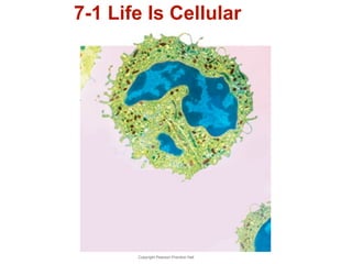
2. The Discovery of the Cell
Cells are the basic units of life.
Copyright Pearson Prentice Hall
3. The Discovery of the Cell
The cell theory states:
•All living things are
composed of cells.
•Cells are the basic units of
structure and function in
living things.
•New cells are produced from
existing cells.
Copyright Pearson Prentice Hall
4. Exploring the Cell
Electron Microscopes
•1000 times more powerful than light microscopes.
•used to visualize nonliving, preserved cells and tissues.
Transmission electron microscopes (TEMs)
Used to study cell structures and large protein molecules
Specimens must be cut into ultra-thin slices
Scanning electron microscopes (SEMs)
Produce three-dimensional images of cells
Specimens do not have to be cut into thin slices
Copyright Pearson Prentice Hall
5. Exploring the Cell
Scanning Electron Micrograph of Neurons
Copyright Pearson Prentice Hall
6. Exploring the Cell
Confocal Light
Microscopes
Confocal light
microscopes scan cells
with a laser beam.
This makes it possible
to build three-dimensional
images of
Copyright Pearson Prentice Hall
cells and their parts.
7. Exploring the Cell
Scanning Probe
Microscopes
Scanning probe
microscopes allow us
to observe single
atoms.
Images are produced
by tracing surfaces of
samples with a fine
probe.
Copyright Pearson Prentice Hall
DNA
8. Prokaryotes and Eukaryotes
Cells come in a variety of shapes and sizes.
All cells:
• are surrounded by a barrier called a cell
membrane.
• at some point contain DNA.
Copyright Pearson Prentice Hall
9. Cells are classified into two categories, depending on
whether they contain a nucleus.
The nucleus is a membrane-enclosed
structure that contains the
cell's genetic material in the form of
DNA.
The nucleus controls many of the cell's activities.
Copyright Pearson Prentice Hall
10. Eukaryotes- cells contain nuclei.
~ ~
nucleus
~ ~
Prokaryotes- cells do not contain
nuclei.
~ ~
DNA
just floats
~ around ~
~
Copyright Pearson Prentice Hall
11. Prokaryotic cells are simpler than eukaryotic cells.
Bacteria are prokaryotes.
Copyright Pearson Prentice Hall
12. Eukaryotic cells are generally larger and more
complex than prokaryotic cells.
Eukaryotic cells generally contain dozens of
structures and internal membranes.
Many eukaryotic cells are highly specialized.
Plants, animals, fungi, and protists
are eukaryotes.
Copyright Pearson Prentice Hall
14. The parts that make up a
eukaryotic cell are known as
organelles.
Cell biologists divide the eukaryotic cell into two
major sections: the nucleus and the cytoplasm.
The Cytoplasm is the fluid outside
the nucleus.
Copyright Pearson Prentice Hall
15. ACTIVE ART go to PHSchool.com and type in web code cbp-3072
Copyright Pearson Prentice Hall
Plant Cell
Nuclear envelope
Ribosome (free)
Ribosome
(attached)
Golgi
apparatus
Mitochondrion
Vacuole
Nucleolus
Nucleus
Smooth
endoplasmic
reticulum
Rough endoplasmic
reticulum
Cell wall
Cell membrane
Chloroplast
17. Draw two eukaryotic cells
PLANT CELL ANIMAL CELL
Nucleus
Cytoplasm
Cell membrane
Draw and label- cell membrane, nucleus, cytoplasm.
Copyright Pearson Prentice Hall
18. Nucleus
Nucleus
The nucleus is the control center of the cell.
The nucleus contains nearly
all the cell's DNA.
DNA contains coded
instructions for making proteins
and other molecules.
Copyright Pearson Prentice Hall
19. Nucleus
Copyright Pearson Prentice Hall
The Nucleus
Nucleolus Nuclear envelope
Nuclear
pores
Chromatin
20. Nucleus
The nucleus is
surrounded by a
nuclear envelope
composed of two
membranes.
The envelope is
dotted with nuclear
pores, which allow
material to move in
and out of the
nucleus.
Nuclear
envelope
Copyright Pearson Prentice Hall
Nuclear
pores
21. Nucleus
The granular material in the
nucleus is called chromatin
(contains DNA and proteins).
Copyright Pearson Prentice Hall
22. Nucleus
When a cell divides, chromatin condenses to form
chromosomes.
Chromosomes contain
the genetic information
that is passed from one
generation of cells to
the next.
Copyright Pearson Prentice Hall
23. Nucleus
Most nuclei also contain a nucleolus.
The nucleolus is where the assembly of
ribosomes begins.
Copyright Pearson Prentice Hall
Nucleolus
24. Draw two eukaryotic cells
PLANT CELL ANIMAL CELL
Chromatin (DNA)
Draw and label- chromatin(DNA)
Copyright Pearson Prentice Hall
25. Ribosomes
Ribosomes
One of the most important jobs carried out in the cell
is making proteins.
Proteins are assembled on
ribosomes found throughout the
cytoplasm.
Copyright Pearson Prentice Hall
26. Ribosomes
Ribosomes produce proteins by following coded
instructions that come from the nucleus.
Cells that are active in protein synthesis are
often packed with ribosomes.
Copyright Pearson Prentice Hall
Ribosome (free)
Ribosome
(attached)
27. Endoplasmic Reticulum
Eukaryotic cells contain an internal membrane
system called the endoplasmic reticulum, or ER.
The endoplasmic reticulum is
where lipids that make the cell
membrane are assembled, along
with proteins and other materials
that are exported from the cell.
Copyright Pearson Prentice Hall
28. Copyright Pearson Prentice Hall
Endoplasmic Reticulum
Ribosomes
Endoplasmic
Reticulum
Ribosomes are found on the
surface of rough ER.
29. Endoplasmic Reticulum
There are two types of ER—rough and smooth.
The portion of the ER involved in protein
synthesis is called rough endoplasmic
reticulum, or rough ER.
Rough ER is abundant in cells that produce
large amounts of protein for export.
Copyright Pearson Prentice Hall
30. Endoplasmic Reticulum
Smooth ER does not have ribosomes on
its surface.
Smooth ER contains collections of
enzymes that perform specialized tasks,
such as synthesis of membrane lipids
and detoxification of drugs.
Copyright Pearson Prentice Hall
31. Draw two eukaryotic cells
PLANT CELL ANIMAL CELL
ribosomes
Endoplasmic
reticulum
Draw and label- ribosomes, endoplasmic reticulum
Copyright Pearson Prentice Hall
32. Golgi Apparatus
Proteins produced in the rough ER move into the
Golgi apparatus.
The Golgi apparatus appears as a stack of
membranes.
Copyright Pearson Prentice Hall
33. Golgi Apparatus
The Golgi apparatus
modifies, sorts, and packages
proteins and other materials from
the endoplasmic reticulum for
storage in the cell or secretion
outside the cell.
From the Golgi apparatus, proteins are then
“shipped” to their final destinations throughout the
cell or outside of the cell.
Copyright Pearson Prentice Hall
34. Draw two eukaryotic cells
PLANT CELL ANIMAL CELL
Golgi apparatus
Draw and label- golgi apparatus
Copyright Pearson Prentice Hall
35. Lysosomes
Lysosomes are filled with enzymes
that can break down large
molecules and old organelles.
Lysosomes are like recycling centers!
Copyright Pearson Prentice Hall
36. Vacuoles
Vacuoles
Some cells contain saclike
structures called vacuoles that
store materials such as water, salts,
proteins, and carbohydrates.
Copyright Pearson Prentice Hall
37. Vacuoles
In many plant cells
there is a single, large
central vacuole filled
with liquid.
The pressure of the
central vacuole allows
plants to support heavy
structures such as
leaves and flowers. Vacuole
Copyright Pearson Prentice Hall
38. Vacuoles
Vacuoles are also found
in some unicellular
organisms and in some
animals.
The paramecium
contains a contractile
vacuole that pumps
excess water out of the
cell.
Copyright Pearson Prentice Hall
Contractile vacuole
39. Draw two eukaryotic cells
PLANT CELL ANIMAL CELL
Vacuole
Draw and label- vacuole
Copyright Pearson Prentice Hall
40. Mitochondria and Chloroplasts
Copyright Pearson Prentice Hall
Mitochondrion =
power
Nearly all eukaryotic cells
contain mitochondria.
Mitochondria
convert food
molecules into a
more usable
energy source.
41. Mitochondria and Chloroplasts
Mitochondria are enclosed by two membranes—
an outer membrane and an inner membrane.
The inner membrane is folded up inside the
organelle.
Copyright Pearson Prentice Hall
42. Mitochondria and Chloroplasts
Chloroplast
Chloroplasts
Plants and some other
organisms contain
GREEN chloroplasts.
Chloroplasts
capture energy
from sunlight
and convert it
into chemical
energy during
photosynthesis.
43. Mitochondria and Chloroplasts
Chloroplasts are surrounded by two membranes.
Chloroplasts contain the green pigment
chlorophyll.
Copyright Pearson Prentice Hall
44. Draw two eukaryotic cells
PLANT CELL ANIMAL CELL
Mitochondria
Draw and label- mitochondria, chloroplasts
Copyright Pearson Prentice Hall
Chloroplasts
46. Cytoskeleton
The cytoskeleton helps the
cell maintain its shape and
can be involved in movement.
Eukaryotic cells are given their shape and
internal organization by the cytoskeleton.
Copyright Pearson Prentice Hall
48. 7-3 Cell Boundaries
All cells are surrounded by a thin, flexible
barrier known as the cell membrane.
Many cells also produce a strong
supporting layer around the membrane
known as a cell wall.
Copyright Pearson Prentice Hall
49. Cell Membrane
Cell Membrane
The cell membrane regulates what
enters and leaves the cell and also
provides protection and support.
Copyright Pearson Prentice Hall
50. Cell Membrane
Copyright Pearson Prentice Hall
Cell Membrane
Outside of
cell
Cell
membrane
Inside of cell
(cytoplasm)
Protein
channel
Proteins
Carbohydrate
chains
Lipid bilayer
51. Cell Membrane
The composition of nearly all cell
membranes is a double-layered sheet
called a lipid bilayer.
Copyright Pearson Prentice Hall
Lipid bilayer
52. Cell Membrane
The lipid bilayer gives cell membranes a
flexible structure that forms a barrier
between the cell and its surroundings.
Copyright Pearson Prentice Hall
53. Cell Membrane
Most cell membranes contain protein
molecules embedded in the lipid bilayer, some
of which have carbohydrate molecules
attached to them.
Copyright Pearson Prentice Hall
Protein
channel
Proteins
Carbohydrate
chains
54. Cell Walls
Cell walls are found in plants,
algae, fungi, and many
prokaryotes.
The cell wall provides support and
protection for the cell.
Copyright Pearson Prentice Hall
55. Cell Walls
Cell Wall
The cell wall lies outside the cell membrane.
Most cell walls are porous enough to allow water,
oxygen, carbon dioxide, and certain other
substances to pass through easily.
Copyright Pearson Prentice Hall
56. Draw two eukaryotic cells
PLANT CELL ANIMAL CELL
Draw and label- cell wall
Copyright Pearson Prentice Hall
Cell wall
57. Diffusion Through Cell Boundaries
Diffusion Through Cell Boundaries
Every living cell exists in a liquid environment.
The cell membrane regulates
movement of dissolved
molecules from the liquid on one side of the
membrane to the liquid on the other side.
Copyright Pearson Prentice Hall
58. Diffusion Through Cell Boundaries
Measuring Concentration
A solution is a mixture of two or more
substances.
The substances dissolved in the solution are
called solutes.
The concentration of a solution
is the mass of solute in a given
volume of solution, or mass/
volume.
Copyright Pearson Prentice Hall
59. Diffusion Through Cell Boundaries
Diffusion– Solutes tend to move
from areas of high concentration
towards areas of low
concentration.
Copyright Pearson Prentice Hall
60. When the solute is evenly spread
through the solution, the system
has reached equilibrium.
It will never become
concentrated in one area
like it was before!
Copyright Pearson Prentice Hall
61. Diffusion Through Cell Boundaries
ACTIVE ART go to PHSchool.com and type in Web Code cbp-3073
Copyright Pearson Prentice Hall
62. Diffusion Through Cell Boundaries
There is a higher concentration
of solute on one side of the
membrane as compared to the
other side of the membrane.
Copyright Pearson Prentice Hall
63. Diffusion Through Cell Boundaries
Solute particles move from the
side of the membrane with a
higher concentration of solute
to the side of the membrane
with a lower concentration of
solute. The solute particles will
continue to diffuse across the
membrane until equilibrium is
reached.
Copyright Pearson Prentice Hall
64. Diffusion Through Cell Boundaries
When equilibrium is
reached, solute
particles continue
to diffuse across
the membrane in
both directions, but
the concentrations
do not change.
Copyright Pearson Prentice Hall
65. Diffusion Through Cell Boundaries
Diffusion depends upon random particle
movements. Therefore, substances
diffuse across membranes
without requiring the cell to use
energy.
Copyright Pearson Prentice Hall
66. Osmosis
Osmosis is the diffusion of
water through a selectively
permeable membrane.
Osmosis is diffusion so it
requires no energy!
Copyright Pearson Prentice Hall
67. Osmosis
ACTIVE ART go to PHSchool.com and type in Web Code cbp-3075
How Osmosis Works
Copyright Pearson Prentice Hall
Movement of
water
Dilute sugar
solution
(Water more
concentrated)
Concentrated
sugar solution
(Water less
concentrated)
Sugar
molecules
Selectively permeable
membrane- only lets
water through
68. Osmosis
Water tends to diffuse from a highly
concentrated region to a less concentrated
region.
If you compare two solutions, the more
concentrated solution is hypertonic
(“above strength”).
The more dilute solution is hypotonic
(“below strength”).
Copyright Pearson Prentice Hall
69. Osmosis
When concentrations of solutions are the
same on both sides of a membrane, the
solutions are isotonic (”same strength”).
Copyright Pearson Prentice Hall
70. Osmosis
Osmotic Pressure
Osmosis exerts a pressure known as osmotic
pressure on the hypertonic side of a selectively
permeable membrane.
Copyright Pearson Prentice Hall
71. Osmosis
The cell is filled with salts, sugars,
proteins, and other molecules so
it is hypertonic(lots of solutes)
compared to fresh water.
This causes water to move into
the cell.
The cell becomes swollen or
bursts.
Copyright Pearson Prentice Hall
72. Osmosis
Cells in large organisms are not in danger
of bursting because they are bathed in
fluids, such as blood, that are isotonic.
Other cells are surrounded by tough cell
walls that prevent the cells from expanding
even under tremendous osmotic pressure.
Copyright Pearson Prentice Hall
73. Facilitated Diffusion
Facilitated Diffusion
Cell membranes have protein channels that act as
carriers, making it easy for certain molecules to
cross.
Copyright Pearson Prentice Hall
74. Facilitated Diffusion
The movement of specific
molecules across cell membranes
through protein channels is
known as facilitated diffusion.
Hundreds of different protein channels
have been found that allow particular
substances to cross different membranes.
Copyright Pearson Prentice Hall
76. Facilitated Diffusion
Although facilitated diffusion is fast and
specific, it is still diffusion.
Therefore, facilitated diffusion will only
occur if there is a higher concentration of
the particular molecules on one side of a
cell membrane as compared to the other
side.
Copyright Pearson Prentice Hall
77. Active Transport
Active Transport
Sometimes cells move materials in the opposite
direction from which the materials would normally
move—that is against a concentration difference.
Active transport is the moving of
substances against diffusion, so
it requires energy.
Copyright Pearson Prentice Hall
78. Active Transport
Molecular Transport
In active transport, small molecules and ions
are carried across membranes by proteins in
the membrane.
Energy use in these systems enables cells
to concentrate substances in a particular
location, even when diffusion might move
them in the opposite direction.
Copyright Pearson Prentice Hall
79. ACTIVE ART go to PHSchool.com and type in Web Code cbp-3076
Copyright Pearson Prentice Hall
Molecule to be carried
Active
Transport
80. Active Transport
Endocytosis and Exocytosis
Large molecules and even solid clumps of material
may undergo active transport by means of the cell
membrane.
Endocytosis is the process of taking material into
the cell by means of infoldings, or pockets, of the
cell membrane.
The pocket breaks loose from the outer portion of
the cell membrane and forms a vacuole within the
cytoplasm.
Copyright Pearson Prentice Hall
81. Active Transport
Two examples of endocytosis are:
• phagocytosis
• pinocytosis
Copyright Pearson Prentice Hall
82. Active Transport
In phagocytosis, extensions of cytoplasm
surround a particle and package it within a
food vacuole. The cell then engulfs it.
Phagocytosis requires a considerable
amount of energy.
Copyright Pearson Prentice Hall
83. Active Transport
In pinocytosis, tiny pockets form along the
cell membrane, fill with liquid, and pinch
off to form vacuoles within the cell.
Copyright Pearson Prentice Hall
84. Active Transport
Exocytosis
Many cells also release large amounts of material
from the cell, in a process called exocytosis.
During exocytosis, the membrane of the vacuole
surrounding the material fuses with the cell
membrane, forcing the contents out of the cell.
Copyright Pearson Prentice Hall
86. Unicellular Organisms
Unicellular Organisms
Unicellular organisms are made up of only
one cell.
Unicellular organisms dominate life on Earth.
Copyright Pearson Prentice Hall
87. Multicellular Organisms
Multicellular Organisms
Organisms that are made up of many cells
are called multicellular.
There is a great variety among multicellular
organisms.
Copyright Pearson Prentice Hall
88. Multicellular Organisms
cell specialization- cells develop
differently to perform different
tasks.
Copyright Pearson Prentice Hall
93. Multicellular Organisms
Specialized Plant Cells
Plants exchange carbon dioxide, oxygen, water
vapor, and other gases through tiny openings
called stomata on the undersides of leaves.
Highly specialized cells, known as guard cells,
regulate this exchange.
Copyright Pearson Prentice Hall
95. Levels of Organization
Levels of Organization
The levels of organization in a
multicellular organism are:
individual cells
tissues
organs
organ systems
Copyright Pearson Prentice Hall
96. Levels of Organization
Levels of Organization
Muscle cell Smooth muscle tissue Stomach Digestive system
Copyright Pearson Prentice Hall
97. Levels of Organization
In multicellular organisms, cells are the first
level of organization.
Copyright Pearson Prentice Hall
98. Levels of Organization
Tissues
Similar cells are grouped into units called tissues.
A tissue is a group of similar
cells that perform a particular
function.
Copyright Pearson Prentice Hall
100. Levels of Organization
Most animals have four main types of
tissue:
muscle
epithelial
nervous
connective
Copyright Pearson Prentice Hall
101. Levels of Organization
Organs
Organs are groups of tissues
that work together to perform a
specific function.
Copyright Pearson Prentice Hall
102. Levels of Organization
Organ Systems
A group of organs
that work together
to perform a
specific function
is called an organ
system.
Copyright Pearson Prentice Hall