SlideShare une entreprise Scribd logo
1  sur  97
Ultrasound & Doppler Ultrasound
in Liver Transplantation
Samir Haffar, MD
US & Doppler US in liver transplantation
 Surgical techniques of LT
 Normal US & Doppler US in LT
 Reversible Doppler findings in post-operative period
 Doppler US of LT complications
 US of LT complications
 Surgical techniques of LT
Types of liver graft
• Full cadaveric graft
• Split cadaveric graft
• Reduced cadaveric graft
• Living donor graft
• Domino transplantation
Liver transplant anatomy
Singh AK et al. RadioGraphics 2010; 30: 339 – 351
Orthotopic liver transplant Living donor liver transplant
Surgical techniques of LT
• Surgical procedure Whole liver, single lobe, segmental
• Gallbladder Normally removed
• PV, HA, & BT End-to-end anastomoses usually
• IVC Piggy-back technique
Avoids clamping IVC during surgery
• Conduit vessels Discrepancies in vessel diameter
Portal vein thrombosis
Increased risk of thrombosis
It is important to know type of surgical procedure performed,
but it is rarely available
Vascular complications are the most frequent
adverse events after LT
Early correction of vascular complications could
permit complete recovery or may save the graft
Timely detection is important
Complications of liver transplantation
• Vascular Hepatic artery Stenosis, thrombosis, PA
Portal vein Stenosis, thrombosis
IVC & HV Stenosis, thrombosis
Splenic artery Splenic artery steal synd
• Biliary Obstruction Stenosis, stones, biliary cast
• Others Abscess
Collections Hematoma, biloma
Neoplasms HCC – PTLPD*
Recurrent disease PSC
Cirrhosis & its complications
*PTLPD: Post Transplant Lympho-Proliferative Disease
Singh AK et al. RadioGraphics 2010; 30: 339 – 351.
Imaging evaluation of LT
Disease Initial study Subsequent study Final invasive study
Vascular Doppler US MR angiography
CT angiography
CEUS
Angiography
Biliary Doppler US MRCP
CT scan
Scintigraphy
ERCP
PTC
Ajay K. Singh RadioGraphics 2010; 30: 339 – 351.
 Normal US & Doppler US in LT
Frequency of Doppler US exam
• First exam 24 – 48 hour after transplantation
Confirm patency of hepatic vessels
• Then Every 3 – 5 days for first 2 weeks
• Additional exam 4 week after transplantation
Piscaglia F, Sidhu P, Brabrand K and Borghi A
EFSUMB – European Course Book – Liver Transplantation – 2011.
No RCT investigates optimal frequency
Each centre decides its own policy
More frequent exam in patients at higher risk
US & Doppler US of LT
• US exam Hepatic parenchyma
Bile ducts
• Doppler exam Hepatic vessels Hepatic artery
Portal vein
Hepatic veins & IVC
Splenic artery
Piscaglia F, Sidhu P, Brabrand K and Borghi A
EFSUMB – European Course Book – Liver Transplantation – 2011.
Normal hepatic artery
Piscaglia F, Sidhu P, Brabrand K and Borghi A
EFSUMB – European Course Book – Liver Transplantation – 2011.
Intercostal approach Epigastric approach
Right hepatic artery Left hepatic artery
Normal hepatic artery
Resistance index (RI) or Pourcelot’s index
RI: S – ED / S
Normal: 0.5 – 0.8
Buscarini E et al. Ultraschall Med 2004 ; 25 : 348 – 55.
Normal hepatic artery
Accleration Time (AT) or Rise Time (RT)
Time in seconds from onset of systole to peak systole
Normal value: ≤ 0.08 second
Normal spectral Doppler of HA
* Buscarini E et al. Ultraschall Med 2004 ; 25 : 348 – 55.
Diameter* 5 ± 1 mm
PSV 70 ± 10 cm/sec
RI 0.50 – 0.80
RT < 0.08 sec
Normal hepatic artery
One or two systolic peaks
Piscaglia F, Sidhu P, Brabrand K and Borghi A
EFSUMB – European Course Book – Liver Transplantation – 2011.
RI Calculated from highest peak, early or late
AT Calculated considering first peak
Normal portal vein
• Diameter Upper limits of normal: 13 – 16 mm
> 20 – 30% increase with food & inspiration
• Flow direction Towards liver (hepatopetal)
Throughout entire cardiac cycle
• Velocity Varies greatly (Max – Mean – Min – TAMV)
Mean velocity: 15 – 18 cm/s
Varies with cardiac & respiration activity
Undulating appearance of waveform
Goyal N et al. Clin Radiology 2009 ; 64 : 1056 – 1066.
Normal portal vein
Normal monophasic waveform with mild phasicity
Sanyal R et al. RadioGraphics 2012 ; 32 : 199 – 211.
Duplex US of main PV
Helical portal vein flow
Donor PV > recipient PV
If not properly recognized, it can produce
the mistaken impression of PV flow reversal
Robinson KA et al. Ultrasound Quarterly 2009 ; 25 : 3 – 13.
Helical portal vein flow
Mimic of hepatofugal flow
Wachsberg RH et al. RadioGraphics 2002 ; 22 : 123 – 140.
Hepatopetal flow within liver confirms that net flow is hepatopetal
Causes of helical portal vein flow
Near bifurcation
• Normal subjects 2%
• Severe liver disease 20%
• TIPS
• Post-liver transplantationDonor PV > recipient PV
• Portal vein stenosis
Robinson KA et al. Ultrasound Quarterly 2009 ; 25 : 3 – 13.
Piggy-back anastomosis of the IVC
Schematic image
Subcostal scan to visualize
the anastomosis
US & color Doppler US
Piscaglia F, Sidhu P, Brabrand K and Borghi A
EFSUMB – European Course Book – Liver Transplantation – 2011.
–
Normal hepatic vein
Normal triphasic waveform flow
Spectral Doppler of hepatic vein
Piscaglia F, Sidhu P, Brabrand K and Borghi A
EFSUMB – European Course Book – Liver Transplantation – 2011.
Normal hepatic vein waveform – 3 components
Kruskal JB et al. RadioGraphics 2004 ; 24 : 657 – 675.
A Atrial systole
S Ventricular systole
D Atrial diastole
A
S D
S wave > D wave
Commonly described as triphasic
 Reversible Doppler findings in
the immediate post-operative period
Reversible Doppler findings in post-op period
• Starry sky pattern
• Pneumobilia
• Pleural effusion
• Hepatic artery Elevated PSV
Decrease RI
Reversed diastolic flow
Tardus parvus waveform
• Portal vein Increased velocity
Pulsatile flow
• Hepatic veins Monophasic flow
• HV-IVC anastomosis Turbulence
Sanyal R et al. RadioGraphics 2012 ; 32 : 199 – 211.
Starry sky pattern
Sanyal R et al. RadioGraphics 2012 ; 32 : 199 – 211.
US on 1st post-operative day US 3 days after OLT
Normal hepatic echotexture
Echogenic portal venules & diminished
parenchymal echogenicity (edema)
Pneumobilia
Sanyal R et al. RadioGraphics 2012 ; 32 : 199 – 211.
US image on first postoperative day after OLT
Normal finding after bilio-enteric anastomosis in OLT
Pleural effusion
Sanyal R et al. RadioGraphics 2012 ; 32 : 199 – 211.
Moderate right pleural effusion, a common finding
Usually resolve within few days
US image on 1st post-operative day after OLT
Elevated PSV in hepatic artery
Sanyal R et al. RadioGraphics 2012 ; 32 : 199 – 211.
1st postoperative day of OLT
PSV: 283 cm/sec – normal RI
Due to anastomotic edema
Perivascular vibration is seen
PSV: 192 cm/sec
Resolution of anastomotic edema
No peri-vascular vibration
3 days after OLT
1st postoperative day of OLT
Sanyal R et al. RadioGraphics 2012 ; 32 : 199 – 211.
Decrease resistance index in hepatic artery
Elevated PSV: 303 cm/sec
Low RI: 0.45
3 days after OLT
Decreased PSV: 118 cm/sec
Increased RI: 0.56
Resolution of narrowing secondary to anastomotic edema
Reversed diastolic flow in hepatic artery
Sanyal R et al. RadioGraphics 2012 ; 32 : 199 – 211.
1st postoperative day of OLT 3 days after OLT
Normal PSV: 55 cm/sec
Reversed or absent diastolic flow
Normal waveform
Improved RI: 0.87
Tardus parvus waveform in hepatic artery
Sanyal R et al. RadioGraphics 2012 ; 32 : 199 – 211.
1st postoperative day of OLT 3 days after OLT
Normalization of tardus parvus
Increased PSV: 53 cm/sec
Improved RI: 0.62
Tardus parvus waveform
Low PSV: 23 cm/sec
Low RI: 0.43
Increased velocity in portal vein
Sanyal R et al. RadioGraphics 2012 ; 32 : 199 – 211.
1st postoperative day of OLT
Increased PV velocity: 149 cm/sec
Decreased portal resistance
3 days after OLT
Improved turbulence
Normal PV: 54 cm/sec
Pulsatile flow in portal vein
Common finding in congestive heart failure
Sanyal R et al. RadioGraphics 2012 ; 32 : 199 – 211.
1st postoperative day of OLT
Pulsatile PV waveform
due to altered hemodynamics
3 days after OLT
Normal monophasic flow
Monophasic flow in hepatic veins
Sanyal R et al. RadioGraphics 2012 ; 32 : 199 – 211.
1st postoperative day of OLT
Monophasic flow in MHV
Graft edema
3 days after OLT
Normal triphasic waveform
Turbulence at HV-IVC anastomosis
Sanyal R et al. RadioGraphics 2012 ; 32 : 199 – 211.
1st postoperative day of OLT
Turbulence at HV-IVC anastomosis
3 days after OLT
Resolution of turbulence
 Doppler US of LT complications
Vascular complications of LT
• Hepatic artery Stenosis – Thrombosis – PSA
• Portal vein Stenosis – Kinking – Thrombosis
• Hepatic veins & IVC Stenosis – Kinking – Thrombosis
• Splenic artery Splenic artery steal syndrome (SASS)
Piscaglia F, Sidhu P, Brabrand K and Borghi A
EFSUMB – European Course Book – Liver Transplantation – 2011.
Presentation of vascular complications after LT
Complication Time of presentation after LT
HA thrombosis First two weeks
Late HAT occurs but less devastating
HA stenosis Few weeks to several months
PV thrombosis Within one month
PV stenosis Usually late (> 6 months)
HV stenosis Usually late (> 6 months)
Sanyal R et al. Indian J Radiol Imag 2014 ; 24 : 360 -366.
Hepatic artery
Thrombosis – Stenosis – Pseudo-aneurysm (PSA)
HA thrombosis – Systematic review
Most severe complications especially when abrupt
• Incidence 2 – 12% of adult recipients
2 – 6% in experienced centers
• Peak occurrence Seventh day following LT
• Re-transplantation Up to 60%
• Mortality 33%
• Doppler US Identify up to 92%
Bekker J, Ploem S, de Jong KP. Am J Transplant 2009 ; 9 : 746 - 57.
Complications of HA thrombosis
Transplanted liver extremely sensitive to arterial ischemia
• Primary liver non-function
• Hepatic infection & abscesses
• BT ischemia Lumen filled with biliary necrotic casts
Irreversible Biliary stenosis with peritoneal spillage
Diffuse & involve all IH branches
Only biliary surgical anastomosis (often)
Piscaglia F, Sidhu P, Brabrand K and Borghi A
EFSUMB – European Course Book – Liver Transplantation – 2011.
Manifestations of HA thrombosis
• General malaise, fullness or fever
• Pain not usually present Denervated transpl liver
• Elevated liver enzymes Elevated γGT & AP
• Liver failure Elevated bilirubin
Prolonged PT
Doppler US in HA thrombosis
Visualize artery in hilum & both lobes
• Fasting examination Arterial buffer vasoconstriction
• Adjust color scale
• Doppler gate near PV
• Wider Doppler gate Normal arterial spikes within PV
Arterial thump in thrombosis
• CEUS 1.2 – 2.4 ml SonoVue IV
Low mechanical index
• Further investigations CECT – angiography
Piscaglia F, Sidhu P, Brabrand K and Borghi A
EFSUMB – European Course Book – Liver Transplantation – 2011.
Doppler US in HA thrombosis
• HA in hilum Absence of visualization
• Intra-hepatic branches RI ˂ 0.50
AT ˃ 0.08 sec
Identification of just one parameter is enough to
prompt rapid further investigations
Hepatic artery thrombosis
Diffuse heterogenous mixed
echogenic change
Gray scale US CT scan
Widespread periportal low
attenuation change
O’Briena J et al. European J Radiol 2010; 74 : 206 – 213.
Doppler US in HA thrombosis
Sanyal R et al. Indian J Radiol Imag 2014 ; 24 : 360 - 366.
Tardus parvus in LHA
Flow in LHA does not exclude HAT
CT angiogram
Thrombosis of main HA
Doppler ultrasound
Hepatic artery stenosis
Horrow M et al. AJR 2007; 189 : 346 – 351.
Flow in portal vein
No flow in hepatic artery
Power Doppler at porta hepatis
Complete occlusion
of hepatic artery
CT angiogram
Biliary cast syndrome
First described in 1975
• Incidence 2.5% after OLT – Less than sludge & stones
• Morphology Hard dark material in biliary ducts
Similar shape to BD
• Symptoms High fever, jaundice, cholestasis
• Characterized Multiple intra-hepatic biliary strictures
Ductal dilatation
Intrahepatic abscesses
Biliary anastomotic leakage
• Treatment Surgery is treatment of choice
Endoscopic techniques successful & safe
Yang Ylet al. World J Gastroenterol 2013 ; 19 (43): 7772 – 77.
Biliary cast syndrome
Ha SI et al. Korean J Gastroenterol 2012 ; 60 : 382 – 385.
Biliary cast
removed endoscopically
Hyperechoic material in CBD
Initially thought as ascariasis
Gray scale US
Longitudinal tubular
filling defect in CBD
ERCP
Biliary cast syndrome
Biliary duct necrosis
Rubens DJ. Ultrasound Clin 2007 ; 2 : 391 – 413.
Linear echogenic debris
with acoustic shadows
Transverse US
Low RI (<0.5)
HA stenosis/thrombosis
Color Doppler US
Extensive BT necrosis
Resulting liver abscess
CT scan
Abscess obscured on US because of shadowing from air in the ducts
Hepatic artery stenosis
Milder effect than thrombosis
• Incidence Up to 10%
• Site Most often at anastomostic site
Consequence of arterial kinking
• Manifestation Bile duct strictures from ischemia
Piscaglia F, Sidhu P, Brabrand K and Borghi A
EFSUMB – European Course Book – Liver Transplantation – 2011.
High degree stenosis leads to biliary ischemia
Doppler US of HA stenosis
• At stenosis Aliasing on color Doppler
PSV ˃ 2m/sec
• Post-stenosis Tardus-parvus wave
RI ˂ 0.5
AT ˃ 0.08 sec
Stenosis could be corrected radiologically or surgically
Piscaglia F, Sidhu P, Brabrand K and Borghi A
EFSUMB – European Course Book – Liver Transplantation – 2011.
Hepatic artery stenosis
Main hepatic artery
Turbulent flow & high PSV (4.7 m/s)
Intra-hepatic artery
Tardus-parvus, low RI & long AT
Angiogram: high degree HA stenosis
Piscaglia F, Sidhu P, Brabrand K and Borghi A
EFSUMB – European Course Book – Liver Transplantation – 2011.
HA pseudo-aneurysm (PSA)
Extra-hepatic PSA considered as surgical urgency
• Incidence Less than 1%
• Symptoms Usually asymptomatic
Rupture with life-threatening hemoperitoneum
• Causes Extrahepatic Infection (mycotic PSA)
Defective reconstruction
Intra-hepatic Percutaneous interventions
Focal infection of parenchyma
• US US diagnosis is difficult
Hypoechoic collection near arterial anastomosis
should be evaluated with CDUS or CEUS
Piscaglia F, Sidhu P, Brabrand K and Borghi A
EFSUMB – European Course Book – Liver Transplantation – 2011.
HA pseudo-aneurysm
Leong L. Biomed Imaging Interv J 2006; 2(2):e17.
High PSV at neck of PATurbulent flow in a lesion near HA
DSA of celiac axis: pseudoaneurysm
O’Briena J et al. European J Radiol 2010; 74 : 206 – 213.
HA pseudo-aneurysm / yin-Yang pattern
Selective arteriography
Gray-scale US
Echopoor lesion in the liver
Color Doppler US
Swirling or “yin-yang” pattern
Splenic artery steal syndrome (SASS)
First described by Manner in 19911
• Clinic Graft ischemia in early postoperative period
non-specific2 Less fulminant than HAT
Elevated liver enzymes first
Then cholestasis, biliary damage, rejection
• Exclude Hepatic arterial thrombosis
Hepatic arterial thrombosis
Acute rejection
• Treatment Splenic artery embolization
1Manner M et al. Transpl Int 1991; 4 : 122 – 124.
2Sanyal R, Shah SN. J Ultrasound Med 2009; 28: 471 – 477.
US Doppler of SASS
• Hepatic artery High RI in IH & EH arteries
RI usually 0.8 & 1 in diastolic reversal
Can be transient post-operative finding
Can be indicator of acute rejection
• Splenic artery Increased flow in splenic artery
• Portal vein High portal vein velocity
Sanyal R, Shah SN. J Ultrasound Med 2009 ; 28 : 471 – 477.
HAT & stenosis are 2 other causes of graft ischemia
Doppler US very sensitive for their diagnosis
Low-resistance or absent flow distal to obstruction
Doppler US of SASS / Hepatic artery
Sanyal R, Shah SN. J Ultrasound Med 2009; 28 : 471 – 477.
Right hepatic artery
Diastolic reversal
Left hepatic artery
Diastolic reversal
Main hepatic artery
Diastolic reversal
Doppler US of SASS / PV & splenic artery
Sanyal R, Shah SN. J Ultrasound Med 2009 ; 28: 471 – 477.
Hyperdynamic splenic artery
PSV: 272 cm/s
Hyperdynamic main PV
Mean PV velocity 115 cm/s
Portal vein
Stenosis – Kinking – Thrombosis
Portal vein
Stenosis – Kinking – Thrombosis
• Incidence 1 – 2% of patients
• Picture Portal hypertension
Hepatic failure
Ascites
or all of these
Do not usually lead to liver failure or graft loss
Doppler US of PV thrombosis
• Ultrasound Internal echoes within PV
• Color Doppler No flow at all – Flow around thrombus
• Differential dg Fresh thrombus vs very slow flow
Benign vs malignant thrombus
• CEUS Fresh flow: absence of flow
Very slow flow: presence of flow
Benign thrombus: no enhancement
Malignant thrombus: arterial enhancement
Piscaglia F, Sidhu P, Brabrand K and Borghi A
EFSUMB – European Course Book – Liver Transplantation – 2011.
Portal vein thrombosis
Echogenic thrombus in main PV with lack of color-filling
Sanyal R et al. Indian J Radiol Imag 2014 ; 24 : 360 – 366.
Portal vein cavernoma
Chronic portal vein thrombosis
Multiple collaterals replacing main portal vein
Bhargava P AJR 2011; 196 : WS15 – WS25.
Diagnosis of malignant PV thrombosis
• “AASLD” guidelines1 PV > 23 mm in diameter
Arterial-like flow on Doppler
Increased serum α-FP
• Piscaglia criteria2 Mass-forming features of thrombus
Disruption of veins walls
Thrombus vascularization (US-CT-MRI)
• FNA CT- or US-guided
1 DeLeve L et al. Vascular disorders of the liver. Hepatology 2009 ; 49 : 1729 – 1764.
Piscaglia F et al. Liver transplantation 2010 ; 16 : 658 – 667.
Malignant PV thrombosis / CEUS
38 pts (15 benigns - 23 malignants) – Conclusive (37/38)
Dănilă M et al. Medical Ultrasonography 2011 ; 13 : 102 – 107.
Gray-scale US
Malignant PVT Arterial phase
Enhancement
Portal phase
Wash-out
Late phase
Wash-out
Contrast-Enhanced US
Malignant PV thrombosis
Piscaglia criteria
Piscaglia F et al. Liver transplantation 2010 ; 16 : 658 – 667.
Low-resistance pulsatile
arterial flow in thrombus
Thrombus occupying part of the left portal vein
Mass-forming features of thrombus
Disruption of veins walls
Doppler US of significant PV stenosis
• Color Doppler Aliasing at stenosis
• Pulsed Doppler PSV ˃ 1 m/s at stenosis
3 – 4 increase in velocity at stenosis
site compared to 2 cm upstream
Piscaglia F, Sidhu P, Brabrand K and Borghi A
EFSUMB – European Course Book – Liver Transplantation – 2011.
Portal vein stenosis
Color aliasing at vascular anastomosis
Sixfold velocity in poststenotic segment relative to prestenotic segment
Significant stenosis
Long axis of main portal vein
Crossin JD et al. RadioGraphics 2003; 23:1093 – 1114.
Portal venous gas in LT
• Causes Common transient finding in early postop period
Serious disease Bowel necrosis
Intra-abdominal abscess
Small-bowel obstruction
Sepsis
• Diagnosis CT scan
Doppler US Useful for PV gas detection
Does not necessarily indicate severe pathology
Abboud B et al. World J Gastroenterol 2009 August 7; 15(29): 3585 – 3590.
Portal vein gas
Acute trans-mural mesenteric infarction
Wiesner W et al. Radiology 2003 ; 226 : 635 – 650.
Intra-hepatic PV gas in periphery of both lobes
CECT scan
Portal vein gaz
Piscaglia F, Sidhu P, Brabrand K and Borghi A
EFSUMB – European Course Book – Liver Transplantation – 2011.
Bright areas in liver parenchyma
Accumulation of gas bubbles
Gray-scale US
Vertical bidirectional spikes
At laparotomy: bowel perforation
Spectral Doppler US
Doppler US is useful for detection of PV gas
Hepatic veins & IVC
Kinking – Stenosis – Thrombosis
Hepatic veins & IVC
Stenosis – Kinking – Thrombosis
• Causes Narrowing of anastomosis
Kinking of vessels
Transplant organ torsion (size mismatch)
• Manifestation Liver failure
Portal hypertension
Ascites & pleural effusion
• Biology Increase liver enzymes & bilirubin
Impairment of hepatic synthetic activities
Piscaglia F, Sidhu P, Brabrand K and Borghi A
EFSUMB – European Course Book – Liver Transplantation – 2011.
Doppler US of significant HV stenosis
• Color Doppler Aliasing at site of anastomosis
Flow reversal in IVC
• Pulsed Doppler Upstream Monophasic wave
PSV ˂ 15 – 20 cm/s
3 – 4 fold velocity increase at stenosis
site compared to few cm upstream
Piscaglia F, Sidhu P, Brabrand K and Borghi A
EFSUMB – European Course Book – Liver Transplantation – 2011.
Monophasic wave seen in absence of stenosis
Serial exams: new appearance of monophasic waveform
HV stenosis at piggyback site
Piscaglia F, Sidhu P, Brabrand K and Borghi A
EFSUMB – European Course Book – Liver Transplantation – 2011.
HV stenosis with aliasing Upstream anastomosis: flattened flow (14 cm/s)
Within piggyback: turbulent flow (100 cm/s)
IVC stenosis
End-to end IVC anastomosis
Crossin JD et al. RadioGraphics 2003; 23: 1093 – 1114.
Gray-scale US
Focal narrowing of
IVC lumen
Spectral Doppler US
Twofold velocity increase
between pre-stenotic &
post-stenotic segment
Turbulence at distal
IVC anastomosis
Color Doppler US
IVC thrombosis
Echogenic thrombus of RHV
extending into IVC
Subcostal oblique US Right paramedian sagittal US
IVC thrombus
Crossin JD et al. RadioGraphics 2003; 23: 1093 – 1114.
Doppler US in hepatic rejection
• Parenchymal texture irregularities not specific
Their presence may have several cause
• Doppler indices unreliable for diagnosis of rejection
Biopsy of transplanted liver required for diagnosis
of rejection
Piscaglia F, Sidhu P, Brabrand K and Borghi A
EFSUMB – European Course Book – Liver Transplantation – 2011.
 US of LT complications
Other complications of LT
• Abscess
• Collection Hematoma, biloma
• Hepatic infarct
• Biliary obstruction Stone, stenosis, biliary cast synd
• Neoplasms Recurrent HCC, PTLPD*
• Recurrent disease PSC
• Cirrhosis & itscomplications
*PTLPD: Post-transplant lympho-proliferative disease
Hepatic abscess
Crossin JD et al. RadioGraphics 2003; 23: 1093 – 1114.
Focal mass in left hepatic lobe
Highly echogenic air & dirty shadowing
Could be mistaken for gas in bowel
Transverse US image Corresponding CECT
Acute and sub-acute fluid collections
Crossin JD et al. RadioGraphics 2003; 23: 1093 – 1114.
Echogenicity similar to adjacent liver
Difficult to detect
Acute hematoma Sub-acute hematoma
Complex septated fluid collection
in hepatorenal space
Biloma after LDLT
Diagnosed by US guided aspiration
Hypoechoic collection in
right hepatic lobe
Tip of needle during US guided
aspiration
Hepatic infract
Echogenic focus with hypoechoic
rim in left hepatic lobe
Biopsy: hepatic infarct
Corresponding CECTMidline transverse US
Crossin JD et al. RadioGraphics 2003; 23: 1093 – 1114.
CBD stone
Dilated CBD
Echogenic calculus distal acoustic shadowing
Right para-median sagittal US
Crossin JD et al. RadioGraphics 2003; 23: 1093 – 1114.
Anastomotic stricture of bile duct
Corresponding ERCP
Normal waveform
Spectral Doppler of HA
CBD stenosis
Sagittal US
Crossin JD et al. RadioGraphics 2003; 23: 1093 – 1114.
CBD stenosis
Recurrent hepato-cellular carcinoma
Crossin JD et al. RadioGraphics 2003; 23: 1093 – 1114.
Median sagittal US Corresponding CECT
Large mass of mixed echogenicity
in right hepatic lobe
Post-transplant lympho-proliferative disease
Crossin JD et al. RadioGraphics 2003; 23:1093 – 1114.
Large heterogeneous mass
displacing porta hepatis
Biopsy: PTLPD
Transverse US image Contrast-enhanced CT
Mass encases vasculature
at porta hepatis
Recurrent sclerosing cholangitis
Right para-median sagittal US
Mural thickening of CBD
Magnified view
Crossin JD et al. RadioGraphics 2003; 23:1093 – 1114.
Adrenal hemorrhage
Crossin JD et al. RadioGraphics 2003; 23: 1093 – 1114.
Corresponding CECT
Hypoechoic nodule in hepatorenal space
Focal adrenal hemorrhage secondary to
intra-operative venous infarction
Transverse US image
Reference
Thank You

Contenu connexe

Tendances

Doppler ultrasound of A-V access for hemodialysis
Doppler ultrasound of A-V access for hemodialysisDoppler ultrasound of A-V access for hemodialysis
Doppler ultrasound of A-V access for hemodialysisSamir Haffar
 
Doppler ultrasound of carotid arteries
Doppler ultrasound of carotid arteriesDoppler ultrasound of carotid arteries
Doppler ultrasound of carotid arteriesSamir Haffar
 
Role of medical imaging in management of arteriovenous fistula Dr. Muhammad B...
Role of medical imaging in management of arteriovenous fistula Dr. Muhammad B...Role of medical imaging in management of arteriovenous fistula Dr. Muhammad B...
Role of medical imaging in management of arteriovenous fistula Dr. Muhammad B...Dr. Muhammad Bin Zulfiqar
 
Venous Doppler upper limb
Venous Doppler upper limb Venous Doppler upper limb
Venous Doppler upper limb Milan Silwal
 
Imaging of portal hypertension
Imaging of portal hypertensionImaging of portal hypertension
Imaging of portal hypertensionDev Lakhera
 
Peripheral arterial doppler
Peripheral  arterial dopplerPeripheral  arterial doppler
Peripheral arterial dopplerAnish Choudhary
 
Part 4 doppler usg of renal artery stenosis in transplant kidney
Part 4 doppler usg of renal artery stenosis in transplant kidneyPart 4 doppler usg of renal artery stenosis in transplant kidney
Part 4 doppler usg of renal artery stenosis in transplant kidneyDr. Muhammad Bin Zulfiqar
 
Radiology in portal hypertension
Radiology in portal hypertensionRadiology in portal hypertension
Radiology in portal hypertensionSunil Kumar
 
HEPATOCELLULAR CARCINOMA RADIOLOGY
HEPATOCELLULAR CARCINOMA RADIOLOGYHEPATOCELLULAR CARCINOMA RADIOLOGY
HEPATOCELLULAR CARCINOMA RADIOLOGYRMLIMS
 
Doppler ultrasound of Budd Chiari syndrome & SOS
Doppler ultrasound of Budd Chiari syndrome & SOSDoppler ultrasound of Budd Chiari syndrome & SOS
Doppler ultrasound of Budd Chiari syndrome & SOSSamir Haffar
 
Doppler ultrasound of the kidneys
Doppler ultrasound of the kidneysDoppler ultrasound of the kidneys
Doppler ultrasound of the kidneysSamir Haffar
 
Imaging of Malignant Liver Lesions
Imaging of Malignant Liver LesionsImaging of Malignant Liver Lesions
Imaging of Malignant Liver LesionsSahil Chaudhry
 
Renal artery doppler study ppt
Renal artery doppler study pptRenal artery doppler study ppt
Renal artery doppler study pptSubash Pathak
 
Doppler ultrasound in deep vein thrombosis
Doppler ultrasound in deep vein thrombosisDoppler ultrasound in deep vein thrombosis
Doppler ultrasound in deep vein thrombosisSamir Haffar
 

Tendances (20)

Doppler ultrasound of A-V access for hemodialysis
Doppler ultrasound of A-V access for hemodialysisDoppler ultrasound of A-V access for hemodialysis
Doppler ultrasound of A-V access for hemodialysis
 
Renal doppler
Renal dopplerRenal doppler
Renal doppler
 
Doppler of the portal system
Doppler of the portal systemDoppler of the portal system
Doppler of the portal system
 
Doppler ultrasound of carotid arteries
Doppler ultrasound of carotid arteriesDoppler ultrasound of carotid arteries
Doppler ultrasound of carotid arteries
 
Role of medical imaging in management of arteriovenous fistula Dr. Muhammad B...
Role of medical imaging in management of arteriovenous fistula Dr. Muhammad B...Role of medical imaging in management of arteriovenous fistula Dr. Muhammad B...
Role of medical imaging in management of arteriovenous fistula Dr. Muhammad B...
 
Venous Doppler upper limb
Venous Doppler upper limb Venous Doppler upper limb
Venous Doppler upper limb
 
Role of color doppler ultrasound in rvhtn
Role of color doppler ultrasound in rvhtnRole of color doppler ultrasound in rvhtn
Role of color doppler ultrasound in rvhtn
 
Imaging of portal hypertension
Imaging of portal hypertensionImaging of portal hypertension
Imaging of portal hypertension
 
Peripheral arterial doppler
Peripheral  arterial dopplerPeripheral  arterial doppler
Peripheral arterial doppler
 
Part 4 doppler usg of renal artery stenosis in transplant kidney
Part 4 doppler usg of renal artery stenosis in transplant kidneyPart 4 doppler usg of renal artery stenosis in transplant kidney
Part 4 doppler usg of renal artery stenosis in transplant kidney
 
Radiology in portal hypertension
Radiology in portal hypertensionRadiology in portal hypertension
Radiology in portal hypertension
 
HEPATOCELLULAR CARCINOMA RADIOLOGY
HEPATOCELLULAR CARCINOMA RADIOLOGYHEPATOCELLULAR CARCINOMA RADIOLOGY
HEPATOCELLULAR CARCINOMA RADIOLOGY
 
Basics of Renal Doppler
Basics of Renal DopplerBasics of Renal Doppler
Basics of Renal Doppler
 
Doppler ultrasound of Budd Chiari syndrome & SOS
Doppler ultrasound of Budd Chiari syndrome & SOSDoppler ultrasound of Budd Chiari syndrome & SOS
Doppler ultrasound of Budd Chiari syndrome & SOS
 
Doppler ultrasound of the kidneys
Doppler ultrasound of the kidneysDoppler ultrasound of the kidneys
Doppler ultrasound of the kidneys
 
Imaging of Malignant Liver Lesions
Imaging of Malignant Liver LesionsImaging of Malignant Liver Lesions
Imaging of Malignant Liver Lesions
 
Renal artery doppler study ppt
Renal artery doppler study pptRenal artery doppler study ppt
Renal artery doppler study ppt
 
Doppler ultrasound in deep vein thrombosis
Doppler ultrasound in deep vein thrombosisDoppler ultrasound in deep vein thrombosis
Doppler ultrasound in deep vein thrombosis
 
CT Angiography Lower Limb
CT Angiography Lower LimbCT Angiography Lower Limb
CT Angiography Lower Limb
 
Imaging in abdominal trauma
Imaging in abdominal traumaImaging in abdominal trauma
Imaging in abdominal trauma
 

En vedette

Liver anatomy - history, lobes, segments (by Armata manus)
Liver anatomy - history, lobes, segments (by Armata manus)Liver anatomy - history, lobes, segments (by Armata manus)
Liver anatomy - history, lobes, segments (by Armata manus)Dmitriy Shamrai
 
Liver anatomy, physiology and imporatance to anesthesia
Liver anatomy, physiology and imporatance to anesthesia Liver anatomy, physiology and imporatance to anesthesia
Liver anatomy, physiology and imporatance to anesthesia Dr. Priyanka Shah
 
Anatomy of liver
Anatomy of liverAnatomy of liver
Anatomy of liverrenjith2015
 
Function of the liver
Function of the liverFunction of the liver
Function of the liverAdil Rahimli
 
A review of liver anatomy and physiology for anesthesiologists
A review of liver anatomy and physiology for anesthesiologistsA review of liver anatomy and physiology for anesthesiologists
A review of liver anatomy and physiology for anesthesiologistsArun Shetty
 
Hepatic physiology & liver function tests
Hepatic physiology & liver function testsHepatic physiology & liver function tests
Hepatic physiology & liver function testsImran Sheikh
 
Functions of human liver
Functions of human liverFunctions of human liver
Functions of human liverBubly Atif
 
Liver ultrasound tips and tricks
Liver ultrasound tips and tricksLiver ultrasound tips and tricks
Liver ultrasound tips and tricksDr-mahmoud Desoky
 
Assessment statements h4
Assessment statements   h4Assessment statements   h4
Assessment statements h4cartlidge
 
Functions Of The Liver
Functions Of The LiverFunctions Of The Liver
Functions Of The LiverStephen Taylor
 

En vedette (15)

Liver
LiverLiver
Liver
 
Liver anatomy - history, lobes, segments (by Armata manus)
Liver anatomy - history, lobes, segments (by Armata manus)Liver anatomy - history, lobes, segments (by Armata manus)
Liver anatomy - history, lobes, segments (by Armata manus)
 
Liver anatomy, physiology and imporatance to anesthesia
Liver anatomy, physiology and imporatance to anesthesia Liver anatomy, physiology and imporatance to anesthesia
Liver anatomy, physiology and imporatance to anesthesia
 
Liver 1
Liver 1Liver 1
Liver 1
 
Anatomy of liver
Anatomy of liverAnatomy of liver
Anatomy of liver
 
Function of the liver
Function of the liverFunction of the liver
Function of the liver
 
A review of liver anatomy and physiology for anesthesiologists
A review of liver anatomy and physiology for anesthesiologistsA review of liver anatomy and physiology for anesthesiologists
A review of liver anatomy and physiology for anesthesiologists
 
Hepatic physiology & liver function tests
Hepatic physiology & liver function testsHepatic physiology & liver function tests
Hepatic physiology & liver function tests
 
Functions of human liver
Functions of human liverFunctions of human liver
Functions of human liver
 
Liver anatomy
Liver anatomyLiver anatomy
Liver anatomy
 
Liver ultrasound tips and tricks
Liver ultrasound tips and tricksLiver ultrasound tips and tricks
Liver ultrasound tips and tricks
 
Liver ultrasound
Liver ultrasoundLiver ultrasound
Liver ultrasound
 
Ultrasound liver
Ultrasound liverUltrasound liver
Ultrasound liver
 
Assessment statements h4
Assessment statements   h4Assessment statements   h4
Assessment statements h4
 
Functions Of The Liver
Functions Of The LiverFunctions Of The Liver
Functions Of The Liver
 

Similaire à Ultrasound & doppler ultrasound in liver transplantation

The diseased liver ..a look in pretransplant evaluation
The diseased liver ..a look in pretransplant evaluationThe diseased liver ..a look in pretransplant evaluation
The diseased liver ..a look in pretransplant evaluationAhmed Bahnassy
 
Hepato portal doppler ultrasound
Hepato portal doppler ultrasoundHepato portal doppler ultrasound
Hepato portal doppler ultrasoundRavi patel
 
Budd chiary syndrome presentation latest.pptx
Budd chiary syndrome presentation latest.pptxBudd chiary syndrome presentation latest.pptx
Budd chiary syndrome presentation latest.pptxkatanchhabra
 
Advanced hfref pearls
Advanced hfref pearlsAdvanced hfref pearls
Advanced hfref pearlsdrucsamal
 
Role of Sonographic Imaging in Nephrology Dr. Muhammad Bin Zulfiqar
Role of Sonographic Imaging in Nephrology Dr. Muhammad Bin ZulfiqarRole of Sonographic Imaging in Nephrology Dr. Muhammad Bin Zulfiqar
Role of Sonographic Imaging in Nephrology Dr. Muhammad Bin ZulfiqarDr. Muhammad Bin Zulfiqar
 
Doppler ultrasound of portal vein thrombosis
Doppler ultrasound of portal vein thrombosisDoppler ultrasound of portal vein thrombosis
Doppler ultrasound of portal vein thrombosisSamir Haffar
 
Multimodality Imaging after Liver Transplant.pptx
Multimodality Imaging after Liver Transplant.pptxMultimodality Imaging after Liver Transplant.pptx
Multimodality Imaging after Liver Transplant.pptxDr. Soe Moe Htoo
 
Shunt quantification and reversibility
Shunt quantification and reversibilityShunt quantification and reversibility
Shunt quantification and reversibilityGOPAL GHOSH
 
AKI IN ICU.pptx
AKI IN ICU.pptxAKI IN ICU.pptx
AKI IN ICU.pptxHarsh shaH
 
2009artandscienceofhemodynamicmonitoringfewphotos (1)
2009artandscienceofhemodynamicmonitoringfewphotos (1)2009artandscienceofhemodynamicmonitoringfewphotos (1)
2009artandscienceofhemodynamicmonitoringfewphotos (1)cjani
 
2009artandscienceofhemodynamicmonitoringfewphotos
2009artandscienceofhemodynamicmonitoringfewphotos2009artandscienceofhemodynamicmonitoringfewphotos
2009artandscienceofhemodynamicmonitoringfewphotoscjani
 
Anaesthesia for liver transplantation
Anaesthesia for liver transplantationAnaesthesia for liver transplantation
Anaesthesia for liver transplantationisakakinada
 
AKI Lecture 2010
AKI Lecture 2010AKI Lecture 2010
AKI Lecture 2010Joel Topf
 
Acute Medicine Skills Part One
Acute Medicine Skills Part OneAcute Medicine Skills Part One
Acute Medicine Skills Part OneRecoveryPackage
 

Similaire à Ultrasound & doppler ultrasound in liver transplantation (20)

The diseased liver ..a look in pretransplant evaluation
The diseased liver ..a look in pretransplant evaluationThe diseased liver ..a look in pretransplant evaluation
The diseased liver ..a look in pretransplant evaluation
 
Doppler of the portal system pathologies
Doppler of the portal system pathologiesDoppler of the portal system pathologies
Doppler of the portal system pathologies
 
Hepato portal doppler ultrasound
Hepato portal doppler ultrasoundHepato portal doppler ultrasound
Hepato portal doppler ultrasound
 
Budd chiary syndrome presentation latest.pptx
Budd chiary syndrome presentation latest.pptxBudd chiary syndrome presentation latest.pptx
Budd chiary syndrome presentation latest.pptx
 
Advanced hfref pearls
Advanced hfref pearlsAdvanced hfref pearls
Advanced hfref pearls
 
Role of Sonographic Imaging in Nephrology Dr. Muhammad Bin Zulfiqar
Role of Sonographic Imaging in Nephrology Dr. Muhammad Bin ZulfiqarRole of Sonographic Imaging in Nephrology Dr. Muhammad Bin Zulfiqar
Role of Sonographic Imaging in Nephrology Dr. Muhammad Bin Zulfiqar
 
Doppler ultrasound of portal vein thrombosis
Doppler ultrasound of portal vein thrombosisDoppler ultrasound of portal vein thrombosis
Doppler ultrasound of portal vein thrombosis
 
Doppler ultrasound of the kidneys 1
Doppler ultrasound of the kidneys 1Doppler ultrasound of the kidneys 1
Doppler ultrasound of the kidneys 1
 
Doppler of the portal system 1
Doppler of the portal system 1Doppler of the portal system 1
Doppler of the portal system 1
 
Multimodality Imaging after Liver Transplant.pptx
Multimodality Imaging after Liver Transplant.pptxMultimodality Imaging after Liver Transplant.pptx
Multimodality Imaging after Liver Transplant.pptx
 
cTGA PPT.pptx
cTGA PPT.pptxcTGA PPT.pptx
cTGA PPT.pptx
 
Indications of renal doppler ultrasound 3
Indications of renal doppler ultrasound 3Indications of renal doppler ultrasound 3
Indications of renal doppler ultrasound 3
 
Shunt quantification and reversibility
Shunt quantification and reversibilityShunt quantification and reversibility
Shunt quantification and reversibility
 
AKI IN ICU.pptx
AKI IN ICU.pptxAKI IN ICU.pptx
AKI IN ICU.pptx
 
Current concepts
Current conceptsCurrent concepts
Current concepts
 
2009artandscienceofhemodynamicmonitoringfewphotos (1)
2009artandscienceofhemodynamicmonitoringfewphotos (1)2009artandscienceofhemodynamicmonitoringfewphotos (1)
2009artandscienceofhemodynamicmonitoringfewphotos (1)
 
2009artandscienceofhemodynamicmonitoringfewphotos
2009artandscienceofhemodynamicmonitoringfewphotos2009artandscienceofhemodynamicmonitoringfewphotos
2009artandscienceofhemodynamicmonitoringfewphotos
 
Anaesthesia for liver transplantation
Anaesthesia for liver transplantationAnaesthesia for liver transplantation
Anaesthesia for liver transplantation
 
AKI Lecture 2010
AKI Lecture 2010AKI Lecture 2010
AKI Lecture 2010
 
Acute Medicine Skills Part One
Acute Medicine Skills Part OneAcute Medicine Skills Part One
Acute Medicine Skills Part One
 

Plus de Samir Haffar

Diagnosis of sliding hiatal hernia
Diagnosis of sliding hiatal herniaDiagnosis of sliding hiatal hernia
Diagnosis of sliding hiatal herniaSamir Haffar
 
Ultrasound of thyroid nodules
Ultrasound of thyroid nodulesUltrasound of thyroid nodules
Ultrasound of thyroid nodulesSamir Haffar
 
Ultrasound of carpal tunnel syndrome
Ultrasound of carpal tunnel syndromeUltrasound of carpal tunnel syndrome
Ultrasound of carpal tunnel syndromeSamir Haffar
 
Assessment of liver fibrosis by us elastography
Assessment of liver fibrosis by us elastographyAssessment of liver fibrosis by us elastography
Assessment of liver fibrosis by us elastographySamir Haffar
 
Doppler ultrasound of visceral arteries
Doppler ultrasound of visceral arteriesDoppler ultrasound of visceral arteries
Doppler ultrasound of visceral arteriesSamir Haffar
 
Ultrasound of groin & anterior abdominal wall hernias
Ultrasound of groin & anterior abdominal wall herniasUltrasound of groin & anterior abdominal wall hernias
Ultrasound of groin & anterior abdominal wall herniasSamir Haffar
 
Extended focus assessment with sonography for trauma
Extended focus assessment with sonography for traumaExtended focus assessment with sonography for trauma
Extended focus assessment with sonography for traumaSamir Haffar
 
Acute appendicitis - Ultrasound first
Acute appendicitis  - Ultrasound firstAcute appendicitis  - Ultrasound first
Acute appendicitis - Ultrasound firstSamir Haffar
 
Carotid intima-media thickness
Carotid intima-media thicknessCarotid intima-media thickness
Carotid intima-media thicknessSamir Haffar
 
Esophageal pH monitoring in pediatrics
Esophageal pH monitoring in pediatricsEsophageal pH monitoring in pediatrics
Esophageal pH monitoring in pediatricsSamir Haffar
 
JNET classification of colo rectal polyps
JNET classification of colo rectal polypsJNET classification of colo rectal polyps
JNET classification of colo rectal polypsSamir Haffar
 
Types of clinical studies
Types of clinical studiesTypes of clinical studies
Types of clinical studiesSamir Haffar
 
MCQs in evidence based practice
MCQs in evidence based practiceMCQs in evidence based practice
MCQs in evidence based practiceSamir Haffar
 
Understanding scientific peer review
Understanding scientific peer reviewUnderstanding scientific peer review
Understanding scientific peer reviewSamir Haffar
 
Artifacts in esophageal high resolution manometry
Artifacts in esophageal high resolution manometryArtifacts in esophageal high resolution manometry
Artifacts in esophageal high resolution manometrySamir Haffar
 
Normal & abnormal swallows in chicago classification version 3.0
Normal & abnormal swallows in chicago classification version 3.0Normal & abnormal swallows in chicago classification version 3.0
Normal & abnormal swallows in chicago classification version 3.0Samir Haffar
 
Indications, examination protocol & results of conventional anorectal manometry
Indications, examination protocol & results of conventional anorectal manometryIndications, examination protocol & results of conventional anorectal manometry
Indications, examination protocol & results of conventional anorectal manometrySamir Haffar
 
Endoanal ultrasound in anal diseases
Endoanal ultrasound in anal diseasesEndoanal ultrasound in anal diseases
Endoanal ultrasound in anal diseasesSamir Haffar
 
Endorectal ultrasound in rectal diseases
Endorectal ultrasound in rectal diseasesEndorectal ultrasound in rectal diseases
Endorectal ultrasound in rectal diseasesSamir Haffar
 
Esophageal motility disorders in Chicago classification v3.0
Esophageal motility disorders in Chicago classification v3.0Esophageal motility disorders in Chicago classification v3.0
Esophageal motility disorders in Chicago classification v3.0Samir Haffar
 

Plus de Samir Haffar (20)

Diagnosis of sliding hiatal hernia
Diagnosis of sliding hiatal herniaDiagnosis of sliding hiatal hernia
Diagnosis of sliding hiatal hernia
 
Ultrasound of thyroid nodules
Ultrasound of thyroid nodulesUltrasound of thyroid nodules
Ultrasound of thyroid nodules
 
Ultrasound of carpal tunnel syndrome
Ultrasound of carpal tunnel syndromeUltrasound of carpal tunnel syndrome
Ultrasound of carpal tunnel syndrome
 
Assessment of liver fibrosis by us elastography
Assessment of liver fibrosis by us elastographyAssessment of liver fibrosis by us elastography
Assessment of liver fibrosis by us elastography
 
Doppler ultrasound of visceral arteries
Doppler ultrasound of visceral arteriesDoppler ultrasound of visceral arteries
Doppler ultrasound of visceral arteries
 
Ultrasound of groin & anterior abdominal wall hernias
Ultrasound of groin & anterior abdominal wall herniasUltrasound of groin & anterior abdominal wall hernias
Ultrasound of groin & anterior abdominal wall hernias
 
Extended focus assessment with sonography for trauma
Extended focus assessment with sonography for traumaExtended focus assessment with sonography for trauma
Extended focus assessment with sonography for trauma
 
Acute appendicitis - Ultrasound first
Acute appendicitis  - Ultrasound firstAcute appendicitis  - Ultrasound first
Acute appendicitis - Ultrasound first
 
Carotid intima-media thickness
Carotid intima-media thicknessCarotid intima-media thickness
Carotid intima-media thickness
 
Esophageal pH monitoring in pediatrics
Esophageal pH monitoring in pediatricsEsophageal pH monitoring in pediatrics
Esophageal pH monitoring in pediatrics
 
JNET classification of colo rectal polyps
JNET classification of colo rectal polypsJNET classification of colo rectal polyps
JNET classification of colo rectal polyps
 
Types of clinical studies
Types of clinical studiesTypes of clinical studies
Types of clinical studies
 
MCQs in evidence based practice
MCQs in evidence based practiceMCQs in evidence based practice
MCQs in evidence based practice
 
Understanding scientific peer review
Understanding scientific peer reviewUnderstanding scientific peer review
Understanding scientific peer review
 
Artifacts in esophageal high resolution manometry
Artifacts in esophageal high resolution manometryArtifacts in esophageal high resolution manometry
Artifacts in esophageal high resolution manometry
 
Normal & abnormal swallows in chicago classification version 3.0
Normal & abnormal swallows in chicago classification version 3.0Normal & abnormal swallows in chicago classification version 3.0
Normal & abnormal swallows in chicago classification version 3.0
 
Indications, examination protocol & results of conventional anorectal manometry
Indications, examination protocol & results of conventional anorectal manometryIndications, examination protocol & results of conventional anorectal manometry
Indications, examination protocol & results of conventional anorectal manometry
 
Endoanal ultrasound in anal diseases
Endoanal ultrasound in anal diseasesEndoanal ultrasound in anal diseases
Endoanal ultrasound in anal diseases
 
Endorectal ultrasound in rectal diseases
Endorectal ultrasound in rectal diseasesEndorectal ultrasound in rectal diseases
Endorectal ultrasound in rectal diseases
 
Esophageal motility disorders in Chicago classification v3.0
Esophageal motility disorders in Chicago classification v3.0Esophageal motility disorders in Chicago classification v3.0
Esophageal motility disorders in Chicago classification v3.0
 

Dernier

Call Girls Tirupati Just Call 9907093804 Top Class Call Girl Service Available
Call Girls Tirupati Just Call 9907093804 Top Class Call Girl Service AvailableCall Girls Tirupati Just Call 9907093804 Top Class Call Girl Service Available
Call Girls Tirupati Just Call 9907093804 Top Class Call Girl Service AvailableDipal Arora
 
Call Girls Aurangabad Just Call 9907093804 Top Class Call Girl Service Available
Call Girls Aurangabad Just Call 9907093804 Top Class Call Girl Service AvailableCall Girls Aurangabad Just Call 9907093804 Top Class Call Girl Service Available
Call Girls Aurangabad Just Call 9907093804 Top Class Call Girl Service AvailableDipal Arora
 
Call Girls Ludhiana Just Call 9907093804 Top Class Call Girl Service Available
Call Girls Ludhiana Just Call 9907093804 Top Class Call Girl Service AvailableCall Girls Ludhiana Just Call 9907093804 Top Class Call Girl Service Available
Call Girls Ludhiana Just Call 9907093804 Top Class Call Girl Service AvailableDipal Arora
 
VIP Mumbai Call Girls Hiranandani Gardens Just Call 9920874524 with A/C Room ...
VIP Mumbai Call Girls Hiranandani Gardens Just Call 9920874524 with A/C Room ...VIP Mumbai Call Girls Hiranandani Gardens Just Call 9920874524 with A/C Room ...
VIP Mumbai Call Girls Hiranandani Gardens Just Call 9920874524 with A/C Room ...Garima Khatri
 
VIP Hyderabad Call Girls Bahadurpally 7877925207 ₹5000 To 25K With AC Room 💚😋
VIP Hyderabad Call Girls Bahadurpally 7877925207 ₹5000 To 25K With AC Room 💚😋VIP Hyderabad Call Girls Bahadurpally 7877925207 ₹5000 To 25K With AC Room 💚😋
VIP Hyderabad Call Girls Bahadurpally 7877925207 ₹5000 To 25K With AC Room 💚😋TANUJA PANDEY
 
(👑VVIP ISHAAN ) Russian Call Girls Service Navi Mumbai🖕9920874524🖕Independent...
(👑VVIP ISHAAN ) Russian Call Girls Service Navi Mumbai🖕9920874524🖕Independent...(👑VVIP ISHAAN ) Russian Call Girls Service Navi Mumbai🖕9920874524🖕Independent...
(👑VVIP ISHAAN ) Russian Call Girls Service Navi Mumbai🖕9920874524🖕Independent...Taniya Sharma
 
Top Rated Hyderabad Call Girls Erragadda ⟟ 6297143586 ⟟ Call Me For Genuine ...
Top Rated  Hyderabad Call Girls Erragadda ⟟ 6297143586 ⟟ Call Me For Genuine ...Top Rated  Hyderabad Call Girls Erragadda ⟟ 6297143586 ⟟ Call Me For Genuine ...
Top Rated Hyderabad Call Girls Erragadda ⟟ 6297143586 ⟟ Call Me For Genuine ...chandars293
 
Call Girls Horamavu WhatsApp Number 7001035870 Meeting With Bangalore Escorts
Call Girls Horamavu WhatsApp Number 7001035870 Meeting With Bangalore EscortsCall Girls Horamavu WhatsApp Number 7001035870 Meeting With Bangalore Escorts
Call Girls Horamavu WhatsApp Number 7001035870 Meeting With Bangalore Escortsvidya singh
 
Best Rate (Guwahati ) Call Girls Guwahati ⟟ 8617370543 ⟟ High Class Call Girl...
Best Rate (Guwahati ) Call Girls Guwahati ⟟ 8617370543 ⟟ High Class Call Girl...Best Rate (Guwahati ) Call Girls Guwahati ⟟ 8617370543 ⟟ High Class Call Girl...
Best Rate (Guwahati ) Call Girls Guwahati ⟟ 8617370543 ⟟ High Class Call Girl...Dipal Arora
 
Call Girls Ooty Just Call 9907093804 Top Class Call Girl Service Available
Call Girls Ooty Just Call 9907093804 Top Class Call Girl Service AvailableCall Girls Ooty Just Call 9907093804 Top Class Call Girl Service Available
Call Girls Ooty Just Call 9907093804 Top Class Call Girl Service AvailableDipal Arora
 
Call Girls Dehradun Just Call 9907093804 Top Class Call Girl Service Available
Call Girls Dehradun Just Call 9907093804 Top Class Call Girl Service AvailableCall Girls Dehradun Just Call 9907093804 Top Class Call Girl Service Available
Call Girls Dehradun Just Call 9907093804 Top Class Call Girl Service AvailableDipal Arora
 
Night 7k to 12k Chennai City Center Call Girls 👉👉 7427069034⭐⭐ 100% Genuine E...
Night 7k to 12k Chennai City Center Call Girls 👉👉 7427069034⭐⭐ 100% Genuine E...Night 7k to 12k Chennai City Center Call Girls 👉👉 7427069034⭐⭐ 100% Genuine E...
Night 7k to 12k Chennai City Center Call Girls 👉👉 7427069034⭐⭐ 100% Genuine E...hotbabesbook
 
Bangalore Call Girl Whatsapp Number 100% Complete Your Sexual Needs
Bangalore Call Girl Whatsapp Number 100% Complete Your Sexual NeedsBangalore Call Girl Whatsapp Number 100% Complete Your Sexual Needs
Bangalore Call Girl Whatsapp Number 100% Complete Your Sexual NeedsGfnyt
 
Book Paid Powai Call Girls Mumbai 𖠋 9930245274 𖠋Low Budget Full Independent H...
Book Paid Powai Call Girls Mumbai 𖠋 9930245274 𖠋Low Budget Full Independent H...Book Paid Powai Call Girls Mumbai 𖠋 9930245274 𖠋Low Budget Full Independent H...
Book Paid Powai Call Girls Mumbai 𖠋 9930245274 𖠋Low Budget Full Independent H...Call Girls in Nagpur High Profile
 
💎VVIP Kolkata Call Girls Parganas🩱7001035870🩱Independent Girl ( Ac Rooms Avai...
💎VVIP Kolkata Call Girls Parganas🩱7001035870🩱Independent Girl ( Ac Rooms Avai...💎VVIP Kolkata Call Girls Parganas🩱7001035870🩱Independent Girl ( Ac Rooms Avai...
💎VVIP Kolkata Call Girls Parganas🩱7001035870🩱Independent Girl ( Ac Rooms Avai...Taniya Sharma
 
Call Girls Bhubaneswar Just Call 9907093804 Top Class Call Girl Service Avail...
Call Girls Bhubaneswar Just Call 9907093804 Top Class Call Girl Service Avail...Call Girls Bhubaneswar Just Call 9907093804 Top Class Call Girl Service Avail...
Call Girls Bhubaneswar Just Call 9907093804 Top Class Call Girl Service Avail...Dipal Arora
 
Call Girls Varanasi Just Call 9907093804 Top Class Call Girl Service Available
Call Girls Varanasi Just Call 9907093804 Top Class Call Girl Service AvailableCall Girls Varanasi Just Call 9907093804 Top Class Call Girl Service Available
Call Girls Varanasi Just Call 9907093804 Top Class Call Girl Service AvailableDipal Arora
 
Call Girls Coimbatore Just Call 9907093804 Top Class Call Girl Service Available
Call Girls Coimbatore Just Call 9907093804 Top Class Call Girl Service AvailableCall Girls Coimbatore Just Call 9907093804 Top Class Call Girl Service Available
Call Girls Coimbatore Just Call 9907093804 Top Class Call Girl Service AvailableDipal Arora
 
Lucknow Call girls - 8800925952 - 24x7 service with hotel room
Lucknow Call girls - 8800925952 - 24x7 service with hotel roomLucknow Call girls - 8800925952 - 24x7 service with hotel room
Lucknow Call girls - 8800925952 - 24x7 service with hotel roomdiscovermytutordmt
 
College Call Girls in Haridwar 9667172968 Short 4000 Night 10000 Best call gi...
College Call Girls in Haridwar 9667172968 Short 4000 Night 10000 Best call gi...College Call Girls in Haridwar 9667172968 Short 4000 Night 10000 Best call gi...
College Call Girls in Haridwar 9667172968 Short 4000 Night 10000 Best call gi...perfect solution
 

Dernier (20)

Call Girls Tirupati Just Call 9907093804 Top Class Call Girl Service Available
Call Girls Tirupati Just Call 9907093804 Top Class Call Girl Service AvailableCall Girls Tirupati Just Call 9907093804 Top Class Call Girl Service Available
Call Girls Tirupati Just Call 9907093804 Top Class Call Girl Service Available
 
Call Girls Aurangabad Just Call 9907093804 Top Class Call Girl Service Available
Call Girls Aurangabad Just Call 9907093804 Top Class Call Girl Service AvailableCall Girls Aurangabad Just Call 9907093804 Top Class Call Girl Service Available
Call Girls Aurangabad Just Call 9907093804 Top Class Call Girl Service Available
 
Call Girls Ludhiana Just Call 9907093804 Top Class Call Girl Service Available
Call Girls Ludhiana Just Call 9907093804 Top Class Call Girl Service AvailableCall Girls Ludhiana Just Call 9907093804 Top Class Call Girl Service Available
Call Girls Ludhiana Just Call 9907093804 Top Class Call Girl Service Available
 
VIP Mumbai Call Girls Hiranandani Gardens Just Call 9920874524 with A/C Room ...
VIP Mumbai Call Girls Hiranandani Gardens Just Call 9920874524 with A/C Room ...VIP Mumbai Call Girls Hiranandani Gardens Just Call 9920874524 with A/C Room ...
VIP Mumbai Call Girls Hiranandani Gardens Just Call 9920874524 with A/C Room ...
 
VIP Hyderabad Call Girls Bahadurpally 7877925207 ₹5000 To 25K With AC Room 💚😋
VIP Hyderabad Call Girls Bahadurpally 7877925207 ₹5000 To 25K With AC Room 💚😋VIP Hyderabad Call Girls Bahadurpally 7877925207 ₹5000 To 25K With AC Room 💚😋
VIP Hyderabad Call Girls Bahadurpally 7877925207 ₹5000 To 25K With AC Room 💚😋
 
(👑VVIP ISHAAN ) Russian Call Girls Service Navi Mumbai🖕9920874524🖕Independent...
(👑VVIP ISHAAN ) Russian Call Girls Service Navi Mumbai🖕9920874524🖕Independent...(👑VVIP ISHAAN ) Russian Call Girls Service Navi Mumbai🖕9920874524🖕Independent...
(👑VVIP ISHAAN ) Russian Call Girls Service Navi Mumbai🖕9920874524🖕Independent...
 
Top Rated Hyderabad Call Girls Erragadda ⟟ 6297143586 ⟟ Call Me For Genuine ...
Top Rated  Hyderabad Call Girls Erragadda ⟟ 6297143586 ⟟ Call Me For Genuine ...Top Rated  Hyderabad Call Girls Erragadda ⟟ 6297143586 ⟟ Call Me For Genuine ...
Top Rated Hyderabad Call Girls Erragadda ⟟ 6297143586 ⟟ Call Me For Genuine ...
 
Call Girls Horamavu WhatsApp Number 7001035870 Meeting With Bangalore Escorts
Call Girls Horamavu WhatsApp Number 7001035870 Meeting With Bangalore EscortsCall Girls Horamavu WhatsApp Number 7001035870 Meeting With Bangalore Escorts
Call Girls Horamavu WhatsApp Number 7001035870 Meeting With Bangalore Escorts
 
Best Rate (Guwahati ) Call Girls Guwahati ⟟ 8617370543 ⟟ High Class Call Girl...
Best Rate (Guwahati ) Call Girls Guwahati ⟟ 8617370543 ⟟ High Class Call Girl...Best Rate (Guwahati ) Call Girls Guwahati ⟟ 8617370543 ⟟ High Class Call Girl...
Best Rate (Guwahati ) Call Girls Guwahati ⟟ 8617370543 ⟟ High Class Call Girl...
 
Call Girls Ooty Just Call 9907093804 Top Class Call Girl Service Available
Call Girls Ooty Just Call 9907093804 Top Class Call Girl Service AvailableCall Girls Ooty Just Call 9907093804 Top Class Call Girl Service Available
Call Girls Ooty Just Call 9907093804 Top Class Call Girl Service Available
 
Call Girls Dehradun Just Call 9907093804 Top Class Call Girl Service Available
Call Girls Dehradun Just Call 9907093804 Top Class Call Girl Service AvailableCall Girls Dehradun Just Call 9907093804 Top Class Call Girl Service Available
Call Girls Dehradun Just Call 9907093804 Top Class Call Girl Service Available
 
Night 7k to 12k Chennai City Center Call Girls 👉👉 7427069034⭐⭐ 100% Genuine E...
Night 7k to 12k Chennai City Center Call Girls 👉👉 7427069034⭐⭐ 100% Genuine E...Night 7k to 12k Chennai City Center Call Girls 👉👉 7427069034⭐⭐ 100% Genuine E...
Night 7k to 12k Chennai City Center Call Girls 👉👉 7427069034⭐⭐ 100% Genuine E...
 
Bangalore Call Girl Whatsapp Number 100% Complete Your Sexual Needs
Bangalore Call Girl Whatsapp Number 100% Complete Your Sexual NeedsBangalore Call Girl Whatsapp Number 100% Complete Your Sexual Needs
Bangalore Call Girl Whatsapp Number 100% Complete Your Sexual Needs
 
Book Paid Powai Call Girls Mumbai 𖠋 9930245274 𖠋Low Budget Full Independent H...
Book Paid Powai Call Girls Mumbai 𖠋 9930245274 𖠋Low Budget Full Independent H...Book Paid Powai Call Girls Mumbai 𖠋 9930245274 𖠋Low Budget Full Independent H...
Book Paid Powai Call Girls Mumbai 𖠋 9930245274 𖠋Low Budget Full Independent H...
 
💎VVIP Kolkata Call Girls Parganas🩱7001035870🩱Independent Girl ( Ac Rooms Avai...
💎VVIP Kolkata Call Girls Parganas🩱7001035870🩱Independent Girl ( Ac Rooms Avai...💎VVIP Kolkata Call Girls Parganas🩱7001035870🩱Independent Girl ( Ac Rooms Avai...
💎VVIP Kolkata Call Girls Parganas🩱7001035870🩱Independent Girl ( Ac Rooms Avai...
 
Call Girls Bhubaneswar Just Call 9907093804 Top Class Call Girl Service Avail...
Call Girls Bhubaneswar Just Call 9907093804 Top Class Call Girl Service Avail...Call Girls Bhubaneswar Just Call 9907093804 Top Class Call Girl Service Avail...
Call Girls Bhubaneswar Just Call 9907093804 Top Class Call Girl Service Avail...
 
Call Girls Varanasi Just Call 9907093804 Top Class Call Girl Service Available
Call Girls Varanasi Just Call 9907093804 Top Class Call Girl Service AvailableCall Girls Varanasi Just Call 9907093804 Top Class Call Girl Service Available
Call Girls Varanasi Just Call 9907093804 Top Class Call Girl Service Available
 
Call Girls Coimbatore Just Call 9907093804 Top Class Call Girl Service Available
Call Girls Coimbatore Just Call 9907093804 Top Class Call Girl Service AvailableCall Girls Coimbatore Just Call 9907093804 Top Class Call Girl Service Available
Call Girls Coimbatore Just Call 9907093804 Top Class Call Girl Service Available
 
Lucknow Call girls - 8800925952 - 24x7 service with hotel room
Lucknow Call girls - 8800925952 - 24x7 service with hotel roomLucknow Call girls - 8800925952 - 24x7 service with hotel room
Lucknow Call girls - 8800925952 - 24x7 service with hotel room
 
College Call Girls in Haridwar 9667172968 Short 4000 Night 10000 Best call gi...
College Call Girls in Haridwar 9667172968 Short 4000 Night 10000 Best call gi...College Call Girls in Haridwar 9667172968 Short 4000 Night 10000 Best call gi...
College Call Girls in Haridwar 9667172968 Short 4000 Night 10000 Best call gi...
 

Ultrasound & doppler ultrasound in liver transplantation

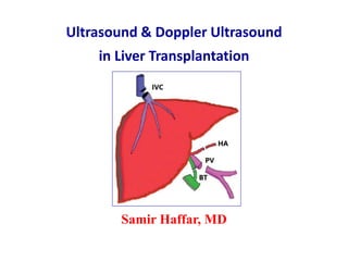
  • 1. Ultrasound & Doppler Ultrasound in Liver Transplantation Samir Haffar, MD
  • 2. US & Doppler US in liver transplantation  Surgical techniques of LT  Normal US & Doppler US in LT  Reversible Doppler findings in post-operative period  Doppler US of LT complications  US of LT complications
  • 4. Types of liver graft • Full cadaveric graft • Split cadaveric graft • Reduced cadaveric graft • Living donor graft • Domino transplantation
  • 5. Liver transplant anatomy Singh AK et al. RadioGraphics 2010; 30: 339 – 351 Orthotopic liver transplant Living donor liver transplant
  • 6. Surgical techniques of LT • Surgical procedure Whole liver, single lobe, segmental • Gallbladder Normally removed • PV, HA, & BT End-to-end anastomoses usually • IVC Piggy-back technique Avoids clamping IVC during surgery • Conduit vessels Discrepancies in vessel diameter Portal vein thrombosis Increased risk of thrombosis It is important to know type of surgical procedure performed, but it is rarely available
  • 7. Vascular complications are the most frequent adverse events after LT Early correction of vascular complications could permit complete recovery or may save the graft Timely detection is important
  • 8. Complications of liver transplantation • Vascular Hepatic artery Stenosis, thrombosis, PA Portal vein Stenosis, thrombosis IVC & HV Stenosis, thrombosis Splenic artery Splenic artery steal synd • Biliary Obstruction Stenosis, stones, biliary cast • Others Abscess Collections Hematoma, biloma Neoplasms HCC – PTLPD* Recurrent disease PSC Cirrhosis & its complications *PTLPD: Post Transplant Lympho-Proliferative Disease Singh AK et al. RadioGraphics 2010; 30: 339 – 351.
  • 9. Imaging evaluation of LT Disease Initial study Subsequent study Final invasive study Vascular Doppler US MR angiography CT angiography CEUS Angiography Biliary Doppler US MRCP CT scan Scintigraphy ERCP PTC Ajay K. Singh RadioGraphics 2010; 30: 339 – 351.
  • 10.  Normal US & Doppler US in LT
  • 11. Frequency of Doppler US exam • First exam 24 – 48 hour after transplantation Confirm patency of hepatic vessels • Then Every 3 – 5 days for first 2 weeks • Additional exam 4 week after transplantation Piscaglia F, Sidhu P, Brabrand K and Borghi A EFSUMB – European Course Book – Liver Transplantation – 2011. No RCT investigates optimal frequency Each centre decides its own policy More frequent exam in patients at higher risk
  • 12. US & Doppler US of LT • US exam Hepatic parenchyma Bile ducts • Doppler exam Hepatic vessels Hepatic artery Portal vein Hepatic veins & IVC Splenic artery Piscaglia F, Sidhu P, Brabrand K and Borghi A EFSUMB – European Course Book – Liver Transplantation – 2011.
  • 13. Normal hepatic artery Piscaglia F, Sidhu P, Brabrand K and Borghi A EFSUMB – European Course Book – Liver Transplantation – 2011. Intercostal approach Epigastric approach Right hepatic artery Left hepatic artery
  • 14. Normal hepatic artery Resistance index (RI) or Pourcelot’s index RI: S – ED / S Normal: 0.5 – 0.8 Buscarini E et al. Ultraschall Med 2004 ; 25 : 348 – 55.
  • 15. Normal hepatic artery Accleration Time (AT) or Rise Time (RT) Time in seconds from onset of systole to peak systole Normal value: ≤ 0.08 second
  • 16. Normal spectral Doppler of HA * Buscarini E et al. Ultraschall Med 2004 ; 25 : 348 – 55. Diameter* 5 ± 1 mm PSV 70 ± 10 cm/sec RI 0.50 – 0.80 RT < 0.08 sec
  • 17. Normal hepatic artery One or two systolic peaks Piscaglia F, Sidhu P, Brabrand K and Borghi A EFSUMB – European Course Book – Liver Transplantation – 2011. RI Calculated from highest peak, early or late AT Calculated considering first peak
  • 18. Normal portal vein • Diameter Upper limits of normal: 13 – 16 mm > 20 – 30% increase with food & inspiration • Flow direction Towards liver (hepatopetal) Throughout entire cardiac cycle • Velocity Varies greatly (Max – Mean – Min – TAMV) Mean velocity: 15 – 18 cm/s Varies with cardiac & respiration activity Undulating appearance of waveform Goyal N et al. Clin Radiology 2009 ; 64 : 1056 – 1066.
  • 19. Normal portal vein Normal monophasic waveform with mild phasicity Sanyal R et al. RadioGraphics 2012 ; 32 : 199 – 211. Duplex US of main PV
  • 20. Helical portal vein flow Donor PV > recipient PV If not properly recognized, it can produce the mistaken impression of PV flow reversal Robinson KA et al. Ultrasound Quarterly 2009 ; 25 : 3 – 13.
  • 21. Helical portal vein flow Mimic of hepatofugal flow Wachsberg RH et al. RadioGraphics 2002 ; 22 : 123 – 140. Hepatopetal flow within liver confirms that net flow is hepatopetal
  • 22. Causes of helical portal vein flow Near bifurcation • Normal subjects 2% • Severe liver disease 20% • TIPS • Post-liver transplantationDonor PV > recipient PV • Portal vein stenosis Robinson KA et al. Ultrasound Quarterly 2009 ; 25 : 3 – 13.
  • 23. Piggy-back anastomosis of the IVC Schematic image Subcostal scan to visualize the anastomosis US & color Doppler US Piscaglia F, Sidhu P, Brabrand K and Borghi A EFSUMB – European Course Book – Liver Transplantation – 2011. –
  • 24. Normal hepatic vein Normal triphasic waveform flow Spectral Doppler of hepatic vein Piscaglia F, Sidhu P, Brabrand K and Borghi A EFSUMB – European Course Book – Liver Transplantation – 2011.
  • 25. Normal hepatic vein waveform – 3 components Kruskal JB et al. RadioGraphics 2004 ; 24 : 657 – 675. A Atrial systole S Ventricular systole D Atrial diastole A S D S wave > D wave Commonly described as triphasic
  • 26.  Reversible Doppler findings in the immediate post-operative period
  • 27. Reversible Doppler findings in post-op period • Starry sky pattern • Pneumobilia • Pleural effusion • Hepatic artery Elevated PSV Decrease RI Reversed diastolic flow Tardus parvus waveform • Portal vein Increased velocity Pulsatile flow • Hepatic veins Monophasic flow • HV-IVC anastomosis Turbulence Sanyal R et al. RadioGraphics 2012 ; 32 : 199 – 211.
  • 28. Starry sky pattern Sanyal R et al. RadioGraphics 2012 ; 32 : 199 – 211. US on 1st post-operative day US 3 days after OLT Normal hepatic echotexture Echogenic portal venules & diminished parenchymal echogenicity (edema)
  • 29. Pneumobilia Sanyal R et al. RadioGraphics 2012 ; 32 : 199 – 211. US image on first postoperative day after OLT Normal finding after bilio-enteric anastomosis in OLT
  • 30. Pleural effusion Sanyal R et al. RadioGraphics 2012 ; 32 : 199 – 211. Moderate right pleural effusion, a common finding Usually resolve within few days US image on 1st post-operative day after OLT
  • 31. Elevated PSV in hepatic artery Sanyal R et al. RadioGraphics 2012 ; 32 : 199 – 211. 1st postoperative day of OLT PSV: 283 cm/sec – normal RI Due to anastomotic edema Perivascular vibration is seen PSV: 192 cm/sec Resolution of anastomotic edema No peri-vascular vibration 3 days after OLT
  • 32. 1st postoperative day of OLT Sanyal R et al. RadioGraphics 2012 ; 32 : 199 – 211. Decrease resistance index in hepatic artery Elevated PSV: 303 cm/sec Low RI: 0.45 3 days after OLT Decreased PSV: 118 cm/sec Increased RI: 0.56 Resolution of narrowing secondary to anastomotic edema
  • 33. Reversed diastolic flow in hepatic artery Sanyal R et al. RadioGraphics 2012 ; 32 : 199 – 211. 1st postoperative day of OLT 3 days after OLT Normal PSV: 55 cm/sec Reversed or absent diastolic flow Normal waveform Improved RI: 0.87
  • 34. Tardus parvus waveform in hepatic artery Sanyal R et al. RadioGraphics 2012 ; 32 : 199 – 211. 1st postoperative day of OLT 3 days after OLT Normalization of tardus parvus Increased PSV: 53 cm/sec Improved RI: 0.62 Tardus parvus waveform Low PSV: 23 cm/sec Low RI: 0.43
  • 35. Increased velocity in portal vein Sanyal R et al. RadioGraphics 2012 ; 32 : 199 – 211. 1st postoperative day of OLT Increased PV velocity: 149 cm/sec Decreased portal resistance 3 days after OLT Improved turbulence Normal PV: 54 cm/sec
  • 36. Pulsatile flow in portal vein Common finding in congestive heart failure Sanyal R et al. RadioGraphics 2012 ; 32 : 199 – 211. 1st postoperative day of OLT Pulsatile PV waveform due to altered hemodynamics 3 days after OLT Normal monophasic flow
  • 37. Monophasic flow in hepatic veins Sanyal R et al. RadioGraphics 2012 ; 32 : 199 – 211. 1st postoperative day of OLT Monophasic flow in MHV Graft edema 3 days after OLT Normal triphasic waveform
  • 38. Turbulence at HV-IVC anastomosis Sanyal R et al. RadioGraphics 2012 ; 32 : 199 – 211. 1st postoperative day of OLT Turbulence at HV-IVC anastomosis 3 days after OLT Resolution of turbulence
  • 39.  Doppler US of LT complications
  • 40. Vascular complications of LT • Hepatic artery Stenosis – Thrombosis – PSA • Portal vein Stenosis – Kinking – Thrombosis • Hepatic veins & IVC Stenosis – Kinking – Thrombosis • Splenic artery Splenic artery steal syndrome (SASS) Piscaglia F, Sidhu P, Brabrand K and Borghi A EFSUMB – European Course Book – Liver Transplantation – 2011.
  • 41. Presentation of vascular complications after LT Complication Time of presentation after LT HA thrombosis First two weeks Late HAT occurs but less devastating HA stenosis Few weeks to several months PV thrombosis Within one month PV stenosis Usually late (> 6 months) HV stenosis Usually late (> 6 months) Sanyal R et al. Indian J Radiol Imag 2014 ; 24 : 360 -366.
  • 42. Hepatic artery Thrombosis – Stenosis – Pseudo-aneurysm (PSA)
  • 43. HA thrombosis – Systematic review Most severe complications especially when abrupt • Incidence 2 – 12% of adult recipients 2 – 6% in experienced centers • Peak occurrence Seventh day following LT • Re-transplantation Up to 60% • Mortality 33% • Doppler US Identify up to 92% Bekker J, Ploem S, de Jong KP. Am J Transplant 2009 ; 9 : 746 - 57.
  • 44. Complications of HA thrombosis Transplanted liver extremely sensitive to arterial ischemia • Primary liver non-function • Hepatic infection & abscesses • BT ischemia Lumen filled with biliary necrotic casts Irreversible Biliary stenosis with peritoneal spillage Diffuse & involve all IH branches Only biliary surgical anastomosis (often) Piscaglia F, Sidhu P, Brabrand K and Borghi A EFSUMB – European Course Book – Liver Transplantation – 2011.
  • 45. Manifestations of HA thrombosis • General malaise, fullness or fever • Pain not usually present Denervated transpl liver • Elevated liver enzymes Elevated γGT & AP • Liver failure Elevated bilirubin Prolonged PT
  • 46. Doppler US in HA thrombosis Visualize artery in hilum & both lobes • Fasting examination Arterial buffer vasoconstriction • Adjust color scale • Doppler gate near PV • Wider Doppler gate Normal arterial spikes within PV Arterial thump in thrombosis • CEUS 1.2 – 2.4 ml SonoVue IV Low mechanical index • Further investigations CECT – angiography Piscaglia F, Sidhu P, Brabrand K and Borghi A EFSUMB – European Course Book – Liver Transplantation – 2011.
  • 47. Doppler US in HA thrombosis • HA in hilum Absence of visualization • Intra-hepatic branches RI ˂ 0.50 AT ˃ 0.08 sec Identification of just one parameter is enough to prompt rapid further investigations
  • 48. Hepatic artery thrombosis Diffuse heterogenous mixed echogenic change Gray scale US CT scan Widespread periportal low attenuation change O’Briena J et al. European J Radiol 2010; 74 : 206 – 213.
  • 49. Doppler US in HA thrombosis Sanyal R et al. Indian J Radiol Imag 2014 ; 24 : 360 - 366. Tardus parvus in LHA Flow in LHA does not exclude HAT CT angiogram Thrombosis of main HA Doppler ultrasound
  • 50. Hepatic artery stenosis Horrow M et al. AJR 2007; 189 : 346 – 351. Flow in portal vein No flow in hepatic artery Power Doppler at porta hepatis Complete occlusion of hepatic artery CT angiogram
  • 51. Biliary cast syndrome First described in 1975 • Incidence 2.5% after OLT – Less than sludge & stones • Morphology Hard dark material in biliary ducts Similar shape to BD • Symptoms High fever, jaundice, cholestasis • Characterized Multiple intra-hepatic biliary strictures Ductal dilatation Intrahepatic abscesses Biliary anastomotic leakage • Treatment Surgery is treatment of choice Endoscopic techniques successful & safe Yang Ylet al. World J Gastroenterol 2013 ; 19 (43): 7772 – 77.
  • 52. Biliary cast syndrome Ha SI et al. Korean J Gastroenterol 2012 ; 60 : 382 – 385. Biliary cast removed endoscopically Hyperechoic material in CBD Initially thought as ascariasis Gray scale US Longitudinal tubular filling defect in CBD ERCP
  • 53. Biliary cast syndrome Biliary duct necrosis Rubens DJ. Ultrasound Clin 2007 ; 2 : 391 – 413. Linear echogenic debris with acoustic shadows Transverse US Low RI (<0.5) HA stenosis/thrombosis Color Doppler US Extensive BT necrosis Resulting liver abscess CT scan Abscess obscured on US because of shadowing from air in the ducts
  • 54. Hepatic artery stenosis Milder effect than thrombosis • Incidence Up to 10% • Site Most often at anastomostic site Consequence of arterial kinking • Manifestation Bile duct strictures from ischemia Piscaglia F, Sidhu P, Brabrand K and Borghi A EFSUMB – European Course Book – Liver Transplantation – 2011. High degree stenosis leads to biliary ischemia
  • 55. Doppler US of HA stenosis • At stenosis Aliasing on color Doppler PSV ˃ 2m/sec • Post-stenosis Tardus-parvus wave RI ˂ 0.5 AT ˃ 0.08 sec Stenosis could be corrected radiologically or surgically Piscaglia F, Sidhu P, Brabrand K and Borghi A EFSUMB – European Course Book – Liver Transplantation – 2011.
  • 56. Hepatic artery stenosis Main hepatic artery Turbulent flow & high PSV (4.7 m/s) Intra-hepatic artery Tardus-parvus, low RI & long AT Angiogram: high degree HA stenosis Piscaglia F, Sidhu P, Brabrand K and Borghi A EFSUMB – European Course Book – Liver Transplantation – 2011.
  • 57. HA pseudo-aneurysm (PSA) Extra-hepatic PSA considered as surgical urgency • Incidence Less than 1% • Symptoms Usually asymptomatic Rupture with life-threatening hemoperitoneum • Causes Extrahepatic Infection (mycotic PSA) Defective reconstruction Intra-hepatic Percutaneous interventions Focal infection of parenchyma • US US diagnosis is difficult Hypoechoic collection near arterial anastomosis should be evaluated with CDUS or CEUS Piscaglia F, Sidhu P, Brabrand K and Borghi A EFSUMB – European Course Book – Liver Transplantation – 2011.
  • 58. HA pseudo-aneurysm Leong L. Biomed Imaging Interv J 2006; 2(2):e17. High PSV at neck of PATurbulent flow in a lesion near HA DSA of celiac axis: pseudoaneurysm
  • 59. O’Briena J et al. European J Radiol 2010; 74 : 206 – 213. HA pseudo-aneurysm / yin-Yang pattern Selective arteriography Gray-scale US Echopoor lesion in the liver Color Doppler US Swirling or “yin-yang” pattern
  • 60. Splenic artery steal syndrome (SASS) First described by Manner in 19911 • Clinic Graft ischemia in early postoperative period non-specific2 Less fulminant than HAT Elevated liver enzymes first Then cholestasis, biliary damage, rejection • Exclude Hepatic arterial thrombosis Hepatic arterial thrombosis Acute rejection • Treatment Splenic artery embolization 1Manner M et al. Transpl Int 1991; 4 : 122 – 124. 2Sanyal R, Shah SN. J Ultrasound Med 2009; 28: 471 – 477.
  • 61. US Doppler of SASS • Hepatic artery High RI in IH & EH arteries RI usually 0.8 & 1 in diastolic reversal Can be transient post-operative finding Can be indicator of acute rejection • Splenic artery Increased flow in splenic artery • Portal vein High portal vein velocity Sanyal R, Shah SN. J Ultrasound Med 2009 ; 28 : 471 – 477. HAT & stenosis are 2 other causes of graft ischemia Doppler US very sensitive for their diagnosis Low-resistance or absent flow distal to obstruction
  • 62. Doppler US of SASS / Hepatic artery Sanyal R, Shah SN. J Ultrasound Med 2009; 28 : 471 – 477. Right hepatic artery Diastolic reversal Left hepatic artery Diastolic reversal Main hepatic artery Diastolic reversal
  • 63. Doppler US of SASS / PV & splenic artery Sanyal R, Shah SN. J Ultrasound Med 2009 ; 28: 471 – 477. Hyperdynamic splenic artery PSV: 272 cm/s Hyperdynamic main PV Mean PV velocity 115 cm/s
  • 64. Portal vein Stenosis – Kinking – Thrombosis
  • 65. Portal vein Stenosis – Kinking – Thrombosis • Incidence 1 – 2% of patients • Picture Portal hypertension Hepatic failure Ascites or all of these Do not usually lead to liver failure or graft loss
  • 66. Doppler US of PV thrombosis • Ultrasound Internal echoes within PV • Color Doppler No flow at all – Flow around thrombus • Differential dg Fresh thrombus vs very slow flow Benign vs malignant thrombus • CEUS Fresh flow: absence of flow Very slow flow: presence of flow Benign thrombus: no enhancement Malignant thrombus: arterial enhancement Piscaglia F, Sidhu P, Brabrand K and Borghi A EFSUMB – European Course Book – Liver Transplantation – 2011.
  • 67. Portal vein thrombosis Echogenic thrombus in main PV with lack of color-filling Sanyal R et al. Indian J Radiol Imag 2014 ; 24 : 360 – 366.
  • 68. Portal vein cavernoma Chronic portal vein thrombosis Multiple collaterals replacing main portal vein Bhargava P AJR 2011; 196 : WS15 – WS25.
  • 69. Diagnosis of malignant PV thrombosis • “AASLD” guidelines1 PV > 23 mm in diameter Arterial-like flow on Doppler Increased serum α-FP • Piscaglia criteria2 Mass-forming features of thrombus Disruption of veins walls Thrombus vascularization (US-CT-MRI) • FNA CT- or US-guided 1 DeLeve L et al. Vascular disorders of the liver. Hepatology 2009 ; 49 : 1729 – 1764. Piscaglia F et al. Liver transplantation 2010 ; 16 : 658 – 667.
  • 70. Malignant PV thrombosis / CEUS 38 pts (15 benigns - 23 malignants) – Conclusive (37/38) Dănilă M et al. Medical Ultrasonography 2011 ; 13 : 102 – 107. Gray-scale US Malignant PVT Arterial phase Enhancement Portal phase Wash-out Late phase Wash-out Contrast-Enhanced US
  • 71. Malignant PV thrombosis Piscaglia criteria Piscaglia F et al. Liver transplantation 2010 ; 16 : 658 – 667. Low-resistance pulsatile arterial flow in thrombus Thrombus occupying part of the left portal vein Mass-forming features of thrombus Disruption of veins walls
  • 72. Doppler US of significant PV stenosis • Color Doppler Aliasing at stenosis • Pulsed Doppler PSV ˃ 1 m/s at stenosis 3 – 4 increase in velocity at stenosis site compared to 2 cm upstream Piscaglia F, Sidhu P, Brabrand K and Borghi A EFSUMB – European Course Book – Liver Transplantation – 2011.
  • 73. Portal vein stenosis Color aliasing at vascular anastomosis Sixfold velocity in poststenotic segment relative to prestenotic segment Significant stenosis Long axis of main portal vein Crossin JD et al. RadioGraphics 2003; 23:1093 – 1114.
  • 74. Portal venous gas in LT • Causes Common transient finding in early postop period Serious disease Bowel necrosis Intra-abdominal abscess Small-bowel obstruction Sepsis • Diagnosis CT scan Doppler US Useful for PV gas detection Does not necessarily indicate severe pathology Abboud B et al. World J Gastroenterol 2009 August 7; 15(29): 3585 – 3590.
  • 75. Portal vein gas Acute trans-mural mesenteric infarction Wiesner W et al. Radiology 2003 ; 226 : 635 – 650. Intra-hepatic PV gas in periphery of both lobes CECT scan
  • 76. Portal vein gaz Piscaglia F, Sidhu P, Brabrand K and Borghi A EFSUMB – European Course Book – Liver Transplantation – 2011. Bright areas in liver parenchyma Accumulation of gas bubbles Gray-scale US Vertical bidirectional spikes At laparotomy: bowel perforation Spectral Doppler US Doppler US is useful for detection of PV gas
  • 77. Hepatic veins & IVC Kinking – Stenosis – Thrombosis
  • 78. Hepatic veins & IVC Stenosis – Kinking – Thrombosis • Causes Narrowing of anastomosis Kinking of vessels Transplant organ torsion (size mismatch) • Manifestation Liver failure Portal hypertension Ascites & pleural effusion • Biology Increase liver enzymes & bilirubin Impairment of hepatic synthetic activities Piscaglia F, Sidhu P, Brabrand K and Borghi A EFSUMB – European Course Book – Liver Transplantation – 2011.
  • 79. Doppler US of significant HV stenosis • Color Doppler Aliasing at site of anastomosis Flow reversal in IVC • Pulsed Doppler Upstream Monophasic wave PSV ˂ 15 – 20 cm/s 3 – 4 fold velocity increase at stenosis site compared to few cm upstream Piscaglia F, Sidhu P, Brabrand K and Borghi A EFSUMB – European Course Book – Liver Transplantation – 2011. Monophasic wave seen in absence of stenosis Serial exams: new appearance of monophasic waveform
  • 80. HV stenosis at piggyback site Piscaglia F, Sidhu P, Brabrand K and Borghi A EFSUMB – European Course Book – Liver Transplantation – 2011. HV stenosis with aliasing Upstream anastomosis: flattened flow (14 cm/s) Within piggyback: turbulent flow (100 cm/s)
  • 81. IVC stenosis End-to end IVC anastomosis Crossin JD et al. RadioGraphics 2003; 23: 1093 – 1114. Gray-scale US Focal narrowing of IVC lumen Spectral Doppler US Twofold velocity increase between pre-stenotic & post-stenotic segment Turbulence at distal IVC anastomosis Color Doppler US
  • 82. IVC thrombosis Echogenic thrombus of RHV extending into IVC Subcostal oblique US Right paramedian sagittal US IVC thrombus Crossin JD et al. RadioGraphics 2003; 23: 1093 – 1114.
  • 83. Doppler US in hepatic rejection • Parenchymal texture irregularities not specific Their presence may have several cause • Doppler indices unreliable for diagnosis of rejection Biopsy of transplanted liver required for diagnosis of rejection Piscaglia F, Sidhu P, Brabrand K and Borghi A EFSUMB – European Course Book – Liver Transplantation – 2011.
  • 84.  US of LT complications
  • 85. Other complications of LT • Abscess • Collection Hematoma, biloma • Hepatic infarct • Biliary obstruction Stone, stenosis, biliary cast synd • Neoplasms Recurrent HCC, PTLPD* • Recurrent disease PSC • Cirrhosis & itscomplications *PTLPD: Post-transplant lympho-proliferative disease
  • 86. Hepatic abscess Crossin JD et al. RadioGraphics 2003; 23: 1093 – 1114. Focal mass in left hepatic lobe Highly echogenic air & dirty shadowing Could be mistaken for gas in bowel Transverse US image Corresponding CECT
  • 87. Acute and sub-acute fluid collections Crossin JD et al. RadioGraphics 2003; 23: 1093 – 1114. Echogenicity similar to adjacent liver Difficult to detect Acute hematoma Sub-acute hematoma Complex septated fluid collection in hepatorenal space
  • 88. Biloma after LDLT Diagnosed by US guided aspiration Hypoechoic collection in right hepatic lobe Tip of needle during US guided aspiration
  • 89. Hepatic infract Echogenic focus with hypoechoic rim in left hepatic lobe Biopsy: hepatic infarct Corresponding CECTMidline transverse US Crossin JD et al. RadioGraphics 2003; 23: 1093 – 1114.
  • 90. CBD stone Dilated CBD Echogenic calculus distal acoustic shadowing Right para-median sagittal US Crossin JD et al. RadioGraphics 2003; 23: 1093 – 1114.
  • 91. Anastomotic stricture of bile duct Corresponding ERCP Normal waveform Spectral Doppler of HA CBD stenosis Sagittal US Crossin JD et al. RadioGraphics 2003; 23: 1093 – 1114. CBD stenosis
  • 92. Recurrent hepato-cellular carcinoma Crossin JD et al. RadioGraphics 2003; 23: 1093 – 1114. Median sagittal US Corresponding CECT Large mass of mixed echogenicity in right hepatic lobe
  • 93. Post-transplant lympho-proliferative disease Crossin JD et al. RadioGraphics 2003; 23:1093 – 1114. Large heterogeneous mass displacing porta hepatis Biopsy: PTLPD Transverse US image Contrast-enhanced CT Mass encases vasculature at porta hepatis
  • 94. Recurrent sclerosing cholangitis Right para-median sagittal US Mural thickening of CBD Magnified view Crossin JD et al. RadioGraphics 2003; 23:1093 – 1114.
  • 95. Adrenal hemorrhage Crossin JD et al. RadioGraphics 2003; 23: 1093 – 1114. Corresponding CECT Hypoechoic nodule in hepatorenal space Focal adrenal hemorrhage secondary to intra-operative venous infarction Transverse US image

Notes de l'éditeur

  1. Low-resistance profile: - Broad systolic peak - Gradual deceleration from systole to diastole - Well-maintained diastolic flow throughout the cardiac cycle.
  2. Undulating: Variation of PV diameter with inspiration: Lack of caliber variation of the splenic and mesenteric vein during respiration is another parameter that has been investigated. In 1 study, this approach had a sensitivity of 80% and specificity of 100% in diagnosing portal hypertension. However, it is a method that has not gained widespread use, likely because of difficulties in measurement accuracy and interobserver variability. PV velocity: Significantly lower mean portal venous velocity was noted in cirrhotic patients (13 ± 3.2 cm/s versus 19.6 ± 2.6 cm/s in controls) by Zironi et al & 15 cm/s was considered as best cut-off value in detection of PHT, showing sensitivity & specificity of 88% and 96%, respectively. 51. Zironi G, Gaiani S, Fenyves D, et al. Value of measurement of mean portal flow velocity by Doppler flowmetry in the diagnosis of portal hypertension. J Hepatol 1992;16:298-303.
  3. Triphasic waveform due to transmitted cardiac activity & Similar to waveform for the jugular vein. Two negative waves and another positive wave.