Short case...Multiple craniocervical anomalies
•
2 j'aime•530 vues

Short case...Multiple craniocervical anomalies

Recommandé
Recommandé
Contenu connexe
Tendances
Tendances (20)
En vedette
En vedette (20)
Similaire à Short case...Multiple craniocervical anomalies
Similaire à Short case...Multiple craniocervical anomalies (20)
Plus de Professor Yasser Metwally
Plus de Professor Yasser Metwally (20)
Dernier
Dernier (20)
Short case...Multiple craniocervical anomalies
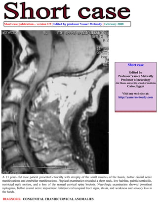
- 1. Short case publication... version 1.9 | Edited by professor Yasser Metwally | February 2008 Short case Edited by Professor Yasser Metwally Professor of neurology Ain Shams university school of medicine Cairo, Egypt Visit my web site at: http://yassermetwally.com A 13 years old male patient presented clinically with atrophy of the small muscles of the hands, bulbar cranial nerve manifestations and cerebellar manifestations. Physical examination revealed a short neck, low hairline, painful torticollis, restricted neck motion, and a loss of the normal cervical spine lordosis. Neurologic examination showed downbeat nystagmus, bulbar cranial nerve impairment, bilateral corticospinal tract signs, ataxia, and weakness and sensory loss in the hands. DIAGNOSIS: CONGENITAL CRANIOCERVICAL ANOMALIES
- 2. Figure 1. Sagittal CT scan reconstructed image (A). Plain x ray image (B) and Precontrast MRI T1 image (C). Notice that the odontoid process is abnormally large, subluxated, and posteriorly located. Also notice basilar invagination, The tip of the odontoid process is in touch of the lower pons. A syringomyelic cavity is seen opposite the second vertebra and extends for only one spinal segment. The cerebellar tonsils are herniated below the level of the foramen magnum. The brain stem is abnormally elongated with herniation of the medullar below the level of the foramen magnum (Type II Chiari malformation). Notice malalignment of the cervical vertebrae. Figure 2. The atlas is abnormally high, almost completely invaginated intracranially (basilar invagination) with complete fusion of the atlas with the occipital bone (atlanto-occipital fusion or assimilation of atlas)
- 3. Figure 3. Precontrast MRI T1 images. Notice that the odontoid process is abnormally large, subluxated, and posteriorly located. Also notice basilar invagination, The tip of the odontoid process is in touch of the lower pons. A syringomyelic cavity is seen opposite the second vertebra and extends for only one spinal segment. The cerebellar tonsils are herniated below the level of the foramen magnum. The brain stem is abnormally elongated with herniation of the medullar below the level of the foramen magnum (Type II Chiari malformation). Interestingly the foramen of magendi (which is situated at the caudal end of the 4th ventricle and can easily be appreciated on the T1 MRI sagittal images) is appreciated as being markedly diminished in volume and transformed into a long slit that opened below the level of the foramen magnum. Notice the extradural space occupying lesion compressing and anteriorly displacing the upper cervical sinal cord. Figure 4. Precontrast MRI T1 images. Notice that the odontoid process is abnormally large, subluxated, and posteriorly located. Also notice basilar invagination, The tip of the odontoid process is in touch of the lower pons. A syringomyelic cavity is seen opposite the second vertebra and extends for only one spinal segment. The cerebellar tonsils are herniated below the level of the foramen magnum. The brain stem is abnormally elongated with herniation of the medullar below the level of the foramen magnum (Type II Chiari malformation). Interestingly the foramen of magendi (which is situated at the caudal end of the 4th ventricle and can easily be appreciated on the T1 MRI sagittal images) is appreciated as being markedly diminished in volume and transformed into a long slit that opened below the level of the foramen magnum. Notice the extradural space occupying lesion compressing and anteriorly displacing the upper cervical sinal cord.
- 4. Figure 5. MRI T2,T1 images. Notice that the odontoid process is abnormally large, subluxated, and posteriorly located. Also notice basilar invagination, The tip of the odontoid process is in touch of the lower pons. A syringomyelic cavity is seen opposite the second vertebra and extends for only one spinal segment. The cerebellar tonsils are herniated below the level of the foramen magnum. The brain stem is abnormally elongated with herniation of the medullar below the level of the foramen magnum (Type II Chiari malformation). Interestingly the foramen of magendi (which is situated at the caudal end of the 4th ventricle and can easily be appreciated on the T1 MRI sagittal images) is appreciated as being markedly diminished in volume and transformed into a long slit that opened below the level of the foramen magnum. Notice the extradural space occupying lesion compressing and anteriorly displacing the upper cervical sinal cord.
- 5. Figure 6. Post contrast MRI T1 image (A) Showing a dumb-bell cystic neurofibroma (Confirmed surgically) at C 2 vertebra compressing and displacing the spinal cord. Addendum A new version of this software is uploaded in my web site every week (every Saturday and remains available till Friday.) To download the current version follow the link quot;http://pdf.yassermetwally.com/short.pdfquot;. You can download the long case version of this short case during the same week from: http://pdf.yassermetwally.com/case.pdf or visit web site: http://pdf.yassermetwally.com To download the software version of the publication (crow.exe) follow the link: http://neurology.yassermetwally.com/ crow.zip At the end of each year, all the publications are compiled on a single CD-ROM, please contact the author to know more details. Screen resolution is better set at 1024*768 pixel screen area for optimum display References 1. Metwally, MYM: Textbook of neurimaging, A CD-ROM publication, (Metwally, MYM editor) WEB-CD agency for electronic publishing, version 9.1a January 2008