
Recommandé
Contenu connexe
Tendances
Tendances (20)
Similaire à virulent characteristics of emerging pathogens
Similaire à virulent characteristics of emerging pathogens (20)
Dernier
Dernier (20)
virulent characteristics of emerging pathogens
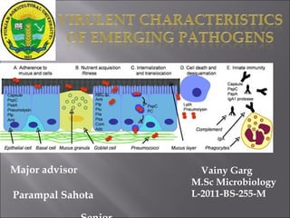
- 1. Major advisor Parampal Sahota Vainy Garg M.Sc Microbiology L-2011-BS-255-M
- 4. Capsule Lipid A Adhesins Invasins Toxins Plasmids Bacteriophages Infecting dose Route of infection Communicability Bacterial appendages
- 7. CAPSULE produce inflammatory cytokines. e.g. Streptococcus pneumoniae, Neisseria meningitidis, and Pseudomonas aeruginosa. differences in capsular polysaccharide chemical structure determine the meningococcal serogroups. CELL WALL : contain toxic components involved in bacterial septic shock, collapse of the circulatory system and multiple organ system failure. Acts via the initiation of an inflammatory response through the stimulation of monocytes and macrophages.
- 8. ADHESINS : Adherence of the pathogen to host surfaces. MECHANISMS OF ADHERENCE TO CELL OR TISSUE SURFACES Possible interactions and forces involved are: hydrophobic interactions electrostatic attractions atomic and molecular vibrations resulting from fluctuating dipoles of similar frequencies Brownian movement recruitment and trapping by biofilm polymers interacting with the bacterial glycocalyx (capsule)
- 9. Receptor-ligand interactions promotes adherence involves protein- protein and protein- carbohydrate interactions. molecules serve as host receptors for microbes include membrane-spanning proteins, surface immunoglobulin, glycolipids, glycoproteins, and extracellular matrix proteins.
- 10. Enterotoxigenic E. coli Type-I fimbriae Intestinal epithelium Diarrhea Uropathogenic E. coli Type I fimbriae Urethral epithelium Urethritis Uropathogenic E. coli P-pili (pap) Upper urinary tract Pyelonephritis Bordetella pertussis Fimbriae (filamentous hemagglutinin) Respiratory epithelium Whooping cough V. cholerae N-methylphenyl- alanine pili Intestinal epithelium Cholera Treponema pallidum Peptide in outer membrane Mucosal epithelium Syphilis
- 11. INVASION : Pathogens gain deeper access into the host to perpetuate the infection cycle. Extracellular invasion : allows pathogens to proliferate tissues, disseminate to other sites in the body, express toxins, and initiate inflammatory responses. E.g. : β hemolytic streptococcus and S. aureus- secrets hyaluronidase streptokinase and staphylokinase, lipase, and nuclease . Intracellular invasion : microbe penetrates the cells of host tissue and survives within this environment. E.g. : Chlamydia spp, Rickettsia spp and Mycobacterium leprae : obligate intracellular lifecycle that requires mammalian cell for growth.
- 12. Invasin Activity Hyaluronidase Degrades hyaluronic of connective tissue Collagenase Dissolves collagen framework of muscles Neuraminidase Degrades neuraminic acid of intestinal mucosa Coagulase Converts fibrinogen to fibrin which causes clotting Kinases Converts plasminogen to plasmin which digests fibrin Leukocidin Disrupts neutrophil membranes and causes discharge of lysosomal granules Streptolysin Repels phagocytes and disrupts phagocyte membrane and causes discharge of lysosomal granules Hemolysins Phospholipases or lecithinases that destroy red blood cells (and other cells) by lysis Lecithinases Destroy lecithin in cell membranes Phospholipases Destroy phospholipids in cell membrane Anthrax EF One component (EF) is an adenylate cyclase which causes increased levels of intracellular cyclic AMP Pertussis AC One toxin component is an adenylate cyclase that acts locally producing an increase in intracellular cyclic AMP Extracellular bacterial proteins that are considered invasins
- 13. Toxigenicity : Exotoxins and Endotoxins Classification of exotoxins: a. Membrane damaging toxins: destroy the host membranes by inducing pore formation or by destabilising cytoplasmic membranes. b. Superantigens: bind non- specifically, activate large numbers of T cells leading to shock. c. A-B component toxins : The B component binds to specific host cell receptors and A is enzymaticcally active portion of the toxin.
- 16. PLASMIDS: Gene coding for virulent characteristics can be plasmidborne. E.g. surface antigens responsible for colonisation of intestinal mucosa by E. coli and enterotoxin production by E. coli and S. aureus. BACTERIOPHAGES : In diphtheria bacilli, gene for toxic production is present in beta or other toxic corynephages. COMMUNICABILITY : Ability of parasite to spread from one host to another. Highly virulent parasite may not exhibit high degree of communicability due to rapid lethal effect on host. E.g. respiratory and intestinal diseases. BACTERIAL APPENDAGES: Bacterial surface antigens such as Vi antigen of S typhi and K antigens of E coli : prevents bacteria from phagocytosis and lytic activity of compliments.
- 17. INFECTING DOSE : Dosage estimated as minimum infecting dose (MID) or minimum lethal dose (MLD) Route of infection : Modes by which different bacteria able to initiate tissue damage. Cholera vibrios infect orally and unable to cause infection subcutaneously.
- 18. • SECRETED OR SURFACE-EXPOSED BACTERIAL PROTEINS IN BACTERIAL- HOST INTERACTIONS. • Proteins secreted by the Type I system cross directly from the cytoplasm to the cell surface, bypassing the general secretory pathway completely. • Type II-secreted proteins use the general secretory pathway to reach the periplasm and traverse the outer membrane through distinct channel proteins. • Type III system : effector molecules move to the external surface of the bacterium, facilitate the pathogen’s ability to survive and replicate.
- 19. colonization of the intestinal tract, and penetrate M cells of Peyer’s patches. replicate in extracellular form within micro- abscesses. form microcolonies and resistant to phagocytosis by macrophages and neutrophils (Fabrega et al 2011).
- 21. six biotypes differentiated by physiochemical and biochemical tests (1A, 1B, 2, 3, 4, and 5) more than 50 serotypes differentiated by antigenic variation in cell wall lipopolysaccharide. virulent biotypes 1B and 2–5 has highly conserved 70-kb virulence plasmid, termed pYV/pCD and certain chromosomal genes.
- 22. The biotype 1A lack pYV plasmid encodes virulence factors {Yersinia adhesin A (YadA) and Ysc-Yop type III secretion system (TTSS)} and chromosomally borne virulence genes {ail, myfA, ystA, ysa, high pathogenicity island- (HPI-)} Biotype 1B carry high-pathogenicity island (HPI),facilitates the uptake and utilization of iron by bacterial cells, promote growth under iron-limiting conditions in host tissues.
- 23. Virulence-associated determinants of pYV-negative strains includes: cell surface lipopolysaccharide SodA (a superoxide dismutase) : bacterial survival in tissues. urease, enhances bacterial resistance to stomach acid and in nitrogen assimilation.
- 24. pYV, antihost plasmid, resist phagocytosis and complement- mediated lysis, to proliferate extracellularly in tissues. virulence factors, outer membrane protein adhesin, YadA, and type III protein secretory apparatus translocates effector proteins (Ysc-Yops) from bacterial cell to the cytoplasm of susceptible host cells.
- 26. Adaptation : Yersinia adapt surface antigenic structures (outer membrane proteins) to colonize in the intestines of humans at temperature of 37◦C through 70-kb virulence plasmid (pYV). Adhesion : allow intimate attachment to the epithelial cells. YadA, a pYV plasmid-encoded protein, adhesion for attachment, induction of disease (e.g., inflammation and necrosis in the liver). mediates adherence to epithelial cells, phagocytes and extracellular matrix components, and protects the bacterium killed by neutrophils.
- 27. Fimbriae present in biotype1A : MR/Y-HA : 8 nm in diameter, agglutinates erythrocytes in the presence of mannose and expressed in vitro at low temperature. MR/K like HA : 4 nm in diameter and mediates mannose resistant hemagglutination of chicken erythrocytes. Y. enterocolitica produces Myf (for mucoid Yersinia fibrillae), bestows mucoid appearance on bacterial colonies. (Sabina et al 2011)
- 28. Invasion : Ail (attachment-invasion locus) : Localized in the OM. Eight transmembrane β-sheets and four cell surface-exposed loops, and the extracellular loop for Ail-mediated binding to host cells. Promotes Yop delivery into the primary target of T3SS--the phagocytic cells and into epithelial cells. Invasin : Located in middle of gene cluster encoding the flagella proteins ~100 kDa, anchored by its amino-terminal region Receptor is β1 integrin, intergrins couple extracellular adhesion events to numerous signaling pathways, and the bacterium is taken up by zipper mechanism.
- 29. Mechanisms of bacterial epithelial cell internalization : “zippering” process : tight enclosing of the bacterial cell by the mammalian cell membrane. involving surface bound bacterial protein invasin (Inv) binding an integrins of the β1 family of mammalian cell surface.
- 30. TOXINS : ystA, ystB : heat-stable enterotoxin. causes Yersinia associated diarrhea. stimulate cGMP synthesis in the intestinal brush border, leading to fluid loss and lack of fluid absorption. Pathogenicity islands in Yersinia : HPI capture the iron molecules for systemic dissemination of the bacteria in the host via yersiniabactin.
- 32. LOCAL AND SYSTEMIC DISSEMINATION Cross the intestinal epithelium through FAE (follicle associated epithelial cell), in the Peyer’s patches of the ileum. Invasin (Inv), a 103 kDa outer membrane protein of Yersinia binds β1 integrins that are expressed apically on M cells. Yersinia surface proteins (Ail, PsaA, and YadA ) account for residual invasion of inv mutants. Yersinia defend attack by resident macrophages by expressing an antiphagocytic strategy. mediated by plasmid encoded type III secretion, of three protein effectors, YopH, T, and E, disrupt cytoskeletal assembly required for phagocytosis process. Extracellular in infected Peyer’s patches and mesenteric lymph nodes and disseminate to cause local and systemic infection.
- 33. When bacteria bind to tissue culture cells, 10 different effector molecules secreted and three injected into cells. YopE and YopH, modify macrophage proteins to destroy the cells abilities to engulf and kill bacteria. immune cells neutralized by effector molecules, enables Yersinia spp to flourish in the reticuloendothelial environment.
- 34. • capture the iron molecules for systemic dissemination in the host. • Yeriniabactin, sub-group of phenolate siderophores and has affinity for ferric iron. •FyuA/Psn-Irp system uses yersiniabactin, a siderophore that remove iron from mammalian proteins. • YbtA, AraC-like regulator required for transcription of fyuA/psn, irp2 and ybtP, downregulate its own transcription. • In the presence of iron, Fur, a cytosolic protein, bind ferrous iron, changes conformation and binds DNA at specific site called Fur box, preventing transcription, downregulates transcription of fyuA/psn, irp2 and other iron-regulated genes.
- 35. classified into 96 serogroups and the O-antigen LPS of A. hydrophila 0:34 strains in adhesion to HEp-2 cells. attaches and enters into host cells through production of flagella, pili and adhesins. multiplication in host tissue by production of siderophores and outer membrane proteins enterotoxins, proteases, phospholipases, and hemolysins cause damage to host cells leading to cell death.
- 36. directed locomotion attachment to gastrointestinal epithelium biofilm formation colonization elaboration of virulence factors infection (Janda et al
- 37. Enterotoxins—cytotonic and cytotoxic. Cytotonic enterotoxins (heat-labile (Alt) and heat- stable (Ast)), donot degenerate crypts and villi of small intestine. Cytotoxic enterotoxin (Act) result in extensive damage to epithelium. Aerolysin, extracellular, soluble, hydrophilic protein exhibiting hemolytic and cytolytic properties. The mature form of Act is 49 kDa in size and involved in hemolytic, cytotoxic, enterotoxic and lethality of mice.
- 38. The capsular gene cluster 17,562 bp long. include 13 genes assembled into three distinct regions. Regions I and III: four- and two-capsule transport genes region II : five genes. type IV pili (bundle-forming pili (Bfp) and Tap (type IV Aeromonas pili)) associated with gastroenteritis. Bfp promote colonization by forming bacterium-to- bacterium linkages.
- 39. The Tap biogenesis gene cluster : four genes (tapABCD) - tapA gene encode subunit protein tapB and tapC genes involved in pilus biogenesis tapD gene encoded type IV prepilin peptidase/N- methyltransferase. Nine lateral flagellar genes (lafA,B,C,E,F,S,T,U,X) : lateral flagella distinct from the polar flagellum and involved in swarming motility
- 40. Entry of water from the external milieu into erythrocytes through the pores, resulting in cell swelling and subsequent lysis. Preincubation of the toxin with cholesterol result in dose- dependent reduction in hemoglobin release from erythrocytes. Act interact with cholesterol on the membranes of erythrocytes aggregation occurred resulting in transmembrane pore formation and cytolysis of erythrocytes.
- 41. production in two precursor forms (pre- protoxin) conversion to an active toxin by removal of a 23- aa-long NH2-terminal signal peptide (protoxin) proteolytic cleavage of the protoxins at their carboxy-terminal end (removal of 4–5 kDa peptide) to form a mature, biologically active toxin. their characteristics of punching holes in the membranes.
- 42. divert some of the metal ions to microbial metabolism. produce siderophores, enterobactin or amonabactin. amonabactin producers -siderophore-dependent and independent means for iron acquisition. enterobactin producers- nonsiderophore heme utilization.
- 43. The ligand exchange step occurs at the cell surface and involves the exchange of iron from a ferric siderophore to an iron-free siderophore bound to the receptor.
- 44. CRYSTAL VIOLET BINDING TEST Virulent plasmid bearing colonies (P+ ) : dark violet by binding of crystal violet dye Plasmidless (P- ) colonies remained white as they could not bind the crystal violet dye. CONGO-RED BINDING TEST Plasmid bearing strains : red colonies Plasmidless strains : colourless to pale pink colonies. Bhaduri et al (1987). Riley and Toma (1989).
- 45. LIPASE TEST egg yolk agar medium. Colonies iridescent and pearl like surrounded by a precipitation ring and a clear zone. AUTOAGGLUTINATION TEST Test organisms grown in MR-VP tubes. Agglutination positive (Agg+ ) : Flocculation of irregularly edged layer of a agglutinated bacteria at the bottom of the tube with clear supernatant fluid. Agg- : smooth round pellet at bottom. Autoagglutination test Laird and Cavanaugh (1989).
- 46. DEOXYRIBONUCLEA SE (DNase) TEST The test and positive control organism (Staphylococcus aureus) were examined for appearance of clear zone around the colonies within 5 min of adding HCl which indicated positive test.
- 47. PROTEASE PRODUCTION The protease production was estimated by the formation of a clear zone caused by casein degradation. HEMOLYSIN PRODUCTION Young test culture (3-4 hour old) was streaked on to the sheep blood agar plates (5%) and observe for hemolysis zone. ESCULIN HYDROLYSIS Protease production Esculin producion
- 48. PYRAZINAMIDASE ACTIVITY Pink colour indicate the presence of pyrazinoic acid and were pyrazinamidase positive (Pyz+ ). Pyz- strains were positive for virulence. SIDEROPHORE PRODUCTION Succinate media with CAB dye was prepared and yellow to orange color zone showed positive for siderophore production. Yellow colored zones showing siderophore production Kandola and Wauters (1985).
- 49. Plasmid based phenotypic tests Aeromonas Yersinia Crystal violet binding test Violet coloured colonies Violet coloured colonies Congo red binding test Red coloured colonies Red coloured colonies Lipase test Clear zone formed around colonies Clear zone formed around colonies Autoagglutination test An irregular edged layer of aggglutinated bacteria which formed flocculate covering at the bottom of tube with clear supernatant fluid. An irregular edged layer of aggglutinated bacteria which formed flocculate covering at the bottom of tube with clear supernatant fluid. Deoxyribonuclease (Dnase) test No clear zone formed. No clear zone formed.
- 50. Aeromonas Yersinia Capsular polysaccharide Positive Positive Cell surface hydrophobicity Positive Positive Protease production Positive Positive Hemolysin production Positive Positive Lipopolysaccharide production Positive Positive Siderophore production Positive Positive Esculin hydrolysis Positive Positive