SlideShare une entreprise Scribd logo
1  sur  87
Copyright © 2005 Pearson Education, Inc. publishing as Benjamin Cummings
The Molecular Basis of Inheritance
• Overview: Life’s Operating Instructions
• In 1953, James Watson and Francis Crick
shook the world
– With an elegant double-helical model for the
structure of deoxyribonucleic acid, or DNA
Figure 16.1
Copyright © 2005 Pearson Education, Inc. publishing as Benjamin Cummings
• Hereditary information
– Is encoded in the chemical language of DNA
and reproduced in all the cells of your body
• It is the DNA program
– That directs the development of many different
types of traits
Copyright © 2005 Pearson Education, Inc. publishing as Benjamin Cummings
Evidence That DNA Can Transform Bacteria
• The role of DNA in heredity
– Was first worked out by studying bacteria and
the viruses that infect them
• Frederick Griffith was studying Streptococcus
pneumoniae
– A bacterium that causes pneumonia in
mammals
• He worked with two strains of the bacterium
– A pathogenic strain and a nonpathogenic strain
Copyright © 2005 Pearson Education, Inc. publishing as Benjamin Cummings
• Griffith found that when he mixed heat-killed
remains of the pathogenic strain
– With living cells of the nonpathogenic strain,
some of these living cells became pathogenic
Bacteria of the “S” (smooth) strain of Streptococcus pneumoniae are pathogenic because they
have a capsule that protects them from an animal’s defense system. Bacteria of the “R” (rough) strain lack a capsule
and are nonpathogenic. Frederick Griffith injected mice with the two strains as shown below:
Griffith concluded that the living R bacteria had been transformed into pathogenic S bacteria by an
unknown, heritable substance from the dead S cells.
EXPERIMENT
RESULTS
CONCLUSION
Living S
(control) cells
Living R
(control) cells
Heat-killed
(control) S cells
Mixture of heat-killed S cells
and living R cells
Mouse dies Mouse healthy Mouse healthy Mouse dies
Living S cells
are found in
blood sample.
Figure 16.2
Copyright © 2005 Pearson Education, Inc. publishing as Benjamin Cummings
• Griffith called the phenomenon transformation
Bacteria of the “S” (smooth) strain of Streptococcus pneumoniae are pathogenic because they
have a capsule that protects them from an animal’s defense system. Bacteria of the “R” (rough) strain lack a capsule
and are nonpathogenic. Frederick Griffith injected mice with the two strains as shown below:
Griffith concluded that the living R bacteria had been transformed into pathogenic S bacteria by an
unknown, heritable substance from the dead S cells.
EXPERIMENT
RESULTS
CONCLUSION
Living S
(control) cells
Living R
(control) cells
Heat-killed
(control) S cells
Mixture of heat-killed S cells
and living R cells
Mouse dies Mouse healthy Mouse healthy Mouse dies
Living S cells
are found in
blood sample.
Figure 16.2
Copyright © 2005 Pearson Education, Inc. publishing as Benjamin Cummings
Evidence That Viral DNA Can Program Cells
• Additional evidence for DNA as the genetic
material
– Came from studies of a virus that infects
bacteria
Copyright © 2005 Pearson Education, Inc. publishing as Benjamin Cummings
• Viruses that infect bacteria, bacteriophages
– Are widely used as tools by researchers in
molecular genetics
Figure 16.3
Phage
head
Tail
Tail fiber
DNA
Bacterial
cell
100nm
Copyright © 2005 Pearson Education, Inc. publishing as Benjamin Cummings
Experiments showing that DNA is the genetic material of a phage (T2)
• The Hershey and Chase experiment
In their famous 1952 experiment, Alfred Hershey and Martha Chase used radioactive sulfur
and phosphorus to trace the fates of the protein and DNA, respectively, of T2 phages that infected bacterial cells.
Radioactivity
(phage protein)
in liquid
Phage
Bacterial cell
Radioactive
protein
Empty
protein shell
Phage
DNA
DNA
Centrifuge
Pellet (bacterial
cells and contents)
Radioactive
DNA
Centrifuge
Pellet
Batch 1: Phages were
grown with radioactive
sulfur (35
S), which was
incorporated into phage
protein (pink).
Batch 2: Phages were
grown with radioactive
phosphorus (32
P), which
was incorporated into
phage DNA (blue).
1 2 3 4Agitated in a blender to
separate phages outside
the bacteria from the
bacterial cells.
Mixed radioactively
labeled phages with
bacteria. The phages
infected the bacterial cells.
Centrifuged the mixture
so that bacteria formed
a pellet at the bottom of
the test tube.
Measured the
radioactivity in
the pellet and
the liquid
Phage proteins remained outside the bacterial cells during infection, while phage DNA entered the cells.
When cultured, bacterial cells with radioactive phage DNA released new phages with some radioactive phosphorus.
Hershey and Chase concluded that DNA, not protein, functions as the T2 phage’s genetic material.
RESULTS
CONCLUSION
EXPERIMENT
Radioactivity
(phage DNA)
in pellet
Figure 16.4
Animation of
experiment
Copyright © 2005 Pearson Education, Inc. publishing as Benjamin Cummings
Additional Evidence That DNA Is the Genetic Materia
• Prior to the 1950s, it was already known that DNA
– Is a polymer of nucleotides, each consisting of
three components: a nitrogenous base, a
sugar, and a phosphate group
Sugar-phosphate
backbone
Nitrogenous
bases
5 end
O–
O P O CH2
5
4O–
H
H
O
H
H
H
3
1
H O
CH3
N
O
N
H
Thymine (T)
O
O P O
O–
CH2
H
H
O
H
H
H
H
N
N
N
H
N
H
H
Adenine (A)
O
O P O
O–
CH2
H
H
O
H
H
H
H
H H
HN
NN
O
Cytosine (C)
O
O P O CH2
5
4O–
H
O
H
H
3
1
OH
2
H
N
N
N H
O
N
N HH
H H
Sugar (deoxyribose)
3 end
Phosphate
Guanine (G)
DNA nucleotide
2
N
Figure 16.5
Copyright © 2005 Pearson Education, Inc. publishing as Benjamin Cummings
• Erwin Chargaff analyzed the base composition of DNA
– From a number of different organisms
• In 1947, Chargaff reported
– That DNA composition varies from one species to
the next
• This evidence of molecular diversity among species
– Made DNA a more credible candidate for the genetic
material
Copyright © 2005 Pearson Education, Inc. publishing as Benjamin Cummings
Building a Structural Model of DNA: Scientific Inquiry
• Maurice Wilkins and Rosalind Franklin
– Were using a technique called X-ray
crystallography to study molecular structure
• Rosalind Franklin
– Produced a picture of the DNA molecule using
this technique
(a) Rosalind Franklin Franklin’s X-ray diffraction
Photograph of DNA
(b)
Figure 16.6 a, b
Copyright © 2005 Pearson Education, Inc. publishing as Benjamin Cummings
Figure 16.7a, c
C
T
A
A
T
CG
GC
A
C G
AT
AT
A T
TA
C
TA
0.34 nm
3.4 nm
(a) Key features of DNA structure
G
1 nm
G
(c) Space-filling model
T
• Watson and Crick deduced that DNA was a
double helix
– Through observations of the X-ray
crystallographic images of DNA
Copyright © 2005 Pearson Education, Inc. publishing as Benjamin Cummings
• Franklin had concluded that DNA
– Was composed of two antiparallel sugar-
phosphate backbones, with the nitrogenous
bases paired in the molecule’s interior
• The nitrogenous bases
– Are paired in specific combinations: adenine
with thymine, and cytosine with guanine
Copyright © 2005 Pearson Education, Inc. publishing as Benjamin Cummings
O
–
O O
OH
O
–
O
O
O
H2C
O
–
O
O
O
H2C
O
–
O
O
O
OH
O
O
O
T A
C
GC
A T
O
O
O
CH2
O
O–
O
O
CH2
CH2
CH2
5 end
Hydrogen bond
3 end
3 end
G
P
P
P
P
O
OH
O–
O
O
O
P
P
O–
O
O
O
P
O–
O
O
O
P
(b) Partial chemical structure
H2C
5 endFigure 16.7b
O
Copyright © 2005 Pearson Education, Inc. publishing as Benjamin Cummings
• Watson and Crick reasoned that there must be
additional specificity of pairing
– Dictated by the structure of the bases
• Each base pair forms a different number of
hydrogen bonds
– Adenine and thymine form two bonds,
cytosine and guanine form three bonds
Copyright © 2005 Pearson Education, Inc. publishing as Benjamin Cummings
N H O CH3
N
N
O
N
N
N
N H
Sugar
Sugar
Adenine (A) Thymine (T)
N
N
N
N
Sugar
O H N
H
NH
N OH
H
N
Sugar
Guanine (G) Cytosine (C)Figure 16.8
H
Copyright © 2005 Pearson Education, Inc. publishing as Benjamin Cummings
• Many proteins work together in DNA replication
and repair (DNA-Protein like Chicken-Egg
debate, which came first?)
• Since the two strands of DNA are
complementary
– Each strand acts as a template for building a
new strand in replication
Copyright © 2005 Pearson Education, Inc. publishing as Benjamin Cummings
• In DNA replication
– The parent molecule unwinds, and two new
daughter strands are built based on base-
pairing rules
(a) The parent molecule has two
complementary strands of DNA.
Each base is paired by hydrogen
bonding with its specific partner,
A with T and G with C.
(b) The first step in replication is
separation of the two DNA
strands.
(c) Each parental strand now
serves as a template that
determines the order of
nucleotides along a new,
complementary strand.
(d) The nucleotides are connected
to form the sugar-phosphate
backbones of the new strands.
Each “daughter” DNA
molecule consists of one parental
strand and one new strand.
A
C
T
A
G
A
C
T
A
G
A
C
T
A
G
A
C
T
A
G
T
G
A
T
C
T
G
A
T
C
A
C
T
A
G
A
C
T
A
G
T
G
A
T
C
T
G
A
T
C
T
G
A
T
C
T
G
A
T
C
Figure 16.9 a–d
Copyright © 2005 Pearson Education, Inc. publishing as Benjamin Cummings
Figure 16.10 a–c
Conservative
model. The two
parental strands
reassociate
after acting as
templates for
new strands,
thus restoring
the parental
double helix.
Semiconservative
model. The two
strands of the
parental molecule
separate,
and each functions
as a template
for synthesis of
a new, comple-
mentary strand.
Dispersive
model. Each
strand of both
daughter mol-
ecules contains
a mixture of
old and newly
synthesized
DNA.
Parent cell
First
replication
Second
replication
• DNA replication is semiconservative
– Each of the two new daughter molecules will
have one old strand, derived from the parent
molecule, and one newly made strand
(a)
(b)
(c)
Copyright © 2005 Pearson Education, Inc. publishing as Benjamin Cummings
DNA Replication: A Closer Look
• The copying of DNA
– Is remarkable in its speed and accuracy
• More than a dozen enzymes and other proteins
– Participate in DNA replication
• The replication of a DNA molecule
– Begins at special sites called origins of
replication, where the two strands are
separated
Copyright © 2005 Pearson Education, Inc. publishing as Benjamin Cummings
• A eukaryotic chromosome
– May have hundreds or even thousands of
replication origins
Replication begins at specific sites
where the two parental strands
separate and form replication
bubbles.
The bubbles expand laterally, as
DNA replication proceeds in both
directions.
Eventually, the replication
bubbles fuse, and synthesis of
the daughter strands is
complete.
1
2
3
Origin of replication
Bubble
Parental (template) strand
Daughter (new) strand
Replication fork
Two daughter DNA molecules
In eukaryotes, DNA replication begins at many sites along the giant
DNA molecule of each chromosome.
In this micrograph, three replication
bubbles are visible along the DNA of
a cultured Chinese hamster cell (TEM).
(b)(a)
0.25 µm
Figure 16.12 a, b
Copyright © 2005 Pearson Education, Inc. publishing as Benjamin Cummings
Figure 16.13
New strand Template strand
5 end 3 end
Sugar A T
Base
C
G
G
C
A
C
T
P
P
P
OH
P P
5 end 3 end
5 end 5 end
A T
C
G
G
C
A
C
T
3 endPyrophosphate
2 P
OH
Phosphate
Elongating a New DNA Strand
• Elongation of new DNA at a replication fork
– Is catalyzed by enzymes called DNA
polymerases, which add nucleotides to the 3′
end of a growing strand
Nucleoside
triphosphate
Copyright © 2005 Pearson Education, Inc. publishing as Benjamin Cummings
• DNA polymerases add nucleotides
– Only to the free 3′ end of a growing strand
• Along one template strand of DNA, the leading
strand
– DNA polymerase III can synthesize a
complementary strand continuously, moving
toward the replication fork
Copyright © 2005 Pearson Education, Inc. publishing as Benjamin Cummings
• To elongate the other new strand of DNA, the
lagging strand
– DNA polymerase III must work in the direction
away from the replication fork
• The lagging strand
– Is synthesized as a series of segments called
Okazaki fragments, which are then joined
together by DNA ligase
Copyright © 2005 Pearson Education, Inc. publishing as Benjamin Cummings
Parental DNA
DNA pol Ill elongates
DNA strands only in the
5 3 direction.
1
Okazaki
fragments
DNA pol III
Template
strand
Lagging strand
3
2
Template
strand DNA ligase
Overall direction of replication
One new strand, the leading strand,
can elongate continuously 5 3
as the replication fork progresses.
2
The other new strand, the
lagging strand must grow in an overall
3 5 direction by addition of short
segments, Okazaki fragments, that grow
5 3 (numbered here in the order
they were made).
3
DNA ligase joins Okazaki
fragments by forming a bond between
their free ends. This results in a
continuous strand.
4
Figure 16.14
3
5
5
3
3
5
2
1
Leading strand
1
• Synthesis of leading and lagging strands
during DNA replication
Copyright © 2005 Pearson Education, Inc. publishing as Benjamin Cummings
Priming DNA Synthesis
• DNA polymerases cannot initiate the synthesis
of a polynucleotide
– They can only add nucleotides to the 3′ end
• The initial nucleotide strand
– Is an RNA or DNA primer
Copyright © 2005 Pearson Education, Inc. publishing as Benjamin Cummings
• Only one primer is needed for synthesis of the
leading strand
– But for synthesis of the lagging strand, each
Okazaki fragment must be primed separately
Copyright © 2005 Pearson Education, Inc. publishing as Benjamin Cummings
Replication Animation****
Overall direction of replication
3
3
3
3
5
3
5
3
5
3
5
3
5
3
5
3
5
3 5
5
1
1
2
1
1
2
5
5
1
2
35
Template
strand
RNA primer
Okazaki
fragment
Figure 16.15
Primase joins RNA nucleotides
into a primer.
1
DNA pol III adds DNA nucleotides to the
primer, forming an Okazaki fragment.
2
After reaching the next
RNA primer (not shown),
DNA pol III falls off.
3
After the second fragment is
primed. DNA pol III adds DNA
nucleotides until it reaches the
first primer and falls off.
4
DNA pol 1 replaces the
RNA with DNA, adding to
the 3 end of fragment 2.
5
DNA ligase forms a bond
between the newest DNA
and the adjacent DNA of
fragment 1.
6 The lagging strand
in this region is now
complete.
7
Copyright © 2005 Pearson Education, Inc. publishing as Benjamin Cummings
Other Proteins That Assist DNA Replication
• Helicase, topoisomerase, single-strand binding
protein
– Are all proteins that assist DNA replication
Table 16.1
Copyright © 2005 Pearson Education, Inc. publishing as Benjamin Cummings
Figure 16.16
Overall direction of replication
Leading
strand
Lagging
strand
Lagging
strand
Leading
strandOVERVIEW
Leading
strand
Replication fork
DNA pol III
Primase
Primer
DNA pol III Lagging
strand
DNA pol I
Parental DNA
5
3
4
3
2
Origin of replication
DNA ligase
1
5
3
Helicase unwinds the
parental double helix.
1
Molecules of single-
strand binding protein
stabilize the unwound
template strands.
2 The leading strand is
synthesized continuously in the
5 3 direction by DNA pol III.
3
Primase begins synthesis
of RNA primer for fifth
Okazaki fragment.
4
DNA pol III is completing synthesis of
the fourth fragment, when it reaches the
RNA primer on the third fragment, it will
dissociate, move to the replication fork,
and add DNA nucleotides to the 3 end
of the fifth fragment primer.
5 DNA pol I removes the primer from the 5 end
of the second fragment, replacing it with DNA
nucleotides that it adds one by one to the 3 end
of the third fragment. The replacement of the
last RNA nucleotide with DNA leaves the sugar-
phosphate backbone with a free 3 end.
6 DNA ligase bonds
the 3 end of the
second fragment to
the 5 end of the first
fragment.
7
Replication Animation #2 Recap
• A summary of DNA replication
Copyright © 2005 Pearson Education, Inc. publishing as Benjamin Cummings
The DNA Replication Machine as a Stationary Complex
• The various proteins that participate in DNA
replication
– Form a single large complex, a DNA
replication “machine”
• The DNA replication machine
– Is probably stationary during the replication
process
Copyright © 2005 Pearson Education, Inc. publishing as Benjamin Cummings
Proofreading and Repairing DNA
• DNA polymerases proofread newly made DNA
– Replacing any incorrect nucleotides
• In mismatch repair of DNA
– Repair enzymes correct errors in base pairing
Copyright © 2005 Pearson Education, Inc. publishing as Benjamin Cummings
Figure 16.17
Nuclease
DNA
polymerase
DNA
ligase
A thymine dimer
distorts the DNA molecule.
1
A nuclease enzyme cuts
the damaged DNA strand
at two points and the
damaged section is
removed.
2
Repair synthesis by
a DNA polymerase
fills in the missing
nucleotides.
3
DNA ligase seals the
Free end of the new DNA
To the old DNA, making the
strand complete.
4
• In nucleotide excision repair
– Enzymes cut out and replace damaged
stretches of DNA
Copyright © 2005 Pearson Education, Inc. publishing as Benjamin Cummings
Replicating the Ends of DNA Molecules
• The ends of eukaryotic chromosomal DNA
– Get shorter with each round of replication
Figure 16.18
End of parental
DNA strands
Leading strand
Lagging strand
Last fragment Previous fragment
RNA primer
Lagging strand
Removal of primers and
replacement with DNA
where a 3 end is available
Primer removed but
cannot be replaced
with DNA because
no 3 end available
for DNA polymerase
Second round
of replication
New leading strand
New lagging strand 5
Further rounds
of replication
Shorter and shorter
daughter molecules
5
3
5
3
5
3
5
3
3
Copyright © 2005 Pearson Education, Inc. publishing as Benjamin Cummings
• Eukaryotic chromosomal DNA molecules
– Have at their ends nucleotide sequences,
called telomeres, that postpone the erosion of
genes near the ends of DNA molecules
Figure 16.19 1 µm
Copyright © 2005 Pearson Education, Inc. publishing as Benjamin Cummings
• If the chromosomes of germ cells became
shorter in every cell cycle
– Essential genes would eventually be missing
from the gametes they produce
• An enzyme called telomerase
– Catalyzes the lengthening of telomeres in
germ cells
Copyright © 2005 Pearson Education, Inc. publishing as Benjamin Cummings
From Gene to Protein
• The DNA inherited by an organism
– Leads to specific traits by dictating the
synthesis of proteins
• The process by which DNA directs protein
synthesis, gene expression
– Includes two stages, called transcription and
translation
Copyright © 2005 Pearson Education, Inc. publishing as Benjamin Cummings
• The ribosome
– Is part of the cellular machinery for translation,
polypeptide synthesis
Figure 17.1
Copyright © 2005 Pearson Education, Inc. publishing as Benjamin Cummings
Genes specify proteins via transcription and translation
• In 1909, British physician Archibald Garrod
– Was the first to suggest that genes dictate
phenotypes through enzymes that catalyze
specific chemical reactions in the cell
• Beadle and Tatum causes bread mold to
mutate with X-rays
– Creating mutants that could not survive on
minimal medium
Copyright © 2005 Pearson Education, Inc. publishing as Benjamin Cummings
• Using genetic crosses
– They determined that their mutants fell into three
classes, each mutated in a different gene
Figure 17.2
Working with the mold Neurospora crassa, George Beadle and Edward Tatum had isolated mutants requiring
arginine in their growth medium and had shown genetically that these mutants fell into three classes, each
defective in a different gene. From other considerations, they suspected that the metabolic pathway of arginine
biosynthesis included the precursors ornithine and citrulline. Their most famous experiment, shown here,
tested both their one gene–one enzyme hypothesis and their postulated arginine pathway. In this experiment,
they grew their three classes of mutants under the four different conditions shown in the Results section below.
The wild-type strain required only the minimal medium for growth. The three classes of mutants had
different growth requirements
EXPERIMENT
RESULTS
Class I
Mutants
Class II
Mutants
Class III
MutantsWild type
Minimal
medium
(MM)
(control)
MM +
Ornithine
MM +
Citrulline
MM +
Arginine
(control)
Copyright © 2005 Pearson Education, Inc. publishing as Benjamin Cummings
CONCLUSION From the growth patterns of the mutants, Beadle and Tatum deduced that each mutant was unable
to carry out one step in the pathway for synthesizing arginine, presumably because it lacked the
necessary enzyme. Because each of their mutants was mutated in a single gene, they concluded that
each mutated gene must normally dictate the production of one enzyme. Their results supported the
one gene–one enzyme hypothesis and also confirmed the arginine pathway.
(Notice that a mutant can grow only if supplied with a compound made after the defective step.)
Class I
Mutants
(mutation
in gene A)
Class II
Mutants
(mutation
in gene B)
Class III
Mutants
(mutation
in gene C)Wild type
Gene A
Gene B
Gene C
Precursor Precursor Precursor Precursor
Ornithine Ornithine Ornithine Ornithine
Citrulline Citrulline Citrulline Citrulline
Arginine Arginine Arginine Arginine
Enzyme
A
Enzyme
B
Enzyme
C
A A A
B B B
C C C
Copyright © 2005 Pearson Education, Inc. publishing as Benjamin Cummings
• Beadle and Tatum developed the “one gene–
one enzyme hypothesis”
– Which states that the function of a gene is to
dictate the production of a specific enzyme
• As researchers learned more about proteins
– They made minor revision to the one gene–
one enzyme hypothesis
• Genes code for polypeptide chains or for RNA
molecules
Copyright © 2005 Pearson Education, Inc. publishing as Benjamin Cummings
Basic Principles of Transcription and Translation
• Transcription
– Is the synthesis of RNA under the direction of
DNA
– Produces messenger RNA (mRNA)
• Translation
– Is the actual synthesis of a polypeptide, which
occurs under the direction of mRNA
– Occurs on ribosomes
http://vcell.ndsu.nodak.edu/animations/
transcription/index.htm - animations
Copyright © 2005 Pearson Education, Inc. publishing as Benjamin Cummings
• In prokaryotes
– Transcription and translation occur together
Figure 17.3a
Prokaryotic cell. In a cell lacking a nucleus, mRNA
produced by transcription is immediately translated
without additional processing.
(a)
TRANSLATION
TRANSCRIPTION
DNA
mRNA
Ribosome
Polypeptide
Copyright © 2005 Pearson Education, Inc. publishing as Benjamin Cummings
Prokaryote/Eukaryote differences animation
• In eukaryotes
– RNA transcripts are modified before becoming
true mRNA
Figure 17.3b
Eukaryotic cell. The nucleus provides a separate
compartment for transcription. The original RNA
transcript, called pre-mRNA, is processed in various
ways before leaving the nucleus as mRNA.
(b)
TRANSCRIPTION
RNA PROCESSING
TRANSLATION
mRNA
DNA
Pre-mRNA
Polypeptide
Ribosome
Nuclear
envelope
Copyright © 2005 Pearson Education, Inc. publishing as Benjamin Cummings
• Cells are governed by a cellular chain of
command
– DNA → RNA → protein
• Genetic information
– Is encoded as a sequence of nonoverlapping
base triplets, or codons
Copyright © 2005 Pearson Education, Inc. publishing as Benjamin Cummings
• During transcription
– The gene determines the sequence of bases
along the length of an mRNA molecule
Figure 17.4
DNA
molecule
Gene 1
Gene 2
Gene 3
DNA strand
(template)
TRANSCRIPTION
mRNA
Protein
TRANSLATION
Amino acid
A C C A A A C C G A G T
U G G U U U G G C U C A
Trp Phe Gly Ser
Codon
3 5
35
Copyright © 2005 Pearson Education, Inc. publishing as Benjamin Cummings
Cracking the Code
• A codon in messenger RNA
– Is either translated into an amino acid or serves as
a translational stop signal
Second mRNA base
U C A G
U
C
A
G
UUU
UUC
UUA
UUG
CUU
CUC
CUA
CUG
AUU
AUC
AUA
AUG
GUU
GUC
GUA
GUG
Met or
start
Phe
Leu
Leu
lle
Val
UCU
UCC
UCA
UCG
CCU
CCC
CCA
CCG
ACU
ACC
ACA
ACG
GCU
GCC
GCA
GCG
Ser
Pro
Thr
Ala
UAU
UAC
UGU
UGC
Tyr Cys
CAU
CAC
CAA
CAG
CGU
CGC
CGA
CGG
AAU
AAC
AAA
AAG
AGU
AGC
AGA
AGG
GAU
GAC
GAA
GAG
GGU
GGC
GGA
GGG
UGG
UAA
UAG Stop
Stop UGA Stop
Trp
His
Gln
Asn
Lys
Asp
Arg
Ser
Arg
Gly
U
C
A
G
U
C
A
G
U
C
A
G
U
C
A
G
FirstmRNAbase(5end)
ThirdmRNAbase(3end)
Glu
Codons must be
read in the correct
reading frame
For the
specified
polypeptide to
be produced
Copyright © 2005 Pearson Education, Inc. publishing as Benjamin Cummings
Molecular Components of Transcription
• RNA synthesis
– Is catalyzed by RNA polymerase, which pries
the DNA strands apart and hooks together the
RNA nucleotides
– Follows the same base-pairing rules as DNA,
except that in RNA, uracil substitutes for
thymine
Copyright © 2005 Pearson Education, Inc. publishing as Benjamin Cummings
Synthesis of an RNA Transcript
• The stages of transcription are
– Initiation
– Elongation
– Termination
Figure 17.7
Promoter
Transcription unit
RNA polymerase
Start point
5
3
3
5
3
5
5
3
5
3
3
5
5
3
3
5
5
5
Rewound
RNA
RNA
transcript
3
3
Completed RNA
transcript
Unwound
DNA
RNA
transcript
Template strand of
DNA
DNA
1 Initiation. After RNA polymerase binds to
the promoter, the DNA strands unwind, and
the polymerase initiates RNA synthesis at the
start point on the template strand.
2 Elongation. The polymerase moves downstream, unwinding the
DNA and elongating the RNA transcript 5  3 . In the wake of
transcription, the DNA strands re-form a double helix.
3 Termination. Eventually, the RNA
transcript is released, and the
polymerase detaches from the DNA.
Copyright © 2005 Pearson Education, Inc. publishing as Benjamin Cummings
Elongation
RNA
polymerase
Non-template
strand of DNA
RNA nucleotides
3 end
C A E G C A
A
U
T A G G T T
A
A
C
G
U
A
T
C
A
T C C A A
T
T
G
G
3
5
5
Newly made
RNA
Direction of transcription
(“downstream”) Template
strand of DNA
Copyright © 2005 Pearson Education, Inc. publishing as Benjamin Cummings
RNA Polymerase Binding and Initiation of Transcription
• Promoters signal the initiation of RNA synthesis
• Transcription factors
– Help eukaryotic RNA polymerase recognize
promoter sequences
Figure 17.8Figure 17.8
TRANSCRIPTION
RNA PROCESSING
TRANSLATION
DNA
Pre-mRNA
mRNA
Ribosome
Polypeptide
T A T AAA A
ATAT T T T
TATA box Start point Template
DNA strand
5
3
3
5
Transcription
factors
5
3
3
5
Promoter
5
3
3
55
RNA polymerase II
Transcription factors
RNA transcript
Transcription initiation complex
Eukaryotic promoters1
Several transcription
factors
2
Additional transcription
factors
3
Copyright © 2005 Pearson Education, Inc. publishing as Benjamin Cummings
Elongation of the RNA Strand
• As RNA polymerase moves along the DNA
– It continues to untwist the double helix,
exposing about 10 to 20 DNA bases at a time
for pairing with RNA nucleotides
• The mechanisms of termination
– Are different in prokaryotes and eukaryotes
Copyright © 2005 Pearson Education, Inc. publishing as Benjamin Cummings
• Eukaryotic cells modify RNA after transcription
• Enzymes in the eukaryotic nucleus
– Modify pre-mRNA in specific ways before the
genetic messages are dispatched to the
cytoplasm
Copyright © 2005 Pearson Education, Inc. publishing as Benjamin Cummings
Alteration of mRNA Ends
• Each end of a pre-mRNA molecule is modified
in a particular way
– The 5′ end receives a modified nucleotide cap
– The 3′ end gets a poly-A tail
Figure 17.9
A modified guanine nucleotide
added to the 5 end
50 to 250 adenine nucleotides
added to the 3 end
Protein-coding segment Polyadenylation signal
Poly-A tail3 UTR
Stop codonStart codon
5 Cap 5 UTR
AAUAAA AAA…AAA
TRANSCRIPTION
RNA PROCESSING
DNA
Pre-mRNA
mRNA
TRANSLATION
Ribosome
Polypeptide
G P P P
5 3
Video clip
Copyright © 2005 Pearson Education, Inc. publishing as Benjamin Cummings
Split Genes and RNA Splicing
• RNA splicing
– Removes introns (supposed “Junk-DNA”) and
joins exons
Figure 17.10
TRANSCRIPTION
RNA PROCESSING
DNA
Pre-mRNA
mRNA
TRANSLATION
Ribosome
Polypeptide
5 Cap
Exon Intron
1
5
30 31
Exon Intron
104 105 146
Exon 3
Poly-A tail
Poly-A tail
Introns cut out and
exons spliced together
Coding
segment
5 Cap
1 146
3 UTR3 UTR
Pre-mRNA
mRNA
Copyright © 2005 Pearson Education, Inc. publishing as Benjamin Cummings
• Is carried out by spliceosomes in some cases
Figure 17.11
RNA transcript (pre-mRNA)
Exon 1 Intron Exon 2
Other proteins
Protein
snRNA
snRNPs
Spliceosome
Spliceosome
components
Cut-out
intron
mRNA
Exon 1 Exon 2
5
5
5
1
2
3
Animation
Ribozymes
Are catalytic
RNA molecules
that function as
enzymes and
can splice RNA
Copyright © 2005 Pearson Education, Inc. publishing as Benjamin Cummings
• Proteins often have a modular architecture
– Consisting of discrete structural and functional
regions called domains
• In many cases
– Different exons code for the different domains in a
protein
Figure 17.12
Gene
DNA
Exon 1 Intron Exon 2 Intron Exon 3
Transcription
RNA processing
Translation
Domain 3
Domain 1
Domain 2
Polypeptide
Copyright © 2005 Pearson Education, Inc. publishing as Benjamin Cummings
• A cell translates an mRNA message into protein
– With the help of transfer RNA (tRNA)
Figure 17.13
TRANSCRIPTION
TRANSLATION
DNA
mRNA
Ribosome
Polypeptide
Polypeptide
Amino
acids
tRNA with
amino acid
attachedRibosome
tRNA
Anticodon
mRNA
Trp
Phe Gly
A
G C
A A A
C
C
G
U G G U U U G G C
Codons5 3
Copyright © 2005 Pearson Education, Inc. publishing as Benjamin Cummings
• Molecules of tRNA are not all identical
– Each carries a specific amino acid on one end
– Each has an anticodon on the other end
(b) Three-dimensional structure
Symbol used
in this book
Amino acid
attachment site
Hydrogen
bonds
Anticodon
Anticodon
A AG
5
3
3 5
(c)
Copyright © 2005 Pearson Education, Inc. publishing as Benjamin Cummings
The Structure and Function of Transfer RNA
A
C
C
• A tRNA molecule
– Consists of a single RNA strand that is only
about 80 nucleotides long
Figure 17.14a
Two-dimensional structure. The four base-paired regions and
three loops are characteristic of all tRNAs, as is the base sequence
of the amino acid attachment site at the 3 end. The anticodon triplet
is unique to each tRNA type. (The asterisks mark bases that have
been chemically modified, a characteristic of tRNA.)
(a)
3
C
C
A
C
G
C
U
U
A
A
GACAC
CU
*
G
C
* *
G U G U
*CU
* G AG
G
U
*
*A
*
A
A G
U
C
A
G
A
C
C
*
C G A G
A G G
G
*
*
GA
CUC*A
U
U
U
A
G
G
C
G
5
Amino acid
attachment site
Hydrogen
bonds
Anticodon
A
Copyright © 2005 Pearson Education, Inc. publishing as Benjamin Cummings
• A specific enzyme called an aminoacyl-tRNA
synthetase
– Joins each amino acid to the correct tRNA
Figure 17.15
Amino acid
ATP
Adenosine
Pyrophosphate
Adenosine
Adenosine
Phosphates
tRNA
P P P
P
P Pi
Pi
Pi
P
AMP
Aminoacyl tRNA
(an “activated
amino acid”)
Aminoacyl-tRNA
synthetase (enzyme)
Active site binds the
amino acid and ATP.
1
ATP loses two P groups
and joins amino acid as AMP.
2
3 Appropriate
tRNA covalently
Bonds to amino
Acid, displacing
AMP.
Activated amino acid
is released by the enzyme.
4
Copyright © 2005 Pearson Education, Inc. publishing as Benjamin Cummings
Ribosomes
• Ribosomes
– Facilitate the specific coupling of tRNA
anticodons with mRNA codons during protein
synthesis
Copyright © 2005 Pearson Education, Inc. publishing as Benjamin Cummings
• The ribosomal subunits
– Are constructed of proteins and RNA
molecules named ribosomal RNA or rRNA
Figure 17.16a
TRANSCRIPTION
TRANSLATION
DNA
mRNA
Ribosome
Polypeptide
Exit tunnel
Growing
polypeptide
tRNA
molecules
E
P A
Large
subunit
Small
subunit
mRNA
Computer model of functioning ribosome. This is a model of a bacterial
ribosome, showing its overall shape. The eukaryotic ribosome is roughly
similar. A ribosomal subunit is an aggregate of ribosomal RNA molecules
and proteins.
(a)
5
3
Copyright © 2005 Pearson Education, Inc. publishing as Benjamin Cummings
• The ribosome has three binding sites for tRNA
– The P site
– The A site
– The E site
Figure 17.16b
E P A
P site (Peptidyl-tRNA
binding site)
E site
(Exit site)
mRNA
binding site
A site (Aminoacyl-
tRNA binding site)
Large
subunit
Small
subunit
Schematic model showing binding sites. A ribosome has an mRNA
binding site and three tRNA binding sites, known as the A, P, and E
sites. This schematic ribosome will appear in later diagrams.
(b)
Copyright © 2005 Pearson Education, Inc. publishing as Benjamin Cummings
Figure 17.16c
Amino end Growing polypeptide
Next amino acid
to be added to
polypeptide chain
tRNA
mRNA
Codons
3
5
Schematic model with mRNA and tRNA. A tRNA fits into a binding site when its anticodon
base-pairs with an mRNA codon. The P site holds the tRNA attached to the growing
polypeptide. The A site holds the tRNA carrying the next amino acid to be added to the
polypeptide chain. Discharged tRNA leaves via the E site.
(c)
Copyright © 2005 Pearson Education, Inc. publishing as Benjamin Cummings
Building a Polypeptide
• We can divide translation into three stages
– Initiation
– Elongation
– Termination
Copyright © 2005 Pearson Education, Inc. publishing as Benjamin Cummings
Ribosome Association and Initiation of Translation
• The initiation stage of translation
– Brings together mRNA, tRNA bearing the first
amino acid of the polypeptide, and two
subunits of a ribosome
Large
ribosomal
subunit
The arrival of a large ribosomal subunit completes
the initiation complex. Proteins called initiation
factors (not shown) are required to bring all the
translation components together. GTP provides
the energy for the assembly. The initiator tRNA is
in the P site; the A site is available to the tRNA
bearing the next amino acid.
2
Initiator tRNA
mRNA
mRNA binding site Small
ribosomal
subunit
Translation initiation complex
P site
GDPGTP
Start codon
A small ribosomal subunit binds to a molecule of
mRNA. In a prokaryotic cell, the mRNA binding site
on this subunit recognizes a specific nucleotide
sequence on the mRNA just upstream of the start
codon. An initiator tRNA, with the anticodon UAC,
base-pairs with the start codon, AUG. This tRNA
carries the amino acid methionine (Met).
1
Met
Met
U A C
A U G
E A
3
5
5
3
35 35
Figure 17.17
Copyright © 2005 Pearson Education, Inc. publishing as Benjamin Cummings
Elongation of the Polypeptide Chain
• In the elongation stage of translation
– Amino acids are added one by one to the
preceding amino acid
Figure 17.18
Amino end
of polypeptide
mRNA
Ribosome ready for
next aminoacyl tRNA
E
P A
E
P A
E
P A
E
P A
GDP
GTP
GTP
GDP
2
2
site site5
3
TRANSCRIPTION
TRANSLATION
DNA
mRNA
Ribosome
Polypeptide
Codon recognition. The anticodon
of an incoming aminoacyl tRNA
base-pairs with the complementary
mRNA codon in the A site. Hydrolysis
of GTP increases the accuracy and
efficiency of this step.
1
Peptide bond formation. An
rRNA molecule of the large
subunit catalyzes the formation
of a peptide bond between the
new amino acid in the A site and
the carboxyl end of the growing
polypeptide in the P site. This step
attaches the polypeptide to the
tRNA in the A site.
2
Translocation. The ribosome
translocates the tRNA in the A
site to the P site. The empty tRNA
in the P site is moved to the E site,
where it is released. The mRNA
moves along with its bound tRNAs,
bringing the next codon to be
translated into the A site.
3
Copyright © 2005 Pearson Education, Inc. publishing as Benjamin Cummings
Termination of Translation
• The final stage of translation is termination
– When the ribosome reaches a stop codon in
the mRNA
Figure 17.19
Release
factor
Free
polypeptide
Stop codon
(UAG, UAA, or UGA)
5
3 3
5
3
5
When a ribosome reaches a stop
codon on mRNA, the A site of the
ribosome accepts a protein called
a release factor instead of tRNA.
1 The release factor hydrolyzes
the bond between the tRNA in
the P site and the last amino
acid of the polypeptide chain.
The polypeptide is thus freed
from the ribosome.
2 3 The two ribosomal subunits
and the other components of
the assembly dissociate.
Protein Synthesis Animation
Copyright © 2005 Pearson Education, Inc. publishing as Benjamin Cummings
Polyribosomes
• A number of ribosomes can translate a single
mRNA molecule simultaneously
– Forming a polyribosome
Figure 17.20a, b
Growing
polypeptides
Completed
polypeptide
Incoming
ribosomal
subunits
Start of
mRNA
(5 end)
End of
mRNA
(3 end)
Polyribosome
An mRNA molecule is generally translated simultaneously
by several ribosomes in clusters called polyribosomes.
(a)
Ribosomes
mRNA
This micrograph shows a large polyribosome in a prokaryotic
cell (TEM).
0.1 µm
(b)
Copyright © 2005 Pearson Education, Inc. publishing as Benjamin Cummings
Protein Folding and Post-Translational Modifications
• After translation
– Proteins may be modified in ways that affect
their three-dimensional shape
Copyright © 2005 Pearson Education, Inc. publishing as Benjamin Cummings
Targeting Polypeptides to Specific Locations
• Two populations of ribosomes are evident in
cells
– Free and bound
• Free ribosomes in the cytosol
– Initiate the synthesis of all proteins
Copyright © 2005 Pearson Education, Inc. publishing as Benjamin Cummings
• Proteins destined for the endomembrane
system or for secretion
– Must be transported into the ER
– Have signal peptides to which a signal-
recognition particle (SRP) binds, enabling the
translation ribosome to bind to the ER
Copyright © 2005 Pearson Education, Inc. publishing as Benjamin Cummings
Figure 17.21
Ribosome
mRNA
Signal
peptide
Signal-
recognition
particle
(SRP) SRP
receptor
protein
Translocation
complex
CYTOSOL
Signal
peptide
removed
ER
membrane
Protein
ERLUMEN
• The signal mechanism for targeting proteins to
the ER
Polypeptide
synthesis begins
on a free
ribosome in
the cytosol.
1 An SRP binds
to the signal
peptide, halting
synthesis
momentarily.
2 The SRP binds to a
receptor protein in the ER
membrane. This receptor
is part of a protein complex
(a translocation complex)
that has a membrane pore
and a signal-cleaving enzyme.
3 The SRP leaves, and
the polypeptide resumes
growing, meanwhile
translocating across the
membrane. (The signal
peptide stays attached
to the membrane.)
4 The signal-
cleaving
enzyme
cuts off the
signal peptide.
5 The rest of
the completed
polypeptide leaves
the ribosome and
folds into its final
conformation.
6
Copyright © 2005 Pearson Education, Inc. publishing as Benjamin Cummings
• RNA plays multiple roles in the cell: a review
• RNA
– Can hydrogen-bond to other nucleic acid
molecules
– Can assume a specific three-dimensional
shape
– Has functional groups that allow it to act as a
catalyst
Copyright © 2005 Pearson Education, Inc. publishing as Benjamin Cummings
• Types of RNA in a Eukaryotic Cell
Table 17.1
Copyright © 2005 Pearson Education, Inc. publishing as Benjamin Cummings
• Comparing gene expression in prokaryotes and
eukaryotes reveals key differences
• Prokaryotic cells lack a nuclear envelope
– Allowing translation to begin while transcription is
still in progress
Figure 17.22
DNA
Polyribosome
mRNA
Direction of
transcription
0.25 mRNA
polymerase
Polyribosome
Ribosome
DNA
mRNA (5 end)
RNA polymerase
Polypeptide
(amino end)
Copyright © 2005 Pearson Education, Inc. publishing as Benjamin Cummings
• In a eukaryotic cell
– The nuclear envelope separates transcription
from translation
– Extensive RNA processing occurs in the
nucleus
Copyright © 2005 Pearson Education, Inc. publishing as Benjamin Cummings
What is a gene? revisiting the question
• A gene
– Is a region of DNA whose final product is either
a polypeptide or an RNA molecule
Copyright © 2005 Pearson Education, Inc. publishing as Benjamin Cummings
• A summary of transcription and translation in a
eukaryotic cell
Figure 17.26
TRANSCRIPTION
RNA is transcribed
from a DNA template.
DNA
RNA
polymerase
RNA
transcript
RNA PROCESSING
In eukaryotes, the
RNA transcript (pre-
mRNA) is spliced and
modified to produce
mRNA, which moves
from the nucleus to the
cytoplasm.
Exon
Poly-A
RNA transcript
(pre-mRNA)
Intron
NUCLEUS
Cap
FORMATION OF
INITIATION COMPLEX
After leaving the
nucleus, mRNA attaches
to the ribosome.
CYTOPLASM
mRNA
Poly-A
Growing
polypeptide
Ribosomal
subunits
Cap
Aminoacyl-tRNA
synthetase
Amino
acid
tRNA
AMINO ACID ACTIVATION
Each amino acid
attaches to its proper tRNA
with the help of a specific
enzyme and ATP.
Activated
amino acid
TRANSLATION
A succession of tRNAs
add their amino acids to
the polypeptide chain
as the mRNA is moved
through the ribosome
one codon at a time.
(When completed, the
polypeptide is released
from the ribosome.)
Anticodon
A CC
A A A
UG GUU UA U G
UACE A
Ribosome
1
Poly-A
5
5
3
Codon
2
3 4
5
Copyright © 2005 Pearson Education, Inc. publishing as Benjamin Cummings
• Point mutations can affect protein structure and
function
• Mutations
– Are changes in the genetic material of a cell
• Point mutations
– Are changes in just one base pair of a gene
Copyright © 2005 Pearson Education, Inc. publishing as Benjamin Cummings
• The change of a single nucleotide in the DNA’s
template strand
– Leads to the production of an abnormal protein
Figure 17.23
In the DNA, the
mutant template
strand has an A where
the wild-type template
has a T.
The mutant mRNA has
a U instead of an A in
one codon.
The mutant (sickle-cell)
hemoglobin has a valine
(Val) instead of a glutamic
acid (Glu).
Mutant hemoglobin DNAWild-type hemoglobin DNA
mRNA mRNA
Normal hemoglobin Sickle-cell hemoglobin
Glu Val
C T T C A T
G A A G U A
3 5 3 5
5 35 3
Copyright © 2005 Pearson Education, Inc. publishing as Benjamin Cummings
Types of Point Mutations
• Point mutations within a gene can be divided
into two general categories
– Base-pair substitutions
– Base-pair insertions or deletions (indels)
Copyright © 2005 Pearson Education, Inc. publishing as Benjamin Cummings
Substitutions
• A base-pair substitution
– Is the replacement of one nucleotide and its
partner with another pair of nucleotides
– Can cause missense or nonsense
Figure 17.24
Wild type
A U G A A G U U U G G C U A A
mRNA
5
Protein Met Lys Phe Gly Stop
Carboxyl end
Amino end
3
A U G A A G U U U G G U U A A
Met Lys Phe Gly
Base-pair substitution
No effect on amino acid sequence
U instead of C
Stop
A U G A A G U U U A G U U A A
Met Lys Phe Ser Stop
A U G U A G U U U G G C U A A
Met Stop
Missense A instead of G
Nonsense
U instead of A
Copyright © 2005 Pearson Education, Inc. publishing as Benjamin Cummings
Insertions and Deletions
• Insertions and deletions
– Are additions or losses of nucleotide pairs in a
gene
– May produce frameshift mutations
Figure 17.25
mRNA
Protein
Wild type
A U G A A G U U U G G C U A A
5
Met Lys Phe Gly
Amino end Carboxyl end
Stop
Base-pair insertion or deletion
Frameshift causing immediate nonsense
A U G U A A G U U U G G C U A
A U G A A G U U G G C U A A
A U G U U U G G C U A A
Met Stop
U
Met Lys Leu Ala
Met Phe Gly
Stop
MissingA A G
Missing
Extra U
Frameshift causing
extensive missense
Insertion or deletion of 3 nucleotides:
no frameshift but extra or missing amino acid
3
Copyright © 2005 Pearson Education, Inc. publishing as Benjamin Cummings
Mutagens
• Spontaneous mutations
– Can occur during DNA replication,
recombination, or repair
• Mutagens
– Are physical or chemical agents that can
cause mutations

Contenu connexe

Tendances

MOLECULAR BASIS OF INHERITANCE -DNA AS GENETIC MATERIAL
MOLECULAR BASIS OF INHERITANCE -DNA AS GENETIC MATERIALMOLECULAR BASIS OF INHERITANCE -DNA AS GENETIC MATERIAL
MOLECULAR BASIS OF INHERITANCE -DNA AS GENETIC MATERIALNilima Patil
 
Chapter 16 molecular inheritance
Chapter 16 molecular inheritanceChapter 16 molecular inheritance
Chapter 16 molecular inheritanceHeather Fogell
 
DNA -Genetic Material
DNA -Genetic MaterialDNA -Genetic Material
DNA -Genetic Materialgueste61bda
 
DNA : a genetic material, replication damage and repair
DNA : a genetic material, replication  damage and repairDNA : a genetic material, replication  damage and repair
DNA : a genetic material, replication damage and repairAnilkumar C
 
Molecular basis of inheritance
Molecular basis of inheritanceMolecular basis of inheritance
Molecular basis of inheritanceDrPritimaGupta
 
The molecular basis of inheritance
The molecular basis of inheritanceThe molecular basis of inheritance
The molecular basis of inheritancetanzeem khan
 
Dna structure and analysis
Dna structure and analysisDna structure and analysis
Dna structure and analysisNuman Sharif
 
Molecular Biology Genetic Material
Molecular Biology Genetic MaterialMolecular Biology Genetic Material
Molecular Biology Genetic MaterialVyankatesh Zambare
 
Chapter12 packet
Chapter12 packetChapter12 packet
Chapter12 packetjfg082
 
Chapter12 spring2012
Chapter12 spring2012Chapter12 spring2012
Chapter12 spring2012CXG050
 
16 molecularinheritance text
16 molecularinheritance text16 molecularinheritance text
16 molecularinheritance textRv26121972
 
Molecular basis of inheritance
Molecular basis of inheritanceMolecular basis of inheritance
Molecular basis of inheritanceV.s. Malik
 
Nucleic acid and its chemistry, dna as genetic material
Nucleic acid and its chemistry, dna as genetic materialNucleic acid and its chemistry, dna as genetic material
Nucleic acid and its chemistry, dna as genetic materialdeepa sundari
 
Genetics : Molecular basis of Inheritance
Genetics : Molecular basis of InheritanceGenetics : Molecular basis of Inheritance
Genetics : Molecular basis of InheritanceEneutron
 
Nature structure and replication of genetic material
Nature structure and replication of genetic materialNature structure and replication of genetic material
Nature structure and replication of genetic materialYashwanth Jv
 

Tendances (20)

MOLECULAR BASIS OF INHERITANCE -DNA AS GENETIC MATERIAL
MOLECULAR BASIS OF INHERITANCE -DNA AS GENETIC MATERIALMOLECULAR BASIS OF INHERITANCE -DNA AS GENETIC MATERIAL
MOLECULAR BASIS OF INHERITANCE -DNA AS GENETIC MATERIAL
 
Chapter 16 molecular inheritance
Chapter 16 molecular inheritanceChapter 16 molecular inheritance
Chapter 16 molecular inheritance
 
DNA -Genetic Material
DNA -Genetic MaterialDNA -Genetic Material
DNA -Genetic Material
 
DNA : a genetic material, replication damage and repair
DNA : a genetic material, replication  damage and repairDNA : a genetic material, replication  damage and repair
DNA : a genetic material, replication damage and repair
 
Molecular basis of inheritance
Molecular basis of inheritanceMolecular basis of inheritance
Molecular basis of inheritance
 
Genetic material
Genetic materialGenetic material
Genetic material
 
The molecular basis of inheritance
The molecular basis of inheritanceThe molecular basis of inheritance
The molecular basis of inheritance
 
Genetic material
Genetic materialGenetic material
Genetic material
 
Dna structure and analysis
Dna structure and analysisDna structure and analysis
Dna structure and analysis
 
Molecular Biology Genetic Material
Molecular Biology Genetic MaterialMolecular Biology Genetic Material
Molecular Biology Genetic Material
 
Chapter12 packet
Chapter12 packetChapter12 packet
Chapter12 packet
 
Chapter12 spring2012
Chapter12 spring2012Chapter12 spring2012
Chapter12 spring2012
 
Molecular basis of inheritance
Molecular basis of inheritanceMolecular basis of inheritance
Molecular basis of inheritance
 
16 molecularinheritance text
16 molecularinheritance text16 molecularinheritance text
16 molecularinheritance text
 
Molecular basis of inheritance
Molecular basis of inheritanceMolecular basis of inheritance
Molecular basis of inheritance
 
Nucleic acid and its chemistry, dna as genetic material
Nucleic acid and its chemistry, dna as genetic materialNucleic acid and its chemistry, dna as genetic material
Nucleic acid and its chemistry, dna as genetic material
 
Genetics : Molecular basis of Inheritance
Genetics : Molecular basis of InheritanceGenetics : Molecular basis of Inheritance
Genetics : Molecular basis of Inheritance
 
The Genetic Material
The Genetic MaterialThe Genetic Material
The Genetic Material
 
The molecular basis of inheritance
The molecular basis of inheritance The molecular basis of inheritance
The molecular basis of inheritance
 
Nature structure and replication of genetic material
Nature structure and replication of genetic materialNature structure and replication of genetic material
Nature structure and replication of genetic material
 

En vedette (20)

4.1 DNA structure
4.1 DNA structure4.1 DNA structure
4.1 DNA structure
 
29 plants ii text
29  plants ii text29  plants ii text
29 plants ii text
 
35 plantstructure text
35  plantstructure text35  plantstructure text
35 plantstructure text
 
23 evolutionofpopulations text
23  evolutionofpopulations text23  evolutionofpopulations text
23 evolutionofpopulations text
 
22 darwin text
22  darwin text22  darwin text
22 darwin text
 
53 communities text
53 communities text53 communities text
53 communities text
 
10 photosynthesis text
10  photosynthesis text10  photosynthesis text
10 photosynthesis text
 
Somatic.specialsenses
Somatic.specialsensesSomatic.specialsenses
Somatic.specialsenses
 
51 behavior text
51 behavior text51 behavior text
51 behavior text
 
Hoofdstuk 20 2008 deel 2
Hoofdstuk 20 2008 deel 2Hoofdstuk 20 2008 deel 2
Hoofdstuk 20 2008 deel 2
 
27 prokaryotes text
27  prokaryotes text27  prokaryotes text
27 prokaryotes text
 
31 fungi text
31  fungi text31  fungi text
31 fungi text
 
Skeletalsystem
SkeletalsystemSkeletalsystem
Skeletalsystem
 
14 mendel text
14  mendel text14  mendel text
14 mendel text
 
09 cellrespiration text
09  cellrespiration text09  cellrespiration text
09 cellrespiration text
 
12 cellcycle text
12  cellcycle text12  cellcycle text
12 cellcycle text
 
Nervoussystem
NervoussystemNervoussystem
Nervoussystem
 
Prokaryotic eukaryoticgenome
Prokaryotic eukaryoticgenomeProkaryotic eukaryoticgenome
Prokaryotic eukaryoticgenome
 
Genomics lecture 3
Genomics lecture 3Genomics lecture 3
Genomics lecture 3
 
32 animaldiversity text
32  animaldiversity text32  animaldiversity text
32 animaldiversity text
 

Similaire à Dn ato protein

Chapter 16
Chapter 16Chapter 16
Chapter 16ktanaka2
 
Chapter16 inheritance-151125150159-lva1-app6891 (1)
Chapter16 inheritance-151125150159-lva1-app6891 (1)Chapter16 inheritance-151125150159-lva1-app6891 (1)
Chapter16 inheritance-151125150159-lva1-app6891 (1)Cleophas Rwemera
 
Molecular biology for post graduates.ppt
Molecular biology for post graduates.pptMolecular biology for post graduates.ppt
Molecular biology for post graduates.pptAhmedKasem39
 
16themolecularbasisofinheritance 130311053514-phpapp02
16themolecularbasisofinheritance 130311053514-phpapp0216themolecularbasisofinheritance 130311053514-phpapp02
16themolecularbasisofinheritance 130311053514-phpapp02Cleophas Rwemera
 
Ch 16: The Molecular Basis of Inheritance
Ch 16: The Molecular Basis of Inheritance Ch 16: The Molecular Basis of Inheritance
Ch 16: The Molecular Basis of Inheritance veneethmathew
 
Biol102 chp16-pp-spr10-100417081028-phpapp02
Biol102 chp16-pp-spr10-100417081028-phpapp02Biol102 chp16-pp-spr10-100417081028-phpapp02
Biol102 chp16-pp-spr10-100417081028-phpapp02Cleophas Rwemera
 
Biol102 chp16-pp-spr10-100417081028-phpapp02
Biol102 chp16-pp-spr10-100417081028-phpapp02Biol102 chp16-pp-spr10-100417081028-phpapp02
Biol102 chp16-pp-spr10-100417081028-phpapp02Cleophas Rwemera
 
Molecular Genetics
Molecular GeneticsMolecular Genetics
Molecular GeneticsJolie Yu
 
Principles of microbial genetics
Principles of microbial geneticsPrinciples of microbial genetics
Principles of microbial geneticsRinaldo John
 
L5 DNA structure and replication.pptx
L5 DNA structure and replication.pptxL5 DNA structure and replication.pptx
L5 DNA structure and replication.pptxAbdulkarim803288
 
Dna History And Replication Review
Dna History And Replication ReviewDna History And Replication Review
Dna History And Replication ReviewCrystal Wood
 
FAMOUS MOLECULAR BIOLOGY EXPERIMENTS
FAMOUS MOLECULAR BIOLOGY EXPERIMENTS FAMOUS MOLECULAR BIOLOGY EXPERIMENTS
FAMOUS MOLECULAR BIOLOGY EXPERIMENTS bhavinikrishnan
 
Unit 1 genetics nucleic acids dna
Unit 1 genetics nucleic acids dnaUnit 1 genetics nucleic acids dna
Unit 1 genetics nucleic acids dnaKhanyisile Masikane
 
Unit 1 genetics nucleic acids dna
Unit 1 genetics nucleic acids dnaUnit 1 genetics nucleic acids dna
Unit 1 genetics nucleic acids dnaGoodness
 
Unit 1 genetics nucleic acids dna
Unit 1 genetics nucleic acids dnaUnit 1 genetics nucleic acids dna
Unit 1 genetics nucleic acids dnaZANELE FORTUNATE
 

Similaire à Dn ato protein (20)

Chapter 16
Chapter 16Chapter 16
Chapter 16
 
Chapter16 inheritance-151125150159-lva1-app6891 (1)
Chapter16 inheritance-151125150159-lva1-app6891 (1)Chapter16 inheritance-151125150159-lva1-app6891 (1)
Chapter16 inheritance-151125150159-lva1-app6891 (1)
 
Molecular biology for post graduates.ppt
Molecular biology for post graduates.pptMolecular biology for post graduates.ppt
Molecular biology for post graduates.ppt
 
16themolecularbasisofinheritance 130311053514-phpapp02
16themolecularbasisofinheritance 130311053514-phpapp0216themolecularbasisofinheritance 130311053514-phpapp02
16themolecularbasisofinheritance 130311053514-phpapp02
 
Ch 16: The Molecular Basis of Inheritance
Ch 16: The Molecular Basis of Inheritance Ch 16: The Molecular Basis of Inheritance
Ch 16: The Molecular Basis of Inheritance
 
Biol102 chp16-pp-spr10-100417081028-phpapp02
Biol102 chp16-pp-spr10-100417081028-phpapp02Biol102 chp16-pp-spr10-100417081028-phpapp02
Biol102 chp16-pp-spr10-100417081028-phpapp02
 
Biol102 chp16-pp-spr10-100417081028-phpapp02
Biol102 chp16-pp-spr10-100417081028-phpapp02Biol102 chp16-pp-spr10-100417081028-phpapp02
Biol102 chp16-pp-spr10-100417081028-phpapp02
 
Molecular Genetics
Molecular GeneticsMolecular Genetics
Molecular Genetics
 
The Discovery of DNA
The Discovery of DNAThe Discovery of DNA
The Discovery of DNA
 
Principles of microbial genetics
Principles of microbial geneticsPrinciples of microbial genetics
Principles of microbial genetics
 
L5 DNA structure and replication.pptx
L5 DNA structure and replication.pptxL5 DNA structure and replication.pptx
L5 DNA structure and replication.pptx
 
Dna History And Replication Review
Dna History And Replication ReviewDna History And Replication Review
Dna History And Replication Review
 
Ch 12
Ch 12Ch 12
Ch 12
 
HistoryofDNA.ppt
HistoryofDNA.pptHistoryofDNA.ppt
HistoryofDNA.ppt
 
HistoryofDNA.ppt
HistoryofDNA.pptHistoryofDNA.ppt
HistoryofDNA.ppt
 
FAMOUS MOLECULAR BIOLOGY EXPERIMENTS
FAMOUS MOLECULAR BIOLOGY EXPERIMENTS FAMOUS MOLECULAR BIOLOGY EXPERIMENTS
FAMOUS MOLECULAR BIOLOGY EXPERIMENTS
 
Unit 1 genetics nucleic acids dna
Unit 1 genetics nucleic acids dnaUnit 1 genetics nucleic acids dna
Unit 1 genetics nucleic acids dna
 
Unit 1 genetics nucleic acids dna
Unit 1 genetics nucleic acids dnaUnit 1 genetics nucleic acids dna
Unit 1 genetics nucleic acids dna
 
Unit 1 genetics nucleic acids dna
Unit 1 genetics nucleic acids dnaUnit 1 genetics nucleic acids dna
Unit 1 genetics nucleic acids dna
 
Unit 1 genetics nucleic acids dna
Unit 1 genetics nucleic acids dnaUnit 1 genetics nucleic acids dna
Unit 1 genetics nucleic acids dna
 

Plus de Andrew McCaskill (14)

Human reproduction and development
Human reproduction and developmentHuman reproduction and development
Human reproduction and development
 
Digestive system
Digestive systemDigestive system
Digestive system
 
Respiratory syst
Respiratory systRespiratory syst
Respiratory syst
 
Cardiovascular system
Cardiovascular systemCardiovascular system
Cardiovascular system
 
37 plantnutrition text
37 plantnutrition text37 plantnutrition text
37 plantnutrition text
 
48 nervous text
48 nervous text48 nervous text
48 nervous text
 
41 animalnutrition text
41  animalnutrition text41  animalnutrition text
41 animalnutrition text
 
46 reproduction text
46 reproduction text46 reproduction text
46 reproduction text
 
44 excretion text
44 excretion text44 excretion text
44 excretion text
 
40 animalform&function text
40 animalform&function text40 animalform&function text
40 animalform&function text
 
28 protists
28  protists28  protists
28 protists
 
24 originofspecies text
24  originofspecies text24  originofspecies text
24 originofspecies text
 
Muscle system
Muscle systemMuscle system
Muscle system
 
Dn atechn genomics-developmemt
Dn atechn genomics-developmemtDn atechn genomics-developmemt
Dn atechn genomics-developmemt
 

Dn ato protein

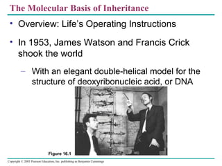
  • 1. Copyright © 2005 Pearson Education, Inc. publishing as Benjamin Cummings The Molecular Basis of Inheritance • Overview: Life’s Operating Instructions • In 1953, James Watson and Francis Crick shook the world – With an elegant double-helical model for the structure of deoxyribonucleic acid, or DNA Figure 16.1
  • 2. Copyright © 2005 Pearson Education, Inc. publishing as Benjamin Cummings • Hereditary information – Is encoded in the chemical language of DNA and reproduced in all the cells of your body • It is the DNA program – That directs the development of many different types of traits
  • 3. Copyright © 2005 Pearson Education, Inc. publishing as Benjamin Cummings Evidence That DNA Can Transform Bacteria • The role of DNA in heredity – Was first worked out by studying bacteria and the viruses that infect them • Frederick Griffith was studying Streptococcus pneumoniae – A bacterium that causes pneumonia in mammals • He worked with two strains of the bacterium – A pathogenic strain and a nonpathogenic strain
  • 4. Copyright © 2005 Pearson Education, Inc. publishing as Benjamin Cummings • Griffith found that when he mixed heat-killed remains of the pathogenic strain – With living cells of the nonpathogenic strain, some of these living cells became pathogenic Bacteria of the “S” (smooth) strain of Streptococcus pneumoniae are pathogenic because they have a capsule that protects them from an animal’s defense system. Bacteria of the “R” (rough) strain lack a capsule and are nonpathogenic. Frederick Griffith injected mice with the two strains as shown below: Griffith concluded that the living R bacteria had been transformed into pathogenic S bacteria by an unknown, heritable substance from the dead S cells. EXPERIMENT RESULTS CONCLUSION Living S (control) cells Living R (control) cells Heat-killed (control) S cells Mixture of heat-killed S cells and living R cells Mouse dies Mouse healthy Mouse healthy Mouse dies Living S cells are found in blood sample. Figure 16.2
  • 5. Copyright © 2005 Pearson Education, Inc. publishing as Benjamin Cummings • Griffith called the phenomenon transformation Bacteria of the “S” (smooth) strain of Streptococcus pneumoniae are pathogenic because they have a capsule that protects them from an animal’s defense system. Bacteria of the “R” (rough) strain lack a capsule and are nonpathogenic. Frederick Griffith injected mice with the two strains as shown below: Griffith concluded that the living R bacteria had been transformed into pathogenic S bacteria by an unknown, heritable substance from the dead S cells. EXPERIMENT RESULTS CONCLUSION Living S (control) cells Living R (control) cells Heat-killed (control) S cells Mixture of heat-killed S cells and living R cells Mouse dies Mouse healthy Mouse healthy Mouse dies Living S cells are found in blood sample. Figure 16.2
  • 6. Copyright © 2005 Pearson Education, Inc. publishing as Benjamin Cummings Evidence That Viral DNA Can Program Cells • Additional evidence for DNA as the genetic material – Came from studies of a virus that infects bacteria
  • 7. Copyright © 2005 Pearson Education, Inc. publishing as Benjamin Cummings • Viruses that infect bacteria, bacteriophages – Are widely used as tools by researchers in molecular genetics Figure 16.3 Phage head Tail Tail fiber DNA Bacterial cell 100nm
  • 8. Copyright © 2005 Pearson Education, Inc. publishing as Benjamin Cummings Experiments showing that DNA is the genetic material of a phage (T2) • The Hershey and Chase experiment In their famous 1952 experiment, Alfred Hershey and Martha Chase used radioactive sulfur and phosphorus to trace the fates of the protein and DNA, respectively, of T2 phages that infected bacterial cells. Radioactivity (phage protein) in liquid Phage Bacterial cell Radioactive protein Empty protein shell Phage DNA DNA Centrifuge Pellet (bacterial cells and contents) Radioactive DNA Centrifuge Pellet Batch 1: Phages were grown with radioactive sulfur (35 S), which was incorporated into phage protein (pink). Batch 2: Phages were grown with radioactive phosphorus (32 P), which was incorporated into phage DNA (blue). 1 2 3 4Agitated in a blender to separate phages outside the bacteria from the bacterial cells. Mixed radioactively labeled phages with bacteria. The phages infected the bacterial cells. Centrifuged the mixture so that bacteria formed a pellet at the bottom of the test tube. Measured the radioactivity in the pellet and the liquid Phage proteins remained outside the bacterial cells during infection, while phage DNA entered the cells. When cultured, bacterial cells with radioactive phage DNA released new phages with some radioactive phosphorus. Hershey and Chase concluded that DNA, not protein, functions as the T2 phage’s genetic material. RESULTS CONCLUSION EXPERIMENT Radioactivity (phage DNA) in pellet Figure 16.4 Animation of experiment
  • 9. Copyright © 2005 Pearson Education, Inc. publishing as Benjamin Cummings Additional Evidence That DNA Is the Genetic Materia • Prior to the 1950s, it was already known that DNA – Is a polymer of nucleotides, each consisting of three components: a nitrogenous base, a sugar, and a phosphate group Sugar-phosphate backbone Nitrogenous bases 5 end O– O P O CH2 5 4O– H H O H H H 3 1 H O CH3 N O N H Thymine (T) O O P O O– CH2 H H O H H H H N N N H N H H Adenine (A) O O P O O– CH2 H H O H H H H H H HN NN O Cytosine (C) O O P O CH2 5 4O– H O H H 3 1 OH 2 H N N N H O N N HH H H Sugar (deoxyribose) 3 end Phosphate Guanine (G) DNA nucleotide 2 N Figure 16.5
  • 10. Copyright © 2005 Pearson Education, Inc. publishing as Benjamin Cummings • Erwin Chargaff analyzed the base composition of DNA – From a number of different organisms • In 1947, Chargaff reported – That DNA composition varies from one species to the next • This evidence of molecular diversity among species – Made DNA a more credible candidate for the genetic material
  • 11. Copyright © 2005 Pearson Education, Inc. publishing as Benjamin Cummings Building a Structural Model of DNA: Scientific Inquiry • Maurice Wilkins and Rosalind Franklin – Were using a technique called X-ray crystallography to study molecular structure • Rosalind Franklin – Produced a picture of the DNA molecule using this technique (a) Rosalind Franklin Franklin’s X-ray diffraction Photograph of DNA (b) Figure 16.6 a, b
  • 12. Copyright © 2005 Pearson Education, Inc. publishing as Benjamin Cummings Figure 16.7a, c C T A A T CG GC A C G AT AT A T TA C TA 0.34 nm 3.4 nm (a) Key features of DNA structure G 1 nm G (c) Space-filling model T • Watson and Crick deduced that DNA was a double helix – Through observations of the X-ray crystallographic images of DNA
  • 13. Copyright © 2005 Pearson Education, Inc. publishing as Benjamin Cummings • Franklin had concluded that DNA – Was composed of two antiparallel sugar- phosphate backbones, with the nitrogenous bases paired in the molecule’s interior • The nitrogenous bases – Are paired in specific combinations: adenine with thymine, and cytosine with guanine
  • 14. Copyright © 2005 Pearson Education, Inc. publishing as Benjamin Cummings O – O O OH O – O O O H2C O – O O O H2C O – O O O OH O O O T A C GC A T O O O CH2 O O– O O CH2 CH2 CH2 5 end Hydrogen bond 3 end 3 end G P P P P O OH O– O O O P P O– O O O P O– O O O P (b) Partial chemical structure H2C 5 endFigure 16.7b O
  • 15. Copyright © 2005 Pearson Education, Inc. publishing as Benjamin Cummings • Watson and Crick reasoned that there must be additional specificity of pairing – Dictated by the structure of the bases • Each base pair forms a different number of hydrogen bonds – Adenine and thymine form two bonds, cytosine and guanine form three bonds
  • 16. Copyright © 2005 Pearson Education, Inc. publishing as Benjamin Cummings N H O CH3 N N O N N N N H Sugar Sugar Adenine (A) Thymine (T) N N N N Sugar O H N H NH N OH H N Sugar Guanine (G) Cytosine (C)Figure 16.8 H
  • 17. Copyright © 2005 Pearson Education, Inc. publishing as Benjamin Cummings • Many proteins work together in DNA replication and repair (DNA-Protein like Chicken-Egg debate, which came first?) • Since the two strands of DNA are complementary – Each strand acts as a template for building a new strand in replication
  • 18. Copyright © 2005 Pearson Education, Inc. publishing as Benjamin Cummings • In DNA replication – The parent molecule unwinds, and two new daughter strands are built based on base- pairing rules (a) The parent molecule has two complementary strands of DNA. Each base is paired by hydrogen bonding with its specific partner, A with T and G with C. (b) The first step in replication is separation of the two DNA strands. (c) Each parental strand now serves as a template that determines the order of nucleotides along a new, complementary strand. (d) The nucleotides are connected to form the sugar-phosphate backbones of the new strands. Each “daughter” DNA molecule consists of one parental strand and one new strand. A C T A G A C T A G A C T A G A C T A G T G A T C T G A T C A C T A G A C T A G T G A T C T G A T C T G A T C T G A T C Figure 16.9 a–d
  • 19. Copyright © 2005 Pearson Education, Inc. publishing as Benjamin Cummings Figure 16.10 a–c Conservative model. The two parental strands reassociate after acting as templates for new strands, thus restoring the parental double helix. Semiconservative model. The two strands of the parental molecule separate, and each functions as a template for synthesis of a new, comple- mentary strand. Dispersive model. Each strand of both daughter mol- ecules contains a mixture of old and newly synthesized DNA. Parent cell First replication Second replication • DNA replication is semiconservative – Each of the two new daughter molecules will have one old strand, derived from the parent molecule, and one newly made strand (a) (b) (c)
  • 20. Copyright © 2005 Pearson Education, Inc. publishing as Benjamin Cummings DNA Replication: A Closer Look • The copying of DNA – Is remarkable in its speed and accuracy • More than a dozen enzymes and other proteins – Participate in DNA replication • The replication of a DNA molecule – Begins at special sites called origins of replication, where the two strands are separated
  • 21. Copyright © 2005 Pearson Education, Inc. publishing as Benjamin Cummings • A eukaryotic chromosome – May have hundreds or even thousands of replication origins Replication begins at specific sites where the two parental strands separate and form replication bubbles. The bubbles expand laterally, as DNA replication proceeds in both directions. Eventually, the replication bubbles fuse, and synthesis of the daughter strands is complete. 1 2 3 Origin of replication Bubble Parental (template) strand Daughter (new) strand Replication fork Two daughter DNA molecules In eukaryotes, DNA replication begins at many sites along the giant DNA molecule of each chromosome. In this micrograph, three replication bubbles are visible along the DNA of a cultured Chinese hamster cell (TEM). (b)(a) 0.25 µm Figure 16.12 a, b
  • 22. Copyright © 2005 Pearson Education, Inc. publishing as Benjamin Cummings Figure 16.13 New strand Template strand 5 end 3 end Sugar A T Base C G G C A C T P P P OH P P 5 end 3 end 5 end 5 end A T C G G C A C T 3 endPyrophosphate 2 P OH Phosphate Elongating a New DNA Strand • Elongation of new DNA at a replication fork – Is catalyzed by enzymes called DNA polymerases, which add nucleotides to the 3′ end of a growing strand Nucleoside triphosphate
  • 23. Copyright © 2005 Pearson Education, Inc. publishing as Benjamin Cummings • DNA polymerases add nucleotides – Only to the free 3′ end of a growing strand • Along one template strand of DNA, the leading strand – DNA polymerase III can synthesize a complementary strand continuously, moving toward the replication fork
  • 24. Copyright © 2005 Pearson Education, Inc. publishing as Benjamin Cummings • To elongate the other new strand of DNA, the lagging strand – DNA polymerase III must work in the direction away from the replication fork • The lagging strand – Is synthesized as a series of segments called Okazaki fragments, which are then joined together by DNA ligase
  • 25. Copyright © 2005 Pearson Education, Inc. publishing as Benjamin Cummings Parental DNA DNA pol Ill elongates DNA strands only in the 5 3 direction. 1 Okazaki fragments DNA pol III Template strand Lagging strand 3 2 Template strand DNA ligase Overall direction of replication One new strand, the leading strand, can elongate continuously 5 3 as the replication fork progresses. 2 The other new strand, the lagging strand must grow in an overall 3 5 direction by addition of short segments, Okazaki fragments, that grow 5 3 (numbered here in the order they were made). 3 DNA ligase joins Okazaki fragments by forming a bond between their free ends. This results in a continuous strand. 4 Figure 16.14 3 5 5 3 3 5 2 1 Leading strand 1 • Synthesis of leading and lagging strands during DNA replication
  • 26. Copyright © 2005 Pearson Education, Inc. publishing as Benjamin Cummings Priming DNA Synthesis • DNA polymerases cannot initiate the synthesis of a polynucleotide – They can only add nucleotides to the 3′ end • The initial nucleotide strand – Is an RNA or DNA primer
  • 27. Copyright © 2005 Pearson Education, Inc. publishing as Benjamin Cummings • Only one primer is needed for synthesis of the leading strand – But for synthesis of the lagging strand, each Okazaki fragment must be primed separately
  • 28. Copyright © 2005 Pearson Education, Inc. publishing as Benjamin Cummings Replication Animation**** Overall direction of replication 3 3 3 3 5 3 5 3 5 3 5 3 5 3 5 3 5 3 5 5 1 1 2 1 1 2 5 5 1 2 35 Template strand RNA primer Okazaki fragment Figure 16.15 Primase joins RNA nucleotides into a primer. 1 DNA pol III adds DNA nucleotides to the primer, forming an Okazaki fragment. 2 After reaching the next RNA primer (not shown), DNA pol III falls off. 3 After the second fragment is primed. DNA pol III adds DNA nucleotides until it reaches the first primer and falls off. 4 DNA pol 1 replaces the RNA with DNA, adding to the 3 end of fragment 2. 5 DNA ligase forms a bond between the newest DNA and the adjacent DNA of fragment 1. 6 The lagging strand in this region is now complete. 7
  • 29. Copyright © 2005 Pearson Education, Inc. publishing as Benjamin Cummings Other Proteins That Assist DNA Replication • Helicase, topoisomerase, single-strand binding protein – Are all proteins that assist DNA replication Table 16.1
  • 30. Copyright © 2005 Pearson Education, Inc. publishing as Benjamin Cummings Figure 16.16 Overall direction of replication Leading strand Lagging strand Lagging strand Leading strandOVERVIEW Leading strand Replication fork DNA pol III Primase Primer DNA pol III Lagging strand DNA pol I Parental DNA 5 3 4 3 2 Origin of replication DNA ligase 1 5 3 Helicase unwinds the parental double helix. 1 Molecules of single- strand binding protein stabilize the unwound template strands. 2 The leading strand is synthesized continuously in the 5 3 direction by DNA pol III. 3 Primase begins synthesis of RNA primer for fifth Okazaki fragment. 4 DNA pol III is completing synthesis of the fourth fragment, when it reaches the RNA primer on the third fragment, it will dissociate, move to the replication fork, and add DNA nucleotides to the 3 end of the fifth fragment primer. 5 DNA pol I removes the primer from the 5 end of the second fragment, replacing it with DNA nucleotides that it adds one by one to the 3 end of the third fragment. The replacement of the last RNA nucleotide with DNA leaves the sugar- phosphate backbone with a free 3 end. 6 DNA ligase bonds the 3 end of the second fragment to the 5 end of the first fragment. 7 Replication Animation #2 Recap • A summary of DNA replication
  • 31. Copyright © 2005 Pearson Education, Inc. publishing as Benjamin Cummings The DNA Replication Machine as a Stationary Complex • The various proteins that participate in DNA replication – Form a single large complex, a DNA replication “machine” • The DNA replication machine – Is probably stationary during the replication process
  • 32. Copyright © 2005 Pearson Education, Inc. publishing as Benjamin Cummings Proofreading and Repairing DNA • DNA polymerases proofread newly made DNA – Replacing any incorrect nucleotides • In mismatch repair of DNA – Repair enzymes correct errors in base pairing
  • 33. Copyright © 2005 Pearson Education, Inc. publishing as Benjamin Cummings Figure 16.17 Nuclease DNA polymerase DNA ligase A thymine dimer distorts the DNA molecule. 1 A nuclease enzyme cuts the damaged DNA strand at two points and the damaged section is removed. 2 Repair synthesis by a DNA polymerase fills in the missing nucleotides. 3 DNA ligase seals the Free end of the new DNA To the old DNA, making the strand complete. 4 • In nucleotide excision repair – Enzymes cut out and replace damaged stretches of DNA
  • 34. Copyright © 2005 Pearson Education, Inc. publishing as Benjamin Cummings Replicating the Ends of DNA Molecules • The ends of eukaryotic chromosomal DNA – Get shorter with each round of replication Figure 16.18 End of parental DNA strands Leading strand Lagging strand Last fragment Previous fragment RNA primer Lagging strand Removal of primers and replacement with DNA where a 3 end is available Primer removed but cannot be replaced with DNA because no 3 end available for DNA polymerase Second round of replication New leading strand New lagging strand 5 Further rounds of replication Shorter and shorter daughter molecules 5 3 5 3 5 3 5 3 3
  • 35. Copyright © 2005 Pearson Education, Inc. publishing as Benjamin Cummings • Eukaryotic chromosomal DNA molecules – Have at their ends nucleotide sequences, called telomeres, that postpone the erosion of genes near the ends of DNA molecules Figure 16.19 1 µm
  • 36. Copyright © 2005 Pearson Education, Inc. publishing as Benjamin Cummings • If the chromosomes of germ cells became shorter in every cell cycle – Essential genes would eventually be missing from the gametes they produce • An enzyme called telomerase – Catalyzes the lengthening of telomeres in germ cells
  • 37. Copyright © 2005 Pearson Education, Inc. publishing as Benjamin Cummings From Gene to Protein • The DNA inherited by an organism – Leads to specific traits by dictating the synthesis of proteins • The process by which DNA directs protein synthesis, gene expression – Includes two stages, called transcription and translation
  • 38. Copyright © 2005 Pearson Education, Inc. publishing as Benjamin Cummings • The ribosome – Is part of the cellular machinery for translation, polypeptide synthesis Figure 17.1
  • 39. Copyright © 2005 Pearson Education, Inc. publishing as Benjamin Cummings Genes specify proteins via transcription and translation • In 1909, British physician Archibald Garrod – Was the first to suggest that genes dictate phenotypes through enzymes that catalyze specific chemical reactions in the cell • Beadle and Tatum causes bread mold to mutate with X-rays – Creating mutants that could not survive on minimal medium
  • 40. Copyright © 2005 Pearson Education, Inc. publishing as Benjamin Cummings • Using genetic crosses – They determined that their mutants fell into three classes, each mutated in a different gene Figure 17.2 Working with the mold Neurospora crassa, George Beadle and Edward Tatum had isolated mutants requiring arginine in their growth medium and had shown genetically that these mutants fell into three classes, each defective in a different gene. From other considerations, they suspected that the metabolic pathway of arginine biosynthesis included the precursors ornithine and citrulline. Their most famous experiment, shown here, tested both their one gene–one enzyme hypothesis and their postulated arginine pathway. In this experiment, they grew their three classes of mutants under the four different conditions shown in the Results section below. The wild-type strain required only the minimal medium for growth. The three classes of mutants had different growth requirements EXPERIMENT RESULTS Class I Mutants Class II Mutants Class III MutantsWild type Minimal medium (MM) (control) MM + Ornithine MM + Citrulline MM + Arginine (control)
  • 41. Copyright © 2005 Pearson Education, Inc. publishing as Benjamin Cummings CONCLUSION From the growth patterns of the mutants, Beadle and Tatum deduced that each mutant was unable to carry out one step in the pathway for synthesizing arginine, presumably because it lacked the necessary enzyme. Because each of their mutants was mutated in a single gene, they concluded that each mutated gene must normally dictate the production of one enzyme. Their results supported the one gene–one enzyme hypothesis and also confirmed the arginine pathway. (Notice that a mutant can grow only if supplied with a compound made after the defective step.) Class I Mutants (mutation in gene A) Class II Mutants (mutation in gene B) Class III Mutants (mutation in gene C)Wild type Gene A Gene B Gene C Precursor Precursor Precursor Precursor Ornithine Ornithine Ornithine Ornithine Citrulline Citrulline Citrulline Citrulline Arginine Arginine Arginine Arginine Enzyme A Enzyme B Enzyme C A A A B B B C C C
  • 42. Copyright © 2005 Pearson Education, Inc. publishing as Benjamin Cummings • Beadle and Tatum developed the “one gene– one enzyme hypothesis” – Which states that the function of a gene is to dictate the production of a specific enzyme • As researchers learned more about proteins – They made minor revision to the one gene– one enzyme hypothesis • Genes code for polypeptide chains or for RNA molecules
  • 43. Copyright © 2005 Pearson Education, Inc. publishing as Benjamin Cummings Basic Principles of Transcription and Translation • Transcription – Is the synthesis of RNA under the direction of DNA – Produces messenger RNA (mRNA) • Translation – Is the actual synthesis of a polypeptide, which occurs under the direction of mRNA – Occurs on ribosomes http://vcell.ndsu.nodak.edu/animations/ transcription/index.htm - animations
  • 44. Copyright © 2005 Pearson Education, Inc. publishing as Benjamin Cummings • In prokaryotes – Transcription and translation occur together Figure 17.3a Prokaryotic cell. In a cell lacking a nucleus, mRNA produced by transcription is immediately translated without additional processing. (a) TRANSLATION TRANSCRIPTION DNA mRNA Ribosome Polypeptide
  • 45. Copyright © 2005 Pearson Education, Inc. publishing as Benjamin Cummings Prokaryote/Eukaryote differences animation • In eukaryotes – RNA transcripts are modified before becoming true mRNA Figure 17.3b Eukaryotic cell. The nucleus provides a separate compartment for transcription. The original RNA transcript, called pre-mRNA, is processed in various ways before leaving the nucleus as mRNA. (b) TRANSCRIPTION RNA PROCESSING TRANSLATION mRNA DNA Pre-mRNA Polypeptide Ribosome Nuclear envelope
  • 46. Copyright © 2005 Pearson Education, Inc. publishing as Benjamin Cummings • Cells are governed by a cellular chain of command – DNA → RNA → protein • Genetic information – Is encoded as a sequence of nonoverlapping base triplets, or codons
  • 47. Copyright © 2005 Pearson Education, Inc. publishing as Benjamin Cummings • During transcription – The gene determines the sequence of bases along the length of an mRNA molecule Figure 17.4 DNA molecule Gene 1 Gene 2 Gene 3 DNA strand (template) TRANSCRIPTION mRNA Protein TRANSLATION Amino acid A C C A A A C C G A G T U G G U U U G G C U C A Trp Phe Gly Ser Codon 3 5 35
  • 48. Copyright © 2005 Pearson Education, Inc. publishing as Benjamin Cummings Cracking the Code • A codon in messenger RNA – Is either translated into an amino acid or serves as a translational stop signal Second mRNA base U C A G U C A G UUU UUC UUA UUG CUU CUC CUA CUG AUU AUC AUA AUG GUU GUC GUA GUG Met or start Phe Leu Leu lle Val UCU UCC UCA UCG CCU CCC CCA CCG ACU ACC ACA ACG GCU GCC GCA GCG Ser Pro Thr Ala UAU UAC UGU UGC Tyr Cys CAU CAC CAA CAG CGU CGC CGA CGG AAU AAC AAA AAG AGU AGC AGA AGG GAU GAC GAA GAG GGU GGC GGA GGG UGG UAA UAG Stop Stop UGA Stop Trp His Gln Asn Lys Asp Arg Ser Arg Gly U C A G U C A G U C A G U C A G FirstmRNAbase(5end) ThirdmRNAbase(3end) Glu Codons must be read in the correct reading frame For the specified polypeptide to be produced
  • 49. Copyright © 2005 Pearson Education, Inc. publishing as Benjamin Cummings Molecular Components of Transcription • RNA synthesis – Is catalyzed by RNA polymerase, which pries the DNA strands apart and hooks together the RNA nucleotides – Follows the same base-pairing rules as DNA, except that in RNA, uracil substitutes for thymine
  • 50. Copyright © 2005 Pearson Education, Inc. publishing as Benjamin Cummings Synthesis of an RNA Transcript • The stages of transcription are – Initiation – Elongation – Termination Figure 17.7 Promoter Transcription unit RNA polymerase Start point 5 3 3 5 3 5 5 3 5 3 3 5 5 3 3 5 5 5 Rewound RNA RNA transcript 3 3 Completed RNA transcript Unwound DNA RNA transcript Template strand of DNA DNA 1 Initiation. After RNA polymerase binds to the promoter, the DNA strands unwind, and the polymerase initiates RNA synthesis at the start point on the template strand. 2 Elongation. The polymerase moves downstream, unwinding the DNA and elongating the RNA transcript 5  3 . In the wake of transcription, the DNA strands re-form a double helix. 3 Termination. Eventually, the RNA transcript is released, and the polymerase detaches from the DNA.
  • 51. Copyright © 2005 Pearson Education, Inc. publishing as Benjamin Cummings Elongation RNA polymerase Non-template strand of DNA RNA nucleotides 3 end C A E G C A A U T A G G T T A A C G U A T C A T C C A A T T G G 3 5 5 Newly made RNA Direction of transcription (“downstream”) Template strand of DNA
  • 52. Copyright © 2005 Pearson Education, Inc. publishing as Benjamin Cummings RNA Polymerase Binding and Initiation of Transcription • Promoters signal the initiation of RNA synthesis • Transcription factors – Help eukaryotic RNA polymerase recognize promoter sequences Figure 17.8Figure 17.8 TRANSCRIPTION RNA PROCESSING TRANSLATION DNA Pre-mRNA mRNA Ribosome Polypeptide T A T AAA A ATAT T T T TATA box Start point Template DNA strand 5 3 3 5 Transcription factors 5 3 3 5 Promoter 5 3 3 55 RNA polymerase II Transcription factors RNA transcript Transcription initiation complex Eukaryotic promoters1 Several transcription factors 2 Additional transcription factors 3
  • 53. Copyright © 2005 Pearson Education, Inc. publishing as Benjamin Cummings Elongation of the RNA Strand • As RNA polymerase moves along the DNA – It continues to untwist the double helix, exposing about 10 to 20 DNA bases at a time for pairing with RNA nucleotides • The mechanisms of termination – Are different in prokaryotes and eukaryotes
  • 54. Copyright © 2005 Pearson Education, Inc. publishing as Benjamin Cummings • Eukaryotic cells modify RNA after transcription • Enzymes in the eukaryotic nucleus – Modify pre-mRNA in specific ways before the genetic messages are dispatched to the cytoplasm
  • 55. Copyright © 2005 Pearson Education, Inc. publishing as Benjamin Cummings Alteration of mRNA Ends • Each end of a pre-mRNA molecule is modified in a particular way – The 5′ end receives a modified nucleotide cap – The 3′ end gets a poly-A tail Figure 17.9 A modified guanine nucleotide added to the 5 end 50 to 250 adenine nucleotides added to the 3 end Protein-coding segment Polyadenylation signal Poly-A tail3 UTR Stop codonStart codon 5 Cap 5 UTR AAUAAA AAA…AAA TRANSCRIPTION RNA PROCESSING DNA Pre-mRNA mRNA TRANSLATION Ribosome Polypeptide G P P P 5 3 Video clip
  • 56. Copyright © 2005 Pearson Education, Inc. publishing as Benjamin Cummings Split Genes and RNA Splicing • RNA splicing – Removes introns (supposed “Junk-DNA”) and joins exons Figure 17.10 TRANSCRIPTION RNA PROCESSING DNA Pre-mRNA mRNA TRANSLATION Ribosome Polypeptide 5 Cap Exon Intron 1 5 30 31 Exon Intron 104 105 146 Exon 3 Poly-A tail Poly-A tail Introns cut out and exons spliced together Coding segment 5 Cap 1 146 3 UTR3 UTR Pre-mRNA mRNA
  • 57. Copyright © 2005 Pearson Education, Inc. publishing as Benjamin Cummings • Is carried out by spliceosomes in some cases Figure 17.11 RNA transcript (pre-mRNA) Exon 1 Intron Exon 2 Other proteins Protein snRNA snRNPs Spliceosome Spliceosome components Cut-out intron mRNA Exon 1 Exon 2 5 5 5 1 2 3 Animation Ribozymes Are catalytic RNA molecules that function as enzymes and can splice RNA
  • 58. Copyright © 2005 Pearson Education, Inc. publishing as Benjamin Cummings • Proteins often have a modular architecture – Consisting of discrete structural and functional regions called domains • In many cases – Different exons code for the different domains in a protein Figure 17.12 Gene DNA Exon 1 Intron Exon 2 Intron Exon 3 Transcription RNA processing Translation Domain 3 Domain 1 Domain 2 Polypeptide
  • 59. Copyright © 2005 Pearson Education, Inc. publishing as Benjamin Cummings • A cell translates an mRNA message into protein – With the help of transfer RNA (tRNA) Figure 17.13 TRANSCRIPTION TRANSLATION DNA mRNA Ribosome Polypeptide Polypeptide Amino acids tRNA with amino acid attachedRibosome tRNA Anticodon mRNA Trp Phe Gly A G C A A A C C G U G G U U U G G C Codons5 3
  • 60. Copyright © 2005 Pearson Education, Inc. publishing as Benjamin Cummings • Molecules of tRNA are not all identical – Each carries a specific amino acid on one end – Each has an anticodon on the other end (b) Three-dimensional structure Symbol used in this book Amino acid attachment site Hydrogen bonds Anticodon Anticodon A AG 5 3 3 5 (c)
  • 61. Copyright © 2005 Pearson Education, Inc. publishing as Benjamin Cummings The Structure and Function of Transfer RNA A C C • A tRNA molecule – Consists of a single RNA strand that is only about 80 nucleotides long Figure 17.14a Two-dimensional structure. The four base-paired regions and three loops are characteristic of all tRNAs, as is the base sequence of the amino acid attachment site at the 3 end. The anticodon triplet is unique to each tRNA type. (The asterisks mark bases that have been chemically modified, a characteristic of tRNA.) (a) 3 C C A C G C U U A A GACAC CU * G C * * G U G U *CU * G AG G U * *A * A A G U C A G A C C * C G A G A G G G * * GA CUC*A U U U A G G C G 5 Amino acid attachment site Hydrogen bonds Anticodon A
  • 62. Copyright © 2005 Pearson Education, Inc. publishing as Benjamin Cummings • A specific enzyme called an aminoacyl-tRNA synthetase – Joins each amino acid to the correct tRNA Figure 17.15 Amino acid ATP Adenosine Pyrophosphate Adenosine Adenosine Phosphates tRNA P P P P P Pi Pi Pi P AMP Aminoacyl tRNA (an “activated amino acid”) Aminoacyl-tRNA synthetase (enzyme) Active site binds the amino acid and ATP. 1 ATP loses two P groups and joins amino acid as AMP. 2 3 Appropriate tRNA covalently Bonds to amino Acid, displacing AMP. Activated amino acid is released by the enzyme. 4
  • 63. Copyright © 2005 Pearson Education, Inc. publishing as Benjamin Cummings Ribosomes • Ribosomes – Facilitate the specific coupling of tRNA anticodons with mRNA codons during protein synthesis
  • 64. Copyright © 2005 Pearson Education, Inc. publishing as Benjamin Cummings • The ribosomal subunits – Are constructed of proteins and RNA molecules named ribosomal RNA or rRNA Figure 17.16a TRANSCRIPTION TRANSLATION DNA mRNA Ribosome Polypeptide Exit tunnel Growing polypeptide tRNA molecules E P A Large subunit Small subunit mRNA Computer model of functioning ribosome. This is a model of a bacterial ribosome, showing its overall shape. The eukaryotic ribosome is roughly similar. A ribosomal subunit is an aggregate of ribosomal RNA molecules and proteins. (a) 5 3
  • 65. Copyright © 2005 Pearson Education, Inc. publishing as Benjamin Cummings • The ribosome has three binding sites for tRNA – The P site – The A site – The E site Figure 17.16b E P A P site (Peptidyl-tRNA binding site) E site (Exit site) mRNA binding site A site (Aminoacyl- tRNA binding site) Large subunit Small subunit Schematic model showing binding sites. A ribosome has an mRNA binding site and three tRNA binding sites, known as the A, P, and E sites. This schematic ribosome will appear in later diagrams. (b)
  • 66. Copyright © 2005 Pearson Education, Inc. publishing as Benjamin Cummings Figure 17.16c Amino end Growing polypeptide Next amino acid to be added to polypeptide chain tRNA mRNA Codons 3 5 Schematic model with mRNA and tRNA. A tRNA fits into a binding site when its anticodon base-pairs with an mRNA codon. The P site holds the tRNA attached to the growing polypeptide. The A site holds the tRNA carrying the next amino acid to be added to the polypeptide chain. Discharged tRNA leaves via the E site. (c)
  • 67. Copyright © 2005 Pearson Education, Inc. publishing as Benjamin Cummings Building a Polypeptide • We can divide translation into three stages – Initiation – Elongation – Termination
  • 68. Copyright © 2005 Pearson Education, Inc. publishing as Benjamin Cummings Ribosome Association and Initiation of Translation • The initiation stage of translation – Brings together mRNA, tRNA bearing the first amino acid of the polypeptide, and two subunits of a ribosome Large ribosomal subunit The arrival of a large ribosomal subunit completes the initiation complex. Proteins called initiation factors (not shown) are required to bring all the translation components together. GTP provides the energy for the assembly. The initiator tRNA is in the P site; the A site is available to the tRNA bearing the next amino acid. 2 Initiator tRNA mRNA mRNA binding site Small ribosomal subunit Translation initiation complex P site GDPGTP Start codon A small ribosomal subunit binds to a molecule of mRNA. In a prokaryotic cell, the mRNA binding site on this subunit recognizes a specific nucleotide sequence on the mRNA just upstream of the start codon. An initiator tRNA, with the anticodon UAC, base-pairs with the start codon, AUG. This tRNA carries the amino acid methionine (Met). 1 Met Met U A C A U G E A 3 5 5 3 35 35 Figure 17.17
  • 69. Copyright © 2005 Pearson Education, Inc. publishing as Benjamin Cummings Elongation of the Polypeptide Chain • In the elongation stage of translation – Amino acids are added one by one to the preceding amino acid Figure 17.18 Amino end of polypeptide mRNA Ribosome ready for next aminoacyl tRNA E P A E P A E P A E P A GDP GTP GTP GDP 2 2 site site5 3 TRANSCRIPTION TRANSLATION DNA mRNA Ribosome Polypeptide Codon recognition. The anticodon of an incoming aminoacyl tRNA base-pairs with the complementary mRNA codon in the A site. Hydrolysis of GTP increases the accuracy and efficiency of this step. 1 Peptide bond formation. An rRNA molecule of the large subunit catalyzes the formation of a peptide bond between the new amino acid in the A site and the carboxyl end of the growing polypeptide in the P site. This step attaches the polypeptide to the tRNA in the A site. 2 Translocation. The ribosome translocates the tRNA in the A site to the P site. The empty tRNA in the P site is moved to the E site, where it is released. The mRNA moves along with its bound tRNAs, bringing the next codon to be translated into the A site. 3
  • 70. Copyright © 2005 Pearson Education, Inc. publishing as Benjamin Cummings Termination of Translation • The final stage of translation is termination – When the ribosome reaches a stop codon in the mRNA Figure 17.19 Release factor Free polypeptide Stop codon (UAG, UAA, or UGA) 5 3 3 5 3 5 When a ribosome reaches a stop codon on mRNA, the A site of the ribosome accepts a protein called a release factor instead of tRNA. 1 The release factor hydrolyzes the bond between the tRNA in the P site and the last amino acid of the polypeptide chain. The polypeptide is thus freed from the ribosome. 2 3 The two ribosomal subunits and the other components of the assembly dissociate. Protein Synthesis Animation
  • 71. Copyright © 2005 Pearson Education, Inc. publishing as Benjamin Cummings Polyribosomes • A number of ribosomes can translate a single mRNA molecule simultaneously – Forming a polyribosome Figure 17.20a, b Growing polypeptides Completed polypeptide Incoming ribosomal subunits Start of mRNA (5 end) End of mRNA (3 end) Polyribosome An mRNA molecule is generally translated simultaneously by several ribosomes in clusters called polyribosomes. (a) Ribosomes mRNA This micrograph shows a large polyribosome in a prokaryotic cell (TEM). 0.1 µm (b)
  • 72. Copyright © 2005 Pearson Education, Inc. publishing as Benjamin Cummings Protein Folding and Post-Translational Modifications • After translation – Proteins may be modified in ways that affect their three-dimensional shape
  • 73. Copyright © 2005 Pearson Education, Inc. publishing as Benjamin Cummings Targeting Polypeptides to Specific Locations • Two populations of ribosomes are evident in cells – Free and bound • Free ribosomes in the cytosol – Initiate the synthesis of all proteins
  • 74. Copyright © 2005 Pearson Education, Inc. publishing as Benjamin Cummings • Proteins destined for the endomembrane system or for secretion – Must be transported into the ER – Have signal peptides to which a signal- recognition particle (SRP) binds, enabling the translation ribosome to bind to the ER
  • 75. Copyright © 2005 Pearson Education, Inc. publishing as Benjamin Cummings Figure 17.21 Ribosome mRNA Signal peptide Signal- recognition particle (SRP) SRP receptor protein Translocation complex CYTOSOL Signal peptide removed ER membrane Protein ERLUMEN • The signal mechanism for targeting proteins to the ER Polypeptide synthesis begins on a free ribosome in the cytosol. 1 An SRP binds to the signal peptide, halting synthesis momentarily. 2 The SRP binds to a receptor protein in the ER membrane. This receptor is part of a protein complex (a translocation complex) that has a membrane pore and a signal-cleaving enzyme. 3 The SRP leaves, and the polypeptide resumes growing, meanwhile translocating across the membrane. (The signal peptide stays attached to the membrane.) 4 The signal- cleaving enzyme cuts off the signal peptide. 5 The rest of the completed polypeptide leaves the ribosome and folds into its final conformation. 6
  • 76. Copyright © 2005 Pearson Education, Inc. publishing as Benjamin Cummings • RNA plays multiple roles in the cell: a review • RNA – Can hydrogen-bond to other nucleic acid molecules – Can assume a specific three-dimensional shape – Has functional groups that allow it to act as a catalyst
  • 77. Copyright © 2005 Pearson Education, Inc. publishing as Benjamin Cummings • Types of RNA in a Eukaryotic Cell Table 17.1
  • 78. Copyright © 2005 Pearson Education, Inc. publishing as Benjamin Cummings • Comparing gene expression in prokaryotes and eukaryotes reveals key differences • Prokaryotic cells lack a nuclear envelope – Allowing translation to begin while transcription is still in progress Figure 17.22 DNA Polyribosome mRNA Direction of transcription 0.25 mRNA polymerase Polyribosome Ribosome DNA mRNA (5 end) RNA polymerase Polypeptide (amino end)
  • 79. Copyright © 2005 Pearson Education, Inc. publishing as Benjamin Cummings • In a eukaryotic cell – The nuclear envelope separates transcription from translation – Extensive RNA processing occurs in the nucleus
  • 80. Copyright © 2005 Pearson Education, Inc. publishing as Benjamin Cummings What is a gene? revisiting the question • A gene – Is a region of DNA whose final product is either a polypeptide or an RNA molecule
  • 81. Copyright © 2005 Pearson Education, Inc. publishing as Benjamin Cummings • A summary of transcription and translation in a eukaryotic cell Figure 17.26 TRANSCRIPTION RNA is transcribed from a DNA template. DNA RNA polymerase RNA transcript RNA PROCESSING In eukaryotes, the RNA transcript (pre- mRNA) is spliced and modified to produce mRNA, which moves from the nucleus to the cytoplasm. Exon Poly-A RNA transcript (pre-mRNA) Intron NUCLEUS Cap FORMATION OF INITIATION COMPLEX After leaving the nucleus, mRNA attaches to the ribosome. CYTOPLASM mRNA Poly-A Growing polypeptide Ribosomal subunits Cap Aminoacyl-tRNA synthetase Amino acid tRNA AMINO ACID ACTIVATION Each amino acid attaches to its proper tRNA with the help of a specific enzyme and ATP. Activated amino acid TRANSLATION A succession of tRNAs add their amino acids to the polypeptide chain as the mRNA is moved through the ribosome one codon at a time. (When completed, the polypeptide is released from the ribosome.) Anticodon A CC A A A UG GUU UA U G UACE A Ribosome 1 Poly-A 5 5 3 Codon 2 3 4 5
  • 82. Copyright © 2005 Pearson Education, Inc. publishing as Benjamin Cummings • Point mutations can affect protein structure and function • Mutations – Are changes in the genetic material of a cell • Point mutations – Are changes in just one base pair of a gene
  • 83. Copyright © 2005 Pearson Education, Inc. publishing as Benjamin Cummings • The change of a single nucleotide in the DNA’s template strand – Leads to the production of an abnormal protein Figure 17.23 In the DNA, the mutant template strand has an A where the wild-type template has a T. The mutant mRNA has a U instead of an A in one codon. The mutant (sickle-cell) hemoglobin has a valine (Val) instead of a glutamic acid (Glu). Mutant hemoglobin DNAWild-type hemoglobin DNA mRNA mRNA Normal hemoglobin Sickle-cell hemoglobin Glu Val C T T C A T G A A G U A 3 5 3 5 5 35 3
  • 84. Copyright © 2005 Pearson Education, Inc. publishing as Benjamin Cummings Types of Point Mutations • Point mutations within a gene can be divided into two general categories – Base-pair substitutions – Base-pair insertions or deletions (indels)
  • 85. Copyright © 2005 Pearson Education, Inc. publishing as Benjamin Cummings Substitutions • A base-pair substitution – Is the replacement of one nucleotide and its partner with another pair of nucleotides – Can cause missense or nonsense Figure 17.24 Wild type A U G A A G U U U G G C U A A mRNA 5 Protein Met Lys Phe Gly Stop Carboxyl end Amino end 3 A U G A A G U U U G G U U A A Met Lys Phe Gly Base-pair substitution No effect on amino acid sequence U instead of C Stop A U G A A G U U U A G U U A A Met Lys Phe Ser Stop A U G U A G U U U G G C U A A Met Stop Missense A instead of G Nonsense U instead of A
  • 86. Copyright © 2005 Pearson Education, Inc. publishing as Benjamin Cummings Insertions and Deletions • Insertions and deletions – Are additions or losses of nucleotide pairs in a gene – May produce frameshift mutations Figure 17.25 mRNA Protein Wild type A U G A A G U U U G G C U A A 5 Met Lys Phe Gly Amino end Carboxyl end Stop Base-pair insertion or deletion Frameshift causing immediate nonsense A U G U A A G U U U G G C U A A U G A A G U U G G C U A A A U G U U U G G C U A A Met Stop U Met Lys Leu Ala Met Phe Gly Stop MissingA A G Missing Extra U Frameshift causing extensive missense Insertion or deletion of 3 nucleotides: no frameshift but extra or missing amino acid 3
  • 87. Copyright © 2005 Pearson Education, Inc. publishing as Benjamin Cummings Mutagens • Spontaneous mutations – Can occur during DNA replication, recombination, or repair • Mutagens – Are physical or chemical agents that can cause mutations