SlideShare une entreprise Scribd logo
1  sur  50
Protein structure , levels of protein structure ,
Domains, Motifs & Folds in protein structure
Presented by Aquib Naseer
M pharm 2nd semester pharmacology
SPER , Jamia Hamdard New delhi
Contents :
• Introduction.
• Amino acids as fundamental units of protein.
• Levels of protein structure :
1. Primary level.
2. Secondary level.
3. Tertiary level.
4. Quaternary
• Folds.
• Motifs.
• Domains.
Introduction :
Proteins are an important class of biological macromolecules which are the
polymers of amino acids.
• Biochemists have distinguished several levels of structural organization of
proteins. They are:
– Primary structure
– Secondary structure
– Tertiary structure
– Quaternary structure
• Methods for Determining Atomic Structures :
1. X-ray crystallography : X-ray crystallography is the most common tool used to
determine protein structure. It provides high resolution of the structure but it does
not give information about protein's conformational flexibility.
2. NMR :Protein NMR gives comparatively lower resolution of protein structure. It is
limited to smaller proteins. However, it can provide information about
conformational changes of a protein in solution.
3. Cryogenic electron microscopy :Cryogenic electron microscopy (cryo-EM) can give
information about both a protein's tertiary and quaternary structure. It is
particularly well-suited to large proteins and symmetrical complexes of protein
subunits.
4. Dual polarisation interferometry :Dual polarisation interferometry provides
complimentary information about surface captured proteins. It assists in
determining structure and conformation changes over time.
The building blocks of proteins are amino acids, which are small organic molecules that consist of
an alpha (central) carbon atom linked to an amino group, a carboxyl group, a hydrogen atom, and
a variable component called a side chain.
Within a protein, multiple amino acids are linked together by peptide bonds (amide bonds between
the -NH2 of one amino acid and the -COOH of another.), thereby forming a long chain.
Peptide bonds are formed by a biochemical reaction that extracts a water molecule as it joins the
amino group of one amino acid to the carboxyl group of a neighboring amino acid.
The linear sequence of amino acids within a protein is considered the primary structure of the
protein
What Are Proteins Made Of ?
v
v
v
vv
Sequences with fewer than 50 amino acids are generally referred to as peptides, while the
terms protein or polypeptide are used for longer sequences.
Charged amino acid side chains can form ionic bonds, and polar amino acids are capable of forming hydrogen
bonds. Hydrophobic side chains interact with each other via weak van der Waals interactions. The vast majority
of bonds formed by these side chains are noncovalent.
The chemistry of amino acid side chains is critical to protein structure because these side chains can bond with
one another to hold a length of protein in a certain shape or conformation.
Proteins are built from a set of only twenty amino acids, each of which has a unique side chain.
Levels of protein structure :
4 levels of protein structure
Protein structureis hierarchical
Quaternary ( multimeric organisation )
Tertiary ( long-range folding )
Secondary ( local folding )
Primary ( sequence )
12
1. Primary structure of proteins :
Often, post-translational modifications, such as glycosylation or phosphorylation, occur which are necessary for
the biological function of the protein.
Proteins are synthesized by a series of steps called transcription (the use of a DNA strand to make a
complimentary messenger RNA strand - mRNA) and translation (the mRNA sequence is used as a template to
guide the synthesis of the chain of amino acids which make up the protein).
The amino acid sequence of a protein is encoded in DNA.
Simple amino acid chain sequence makes up the primary structure of the protein.
Importance of primary structure :-
 To predict secondary & tertiary structures from sequence homologies
with related proteins  structure prediction.
 Many genetic diseases result from abnormal amino acid sequences.
 To understand the molecular mechanism of action of proteins.
 To trace evolutionary paths.
Methods of aminoacid sequencedetermination:-
 End group analysis – Edman degradation
 Gene sequencing method
15
Folds in proteins :
Molecular chaperones are a class of proteins that aid in the correct folding of other
proteins .
It is the physical process by which a polypeptide folds into its characteristic and
functional three-dimensional structure from random coil.
Each protein exists as an unfolded polypeptide or random coil when translated from a
sequence of mRNA to a linear chain of amino acids. This polypeptide lacks any stable
(long-lasting) three-dimensional structure .
As the polypeptide chain is being synthesized by a ribosome, the linear chain begins to fold into
its three-dimensional structure by molecular chaperones. Folding begins to occur even during
translation of the polypeptide chain. Amino acids interact with each other to produce a well-defined
three-dimensional structure, the folded protein, known as the native state.
Protein before and after folding.
Role of folds in different disease pathology :
At least eight neurodegenerative disorders are caused by an expanded polyglutamine tract in the various
disease proteins, including Huntington’s disease and spinocerebellar ataxia type 3. Polyglutamine expansion
causes the disease protein to misfold and aggregate, ultimately leading to neuronal death
Variants of the serpin (superfamily of proteins), antitrypsin, misfold and accumulate in the endoplasmic
reticulum of hepatocyte cells, leading to a plasma deficiency and hepatocyte damage due to the accumulation
of aggregated protein, leading to cirrhosis and emphysema.
We study two protein families involved in misfolding disorders: the serine proteinase inhibitor (serpin) and
polyglutamine family of proteins.
Most proteins have no trouble folding quickly and efficiently to their native state. However, an increasing
number of diseases such as emphysema, Alzheimer’s and Huntington’s disease are associated with the failure of
proteins to fold correctly.
2. Secondary structure :
• The primary structure of a protein — its amino acid
sequence — drives the folding and intramolecular bonding
of the linear amino acid chain, which ultimately determines
the protein's unique three-dimensional shape.
• Hydrogen bonding between amino groups and carboxyl
groups in neighboring regions of the protein chain
sometimes causes certain patterns of folding to occur.
• Linus Pauling proposed some essential features of peptide units and
polypeptide backbone. They are:
– The amide group is rigid and planar as a result of resonance. So rotation
about C-N bond is not feasible.
– Rotation can take place only about N- Cα and Cα – C bonds.
– Trans-configuration is more stable than cis- for R groups at Cα
• From these conclusions Pauling postulated 2 ordered structures :
• α helix and
• β sheet
• These stable folding patterns make up the secondary structure of a protein.
ALPHA HELIX :
• Spiral structure
• Tightly packed, coiled polypeptide
backbone core.
• Side chain extend outwards
• Stabilized by H bonding b/w
carbonyl oxygen and amide
hydrogen.
• Amino acids per turn – 3.6
• Pitch is 5.4 A
• Alpha helical segments are found in
many globular proteins like
myoglobins, troponin- C etc.
BETA PLEATED SHEET :
• Formed when 2 or more polypeptides line up side by
side.
• Individual polypeptide - β strand
• Each β strand is fully extended.
• They are stabilized by H bond b/w N-H and carbonyl grps of
adjacent chains.
•BETA PLEATED SHEET
• 2 types
• Parallel Anti -Parallel
• N C N C
• N C N C
Secondary structure :
v
Examples :
Tertiary structure :
Hydrophilic amino acids that can hydrogen-bond to water are at the surface of soluble proteins.
In soluble proteins, hydrophobic side chains are found in the interior of a protein; thus, domains pack together
so as to minimize the exposure of hydrophobic side chains to a water interface.
It is formed spontaneously and stabilized both by side chain interactions and, in extracellular proteins, by
disulfide bonds. This folding brings distant sequences in a linear polypeptide together into a stable structure.
Tertiary structure is the complete three-dimensional (3-D) structure of a polypeptide.
3. Tertiary structure :
Tertiary structure :
The most stable structure under any given physiologic condition is called the native
conformation of a protein. There are four side chain interactions that stabilize the native
conformation.
• Hydrophobic interactions: Hydrophobic side chains are repelled by water and forced together at
the interior of proteins to escape the aqueous environment.
• Van der Waals forces: A nonspecific attraction develops based on the proximity of interacting
atoms; if the shape of the side chain allows a good fit between surfaces, an attractive force
develops. A poor fit gives either repulsion or no force. Note: Both hydrophobic interactions and
the shape of side chains are major factors in determining tertiary structure.
• Electrostatic bonds: Oppositely charged side chains can attract each other, forming salt bridges.
They also play a role in the binding of substrates and allosteric effectors and in the association of
the protein with other protein molecules (see Quaternary Structure, below). In addition, they can
bind large amounts of water to solubilize the protein when located on the surface.
• Hydrogen bonds: Polar groups can share a partial positive charge between a hydrogen donor and
a hydrogen acceptor to form a weak bond.
• Based upon their tertiary structure, proteins are often divided into
1. Fibrous types
2. Globular types.
• Fibrous proteins, like α-keratin, have elongated rope-like structures that are strong and hydrophobic
proteins, having polypeptide chains arranged in long strands or sheets.
• Globular proteins, like the plasma proteins and the immunoglobulins, are more spherical and hydrophilic.
• The two groups are structurally distinct: fibrous proteins usually consist largely of a single type of secondary
structure; globular proteins often contain several types of secondary structure.
2 subtypes of tertiary structure :
Quaternary structure of proteins :
The quaternary structure of a protein is the association of several protein chains or subunits into
a closely packed arrangement.
Each of the subunits has its own
• Primary ,
• Secondary , and
• Tertiary structure.
The subunits are held together by hydrogen bonds and van der Waals forces between nonpolar
side chains.
The subunits in a quaternary structure must be specifically arranged for the entire protein to
function properly.
Any alteration in the structure of the subunits or how they are associated causes marked changes
in biological activity.
Protein Molecular weight Number of subunits Function
Alcohal dehydrogenase 80,000 4 Enzymatic reaction in fermentation
Aldolase 150,000 4 Enzymatic reaction in glycolysis
Fumarase 194,000 4
Enzymatic reaction in citric acid cycle
Haemoglobin 65,000 4 Oxygen transport in blood
Insulin 11,400 2 Hormone that regulates metabolism of
glucose
Examples of quaternary structure :
Sub units :
In structural biology, a protein subunit is a single protein molecule that assembles (or
"coassembles") with other protein molecules to form a protein complex.
Some naturally occurring proteins have a relatively small number of subunits and
therefore described as oligomeric, for example hemoglobin or DNA polymerase.
Others may consist of a very large number of subunits and therefore described as
multimeric, for example microtubules and other cytoskeleton proteins.
The subunits of a multimeric protein may be identical, homologous or totally dissimilar
and dedicated to disparate tasks.
In some protein assemblies, one subunit may be a "catalytic subunit" that enzymatically
catalyzes a reaction, whereas a "regulatory subunit" will facilitate or inhibit the activity.
• A subunit is often named with a Greek or Roman letter, and the numbers of this type of subunit in a protein is
indicated by a subscript.
• For example, ATP synthase has a type of subunit called α. Three of these are present in the ATP synthase
molecule, and is therefore designated α3
Haemoglobin molecule
How protein structure
determination and drug
discovery are interrelated:
Motifs (supersecondary structures):
• A motif is a recognizable folding pattern involving two or more elements of secondary structure and the
connection(s) between them. Or
• “The connectivity between secondary structure elements and the type of secondary structure elements
involved define the level of structural organization called structural motifs”.
• A motif is is simply a folding pattern.
• Motifs do not allow us to predict the biological functions: they are found in proteins and enzymes with
dissimilar functions.
• In proteins, a structural motif describes the connectivity between secondary structural elements.
Types of motifs :
• A motif can be very simple, such as two elements of secondary structure folded against each other, and
represent only a small part of a protein. An example is a β-α-β loop .
• A motif can also be a very complex structure involving scores of protein segments folded together, such as the
β barrel .
Motif mediated protein-protein interactions as drug targets :
In addition, numerous oncogenic proteins either contain a motif, or recognise motif interaction sequences for
which inhibition is a potential cancer treatment .
It has also been recognized that several viruses, e.g., Ebola and Rabies viruses, hijack the cell machinery using
modified domain motifs interactions.
For instance, Liddle’s, Noonan’s and Usher’s hereditary syndromes can be caused by mutations in the recognition
motif (PDZ recognition motif respectively) leading to the deregulation of important signalling pathways.
There are several diseases and syndromes related to the disruption of specific DMI (drug mediated interaction)
motifs.
Domains :
• A protein domain is a conserved part of a given protein sequence and (tertiary) structure that can evolve ,
function, and exist independently of the rest of the protein chain.
• Each domain forms a compact three-dimensional structure and often can be independently stable and folded.
• A domain usually contains between 40 and 350 amino acids, and it is the modular unit from which many larger
proteins are constructed.
• The different domains of a protein are often associated with different functions.
• E.g the Src protein kinase, which functions in signaling pathways inside vertebrate cells.
• This protein has four domains: the SH2 and SH3 domains have regulatory roles, while the two remaining
domains are responsible for the kinase catalytic activity.
• They explain how proteins can form molecular switches that transmit
information throughout cells.
• The central core of a domain can be constructed from α helices, from β sheets,
or from various combinations of these two fundamental folding elements. Each
different combination is known as a protein fold.
Predicting drug targets based on protein domains :
Most recently, it is reported that there are some interactions between compound
substructures and protein domains, and a set of chemical substructures shared by drugs are
able to bind to a set of protein domains.
Therefore, it is suspected that the specificity of drug–protein interactions is possibly
determined by drug– domain interactions even though the drugs do not physically bind to
protein domains.
It has been found that protein–protein interactions are dominated by domain–domain
interactions, which in turn could be used to predict new protein–protein interactions.
Thank you
References :
• https://www.nature.com/scitable/topicpage/protein-structure-
14122136
• https://www.particlesciences.com/news/technical-
briefs/2009/protein-structure.html
• https://www.sciencedirect.com/topics/biochemistry-genetics-and-
molecular-biology/tertiary-structure
• https://proteinstructures.com/Structure/Structure/protein-fold.html
• Leninger’s biochemistry , structure of protein & amino acids.

Contenu connexe

Tendances

Zinc finger protein in drug discovery process
Zinc finger protein in drug discovery processZinc finger protein in drug discovery process
Zinc finger protein in drug discovery processSanchit Dhankhar
 
NMR of protein
NMR of proteinNMR of protein
NMR of proteinJiya Ali
 
Protein micro array
Protein micro arrayProtein micro array
Protein micro arraykrupa sagar
 
molecular docking its types and de novo drug design and application and softw...
molecular docking its types and de novo drug design and application and softw...molecular docking its types and de novo drug design and application and softw...
molecular docking its types and de novo drug design and application and softw...GAUTAM KHUNE
 
Applications of proteomic sciences
Applications of proteomic sciencesApplications of proteomic sciences
Applications of proteomic sciencessukanyakk
 
Role of bioinformatics of drug designing
Role of bioinformatics of drug designingRole of bioinformatics of drug designing
Role of bioinformatics of drug designingDr NEETHU ASOKAN
 
Virtual Screening in Drug Discovery
Virtual Screening in Drug DiscoveryVirtual Screening in Drug Discovery
Virtual Screening in Drug DiscoveryAbhik Seal
 
Protein Structure Determination
Protein Structure DeterminationProtein Structure Determination
Protein Structure DeterminationAmjad Ibrahim
 
Computational predictiction of prrotein structure
Computational predictiction of prrotein structureComputational predictiction of prrotein structure
Computational predictiction of prrotein structureArchita Srivastava
 
Role of Target Identification and Target Validation in Drug Discovery Process
Role of Target Identification and Target Validation in Drug Discovery ProcessRole of Target Identification and Target Validation in Drug Discovery Process
Role of Target Identification and Target Validation in Drug Discovery ProcessPallavi Duggal
 

Tendances (20)

Zinc finger protein in drug discovery process
Zinc finger protein in drug discovery processZinc finger protein in drug discovery process
Zinc finger protein in drug discovery process
 
NMR of protein
NMR of proteinNMR of protein
NMR of protein
 
Motifs and domains
Motifs and domainsMotifs and domains
Motifs and domains
 
Homology modeling
Homology modelingHomology modeling
Homology modeling
 
Protein micro array
Protein micro arrayProtein micro array
Protein micro array
 
Nucleic acid microarray
Nucleic acid microarrayNucleic acid microarray
Nucleic acid microarray
 
Role of genomics proteomics, and bioinformatics.
Role of genomics proteomics, and bioinformatics.Role of genomics proteomics, and bioinformatics.
Role of genomics proteomics, and bioinformatics.
 
Homology modelling
Homology modellingHomology modelling
Homology modelling
 
molecular docking its types and de novo drug design and application and softw...
molecular docking its types and de novo drug design and application and softw...molecular docking its types and de novo drug design and application and softw...
molecular docking its types and de novo drug design and application and softw...
 
Protein Structure Prediction
Protein Structure PredictionProtein Structure Prediction
Protein Structure Prediction
 
Applications of proteomic sciences
Applications of proteomic sciencesApplications of proteomic sciences
Applications of proteomic sciences
 
Role of bioinformatics of drug designing
Role of bioinformatics of drug designingRole of bioinformatics of drug designing
Role of bioinformatics of drug designing
 
Denovo Drug Design
Denovo Drug DesignDenovo Drug Design
Denovo Drug Design
 
Virtual Screening in Drug Discovery
Virtual Screening in Drug DiscoveryVirtual Screening in Drug Discovery
Virtual Screening in Drug Discovery
 
SiRNA
SiRNA SiRNA
SiRNA
 
Protein Structure Determination
Protein Structure DeterminationProtein Structure Determination
Protein Structure Determination
 
Computational predictiction of prrotein structure
Computational predictiction of prrotein structureComputational predictiction of prrotein structure
Computational predictiction of prrotein structure
 
antisense technology
antisense technologyantisense technology
antisense technology
 
Role of Target Identification and Target Validation in Drug Discovery Process
Role of Target Identification and Target Validation in Drug Discovery ProcessRole of Target Identification and Target Validation in Drug Discovery Process
Role of Target Identification and Target Validation in Drug Discovery Process
 
PROTEIN MICROARRAYS
PROTEIN MICROARRAYSPROTEIN MICROARRAYS
PROTEIN MICROARRAYS
 

Similaire à Protein Structure Levels, Domains, Motifs & Folds

Protein structural organisation
Protein structural organisationProtein structural organisation
Protein structural organisationDr.M.Prasad Naidu
 
Proteins Structure
Proteins StructureProteins Structure
Proteins StructureAnkur Patel
 
presentation. (1).pptx
presentation. (1).pptxpresentation. (1).pptx
presentation. (1).pptxMISSCOM1
 
structure of proteins and its type I PPT
structure of proteins and its type I PPTstructure of proteins and its type I PPT
structure of proteins and its type I PPTvishalbhati28
 
Proteins-Classification ,Structure of protein, properties and biological impo...
Proteins-Classification ,Structure of protein, properties and biological impo...Proteins-Classification ,Structure of protein, properties and biological impo...
Proteins-Classification ,Structure of protein, properties and biological impo...SoniaBajaj10
 
structure of proteins
structure of proteinsstructure of proteins
structure of proteinsAtheer Ahmed
 
structure of protins
structure of protins structure of protins
structure of protins Atheer Ahmed
 
Structural level of organization of proteins
Structural level of organization of proteinsStructural level of organization of proteins
Structural level of organization of proteinsIndrajaDoradla
 
Proteins as drug target
Proteins as drug targetProteins as drug target
Proteins as drug targetRavish Yadav
 
Chapters 3,4,5
Chapters 3,4,5Chapters 3,4,5
Chapters 3,4,5obanbrahma
 
Protein Structure and levels of protein structure
Protein Structure and levels of protein structureProtein Structure and levels of protein structure
Protein Structure and levels of protein structurePankajGurra1
 
Protein folding & its relation to function; biochemistry - April 2014
Protein folding & its relation to function; biochemistry - April 2014Protein folding & its relation to function; biochemistry - April 2014
Protein folding & its relation to function; biochemistry - April 2014Kareem Alnakeeb
 
Protein Chemistry and Functions
Protein Chemistry and FunctionsProtein Chemistry and Functions
Protein Chemistry and FunctionsjigishaP
 

Similaire à Protein Structure Levels, Domains, Motifs & Folds (20)

Protein structural organisation
Protein structural organisationProtein structural organisation
Protein structural organisation
 
Proteins Structure
Proteins StructureProteins Structure
Proteins Structure
 
219103 lecture 8
219103 lecture 8219103 lecture 8
219103 lecture 8
 
presentation. (1).pptx
presentation. (1).pptxpresentation. (1).pptx
presentation. (1).pptx
 
structure of proteins and its type I PPT
structure of proteins and its type I PPTstructure of proteins and its type I PPT
structure of proteins and its type I PPT
 
Proteins-Classification ,Structure of protein, properties and biological impo...
Proteins-Classification ,Structure of protein, properties and biological impo...Proteins-Classification ,Structure of protein, properties and biological impo...
Proteins-Classification ,Structure of protein, properties and biological impo...
 
Protein stability
Protein stabilityProtein stability
Protein stability
 
structure of proteins
structure of proteinsstructure of proteins
structure of proteins
 
structure of protins
structure of protins structure of protins
structure of protins
 
Proteins.pdf
Proteins.pdfProteins.pdf
Proteins.pdf
 
Proteins
ProteinsProteins
Proteins
 
Structural level of organization of proteins
Structural level of organization of proteinsStructural level of organization of proteins
Structural level of organization of proteins
 
Proteins as drug target
Proteins as drug targetProteins as drug target
Proteins as drug target
 
Chapters 3,4,5
Chapters 3,4,5Chapters 3,4,5
Chapters 3,4,5
 
Structure of protein
Structure of proteinStructure of protein
Structure of protein
 
Protein Structure and levels of protein structure
Protein Structure and levels of protein structureProtein Structure and levels of protein structure
Protein Structure and levels of protein structure
 
Protein structure basics
Protein structure basicsProtein structure basics
Protein structure basics
 
Protein folding & its relation to function; biochemistry - April 2014
Protein folding & its relation to function; biochemistry - April 2014Protein folding & its relation to function; biochemistry - April 2014
Protein folding & its relation to function; biochemistry - April 2014
 
Protein Chemistry and Functions
Protein Chemistry and FunctionsProtein Chemistry and Functions
Protein Chemistry and Functions
 
Protein structure
Protein structureProtein structure
Protein structure
 

Plus de Aaqib Naseer

Pre clinical screening of anti fertility drugs
Pre clinical screening of anti fertility drugsPre clinical screening of anti fertility drugs
Pre clinical screening of anti fertility drugsAaqib Naseer
 
General anesthetics
General anestheticsGeneral anesthetics
General anestheticsAaqib Naseer
 
Tyrosine kinase receptors & Nuclear receptors
Tyrosine kinase receptors & Nuclear receptorsTyrosine kinase receptors & Nuclear receptors
Tyrosine kinase receptors & Nuclear receptorsAaqib Naseer
 
Pre clinical Screening of anti depressants
Pre clinical Screening of anti depressants Pre clinical Screening of anti depressants
Pre clinical Screening of anti depressants Aaqib Naseer
 
Humanisation of antibodies & immunotherapeutics in clinical practice
Humanisation of antibodies  & immunotherapeutics in clinical practice Humanisation of antibodies  & immunotherapeutics in clinical practice
Humanisation of antibodies & immunotherapeutics in clinical practice Aaqib Naseer
 

Plus de Aaqib Naseer (7)

Drug repurposing
Drug repurposingDrug repurposing
Drug repurposing
 
Pre clinical screening of anti fertility drugs
Pre clinical screening of anti fertility drugsPre clinical screening of anti fertility drugs
Pre clinical screening of anti fertility drugs
 
General anesthetics
General anestheticsGeneral anesthetics
General anesthetics
 
Tyrosine kinase receptors & Nuclear receptors
Tyrosine kinase receptors & Nuclear receptorsTyrosine kinase receptors & Nuclear receptors
Tyrosine kinase receptors & Nuclear receptors
 
Pre clinical Screening of anti depressants
Pre clinical Screening of anti depressants Pre clinical Screening of anti depressants
Pre clinical Screening of anti depressants
 
Humanisation of antibodies & immunotherapeutics in clinical practice
Humanisation of antibodies  & immunotherapeutics in clinical practice Humanisation of antibodies  & immunotherapeutics in clinical practice
Humanisation of antibodies & immunotherapeutics in clinical practice
 
Epilepsy
EpilepsyEpilepsy
Epilepsy
 

Dernier

Discovery of an Accretion Streamer and a Slow Wide-angle Outflow around FUOri...
Discovery of an Accretion Streamer and a Slow Wide-angle Outflow around FUOri...Discovery of an Accretion Streamer and a Slow Wide-angle Outflow around FUOri...
Discovery of an Accretion Streamer and a Slow Wide-angle Outflow around FUOri...Sérgio Sacani
 
Formation of low mass protostars and their circumstellar disks
Formation of low mass protostars and their circumstellar disksFormation of low mass protostars and their circumstellar disks
Formation of low mass protostars and their circumstellar disksSérgio Sacani
 
Unlocking the Potential: Deep dive into ocean of Ceramic Magnets.pptx
Unlocking  the Potential: Deep dive into ocean of Ceramic Magnets.pptxUnlocking  the Potential: Deep dive into ocean of Ceramic Magnets.pptx
Unlocking the Potential: Deep dive into ocean of Ceramic Magnets.pptxanandsmhk
 
Recombination DNA Technology (Nucleic Acid Hybridization )
Recombination DNA Technology (Nucleic Acid Hybridization )Recombination DNA Technology (Nucleic Acid Hybridization )
Recombination DNA Technology (Nucleic Acid Hybridization )aarthirajkumar25
 
Biopesticide (2).pptx .This slides helps to know the different types of biop...
Biopesticide (2).pptx  .This slides helps to know the different types of biop...Biopesticide (2).pptx  .This slides helps to know the different types of biop...
Biopesticide (2).pptx .This slides helps to know the different types of biop...RohitNehra6
 
Lucknow 💋 Russian Call Girls Lucknow Finest Escorts Service 8923113531 Availa...
Lucknow 💋 Russian Call Girls Lucknow Finest Escorts Service 8923113531 Availa...Lucknow 💋 Russian Call Girls Lucknow Finest Escorts Service 8923113531 Availa...
Lucknow 💋 Russian Call Girls Lucknow Finest Escorts Service 8923113531 Availa...anilsa9823
 
GBSN - Microbiology (Unit 1)
GBSN - Microbiology (Unit 1)GBSN - Microbiology (Unit 1)
GBSN - Microbiology (Unit 1)Areesha Ahmad
 
Raman spectroscopy.pptx M Pharm, M Sc, Advanced Spectral Analysis
Raman spectroscopy.pptx M Pharm, M Sc, Advanced Spectral AnalysisRaman spectroscopy.pptx M Pharm, M Sc, Advanced Spectral Analysis
Raman spectroscopy.pptx M Pharm, M Sc, Advanced Spectral AnalysisDiwakar Mishra
 
Chromatin Structure | EUCHROMATIN | HETEROCHROMATIN
Chromatin Structure | EUCHROMATIN | HETEROCHROMATINChromatin Structure | EUCHROMATIN | HETEROCHROMATIN
Chromatin Structure | EUCHROMATIN | HETEROCHROMATINsankalpkumarsahoo174
 
Chemistry 4th semester series (krishna).pdf
Chemistry 4th semester series (krishna).pdfChemistry 4th semester series (krishna).pdf
Chemistry 4th semester series (krishna).pdfSumit Kumar yadav
 
All-domain Anomaly Resolution Office U.S. Department of Defense (U) Case: “Eg...
All-domain Anomaly Resolution Office U.S. Department of Defense (U) Case: “Eg...All-domain Anomaly Resolution Office U.S. Department of Defense (U) Case: “Eg...
All-domain Anomaly Resolution Office U.S. Department of Defense (U) Case: “Eg...Sérgio Sacani
 
Labelling Requirements and Label Claims for Dietary Supplements and Recommend...
Labelling Requirements and Label Claims for Dietary Supplements and Recommend...Labelling Requirements and Label Claims for Dietary Supplements and Recommend...
Labelling Requirements and Label Claims for Dietary Supplements and Recommend...Lokesh Kothari
 
Hubble Asteroid Hunter III. Physical properties of newly found asteroids
Hubble Asteroid Hunter III. Physical properties of newly found asteroidsHubble Asteroid Hunter III. Physical properties of newly found asteroids
Hubble Asteroid Hunter III. Physical properties of newly found asteroidsSérgio Sacani
 
Pests of cotton_Sucking_Pests_Dr.UPR.pdf
Pests of cotton_Sucking_Pests_Dr.UPR.pdfPests of cotton_Sucking_Pests_Dr.UPR.pdf
Pests of cotton_Sucking_Pests_Dr.UPR.pdfPirithiRaju
 
9654467111 Call Girls In Raj Nagar Delhi Short 1500 Night 6000
9654467111 Call Girls In Raj Nagar Delhi Short 1500 Night 60009654467111 Call Girls In Raj Nagar Delhi Short 1500 Night 6000
9654467111 Call Girls In Raj Nagar Delhi Short 1500 Night 6000Sapana Sha
 
Zoology 4th semester series (krishna).pdf
Zoology 4th semester series (krishna).pdfZoology 4th semester series (krishna).pdf
Zoology 4th semester series (krishna).pdfSumit Kumar yadav
 
Nightside clouds and disequilibrium chemistry on the hot Jupiter WASP-43b
Nightside clouds and disequilibrium chemistry on the hot Jupiter WASP-43bNightside clouds and disequilibrium chemistry on the hot Jupiter WASP-43b
Nightside clouds and disequilibrium chemistry on the hot Jupiter WASP-43bSérgio Sacani
 
Hire 💕 9907093804 Hooghly Call Girls Service Call Girls Agency
Hire 💕 9907093804 Hooghly Call Girls Service Call Girls AgencyHire 💕 9907093804 Hooghly Call Girls Service Call Girls Agency
Hire 💕 9907093804 Hooghly Call Girls Service Call Girls AgencySheetal Arora
 
PossibleEoarcheanRecordsoftheGeomagneticFieldPreservedintheIsuaSupracrustalBe...
PossibleEoarcheanRecordsoftheGeomagneticFieldPreservedintheIsuaSupracrustalBe...PossibleEoarcheanRecordsoftheGeomagneticFieldPreservedintheIsuaSupracrustalBe...
PossibleEoarcheanRecordsoftheGeomagneticFieldPreservedintheIsuaSupracrustalBe...Sérgio Sacani
 
GFP in rDNA Technology (Biotechnology).pptx
GFP in rDNA Technology (Biotechnology).pptxGFP in rDNA Technology (Biotechnology).pptx
GFP in rDNA Technology (Biotechnology).pptxAleenaTreesaSaji
 

Dernier (20)

Discovery of an Accretion Streamer and a Slow Wide-angle Outflow around FUOri...
Discovery of an Accretion Streamer and a Slow Wide-angle Outflow around FUOri...Discovery of an Accretion Streamer and a Slow Wide-angle Outflow around FUOri...
Discovery of an Accretion Streamer and a Slow Wide-angle Outflow around FUOri...
 
Formation of low mass protostars and their circumstellar disks
Formation of low mass protostars and their circumstellar disksFormation of low mass protostars and their circumstellar disks
Formation of low mass protostars and their circumstellar disks
 
Unlocking the Potential: Deep dive into ocean of Ceramic Magnets.pptx
Unlocking  the Potential: Deep dive into ocean of Ceramic Magnets.pptxUnlocking  the Potential: Deep dive into ocean of Ceramic Magnets.pptx
Unlocking the Potential: Deep dive into ocean of Ceramic Magnets.pptx
 
Recombination DNA Technology (Nucleic Acid Hybridization )
Recombination DNA Technology (Nucleic Acid Hybridization )Recombination DNA Technology (Nucleic Acid Hybridization )
Recombination DNA Technology (Nucleic Acid Hybridization )
 
Biopesticide (2).pptx .This slides helps to know the different types of biop...
Biopesticide (2).pptx  .This slides helps to know the different types of biop...Biopesticide (2).pptx  .This slides helps to know the different types of biop...
Biopesticide (2).pptx .This slides helps to know the different types of biop...
 
Lucknow 💋 Russian Call Girls Lucknow Finest Escorts Service 8923113531 Availa...
Lucknow 💋 Russian Call Girls Lucknow Finest Escorts Service 8923113531 Availa...Lucknow 💋 Russian Call Girls Lucknow Finest Escorts Service 8923113531 Availa...
Lucknow 💋 Russian Call Girls Lucknow Finest Escorts Service 8923113531 Availa...
 
GBSN - Microbiology (Unit 1)
GBSN - Microbiology (Unit 1)GBSN - Microbiology (Unit 1)
GBSN - Microbiology (Unit 1)
 
Raman spectroscopy.pptx M Pharm, M Sc, Advanced Spectral Analysis
Raman spectroscopy.pptx M Pharm, M Sc, Advanced Spectral AnalysisRaman spectroscopy.pptx M Pharm, M Sc, Advanced Spectral Analysis
Raman spectroscopy.pptx M Pharm, M Sc, Advanced Spectral Analysis
 
Chromatin Structure | EUCHROMATIN | HETEROCHROMATIN
Chromatin Structure | EUCHROMATIN | HETEROCHROMATINChromatin Structure | EUCHROMATIN | HETEROCHROMATIN
Chromatin Structure | EUCHROMATIN | HETEROCHROMATIN
 
Chemistry 4th semester series (krishna).pdf
Chemistry 4th semester series (krishna).pdfChemistry 4th semester series (krishna).pdf
Chemistry 4th semester series (krishna).pdf
 
All-domain Anomaly Resolution Office U.S. Department of Defense (U) Case: “Eg...
All-domain Anomaly Resolution Office U.S. Department of Defense (U) Case: “Eg...All-domain Anomaly Resolution Office U.S. Department of Defense (U) Case: “Eg...
All-domain Anomaly Resolution Office U.S. Department of Defense (U) Case: “Eg...
 
Labelling Requirements and Label Claims for Dietary Supplements and Recommend...
Labelling Requirements and Label Claims for Dietary Supplements and Recommend...Labelling Requirements and Label Claims for Dietary Supplements and Recommend...
Labelling Requirements and Label Claims for Dietary Supplements and Recommend...
 
Hubble Asteroid Hunter III. Physical properties of newly found asteroids
Hubble Asteroid Hunter III. Physical properties of newly found asteroidsHubble Asteroid Hunter III. Physical properties of newly found asteroids
Hubble Asteroid Hunter III. Physical properties of newly found asteroids
 
Pests of cotton_Sucking_Pests_Dr.UPR.pdf
Pests of cotton_Sucking_Pests_Dr.UPR.pdfPests of cotton_Sucking_Pests_Dr.UPR.pdf
Pests of cotton_Sucking_Pests_Dr.UPR.pdf
 
9654467111 Call Girls In Raj Nagar Delhi Short 1500 Night 6000
9654467111 Call Girls In Raj Nagar Delhi Short 1500 Night 60009654467111 Call Girls In Raj Nagar Delhi Short 1500 Night 6000
9654467111 Call Girls In Raj Nagar Delhi Short 1500 Night 6000
 
Zoology 4th semester series (krishna).pdf
Zoology 4th semester series (krishna).pdfZoology 4th semester series (krishna).pdf
Zoology 4th semester series (krishna).pdf
 
Nightside clouds and disequilibrium chemistry on the hot Jupiter WASP-43b
Nightside clouds and disequilibrium chemistry on the hot Jupiter WASP-43bNightside clouds and disequilibrium chemistry on the hot Jupiter WASP-43b
Nightside clouds and disequilibrium chemistry on the hot Jupiter WASP-43b
 
Hire 💕 9907093804 Hooghly Call Girls Service Call Girls Agency
Hire 💕 9907093804 Hooghly Call Girls Service Call Girls AgencyHire 💕 9907093804 Hooghly Call Girls Service Call Girls Agency
Hire 💕 9907093804 Hooghly Call Girls Service Call Girls Agency
 
PossibleEoarcheanRecordsoftheGeomagneticFieldPreservedintheIsuaSupracrustalBe...
PossibleEoarcheanRecordsoftheGeomagneticFieldPreservedintheIsuaSupracrustalBe...PossibleEoarcheanRecordsoftheGeomagneticFieldPreservedintheIsuaSupracrustalBe...
PossibleEoarcheanRecordsoftheGeomagneticFieldPreservedintheIsuaSupracrustalBe...
 
GFP in rDNA Technology (Biotechnology).pptx
GFP in rDNA Technology (Biotechnology).pptxGFP in rDNA Technology (Biotechnology).pptx
GFP in rDNA Technology (Biotechnology).pptx
 

Protein Structure Levels, Domains, Motifs & Folds

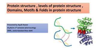
  • 1. Protein structure , levels of protein structure , Domains, Motifs & Folds in protein structure Presented by Aquib Naseer M pharm 2nd semester pharmacology SPER , Jamia Hamdard New delhi
  • 2. Contents : • Introduction. • Amino acids as fundamental units of protein. • Levels of protein structure : 1. Primary level. 2. Secondary level. 3. Tertiary level. 4. Quaternary • Folds. • Motifs. • Domains.
  • 3. Introduction : Proteins are an important class of biological macromolecules which are the polymers of amino acids. • Biochemists have distinguished several levels of structural organization of proteins. They are: – Primary structure – Secondary structure – Tertiary structure – Quaternary structure
  • 4. • Methods for Determining Atomic Structures : 1. X-ray crystallography : X-ray crystallography is the most common tool used to determine protein structure. It provides high resolution of the structure but it does not give information about protein's conformational flexibility. 2. NMR :Protein NMR gives comparatively lower resolution of protein structure. It is limited to smaller proteins. However, it can provide information about conformational changes of a protein in solution. 3. Cryogenic electron microscopy :Cryogenic electron microscopy (cryo-EM) can give information about both a protein's tertiary and quaternary structure. It is particularly well-suited to large proteins and symmetrical complexes of protein subunits. 4. Dual polarisation interferometry :Dual polarisation interferometry provides complimentary information about surface captured proteins. It assists in determining structure and conformation changes over time.
  • 5. The building blocks of proteins are amino acids, which are small organic molecules that consist of an alpha (central) carbon atom linked to an amino group, a carboxyl group, a hydrogen atom, and a variable component called a side chain. Within a protein, multiple amino acids are linked together by peptide bonds (amide bonds between the -NH2 of one amino acid and the -COOH of another.), thereby forming a long chain. Peptide bonds are formed by a biochemical reaction that extracts a water molecule as it joins the amino group of one amino acid to the carboxyl group of a neighboring amino acid. The linear sequence of amino acids within a protein is considered the primary structure of the protein What Are Proteins Made Of ?
  • 6.
  • 7. v
  • 9.
  • 10. Sequences with fewer than 50 amino acids are generally referred to as peptides, while the terms protein or polypeptide are used for longer sequences. Charged amino acid side chains can form ionic bonds, and polar amino acids are capable of forming hydrogen bonds. Hydrophobic side chains interact with each other via weak van der Waals interactions. The vast majority of bonds formed by these side chains are noncovalent. The chemistry of amino acid side chains is critical to protein structure because these side chains can bond with one another to hold a length of protein in a certain shape or conformation. Proteins are built from a set of only twenty amino acids, each of which has a unique side chain.
  • 11. Levels of protein structure : 4 levels of protein structure
  • 12. Protein structureis hierarchical Quaternary ( multimeric organisation ) Tertiary ( long-range folding ) Secondary ( local folding ) Primary ( sequence ) 12
  • 13. 1. Primary structure of proteins : Often, post-translational modifications, such as glycosylation or phosphorylation, occur which are necessary for the biological function of the protein. Proteins are synthesized by a series of steps called transcription (the use of a DNA strand to make a complimentary messenger RNA strand - mRNA) and translation (the mRNA sequence is used as a template to guide the synthesis of the chain of amino acids which make up the protein). The amino acid sequence of a protein is encoded in DNA. Simple amino acid chain sequence makes up the primary structure of the protein.
  • 14.
  • 15. Importance of primary structure :-  To predict secondary & tertiary structures from sequence homologies with related proteins  structure prediction.  Many genetic diseases result from abnormal amino acid sequences.  To understand the molecular mechanism of action of proteins.  To trace evolutionary paths. Methods of aminoacid sequencedetermination:-  End group analysis – Edman degradation  Gene sequencing method 15
  • 16. Folds in proteins : Molecular chaperones are a class of proteins that aid in the correct folding of other proteins . It is the physical process by which a polypeptide folds into its characteristic and functional three-dimensional structure from random coil. Each protein exists as an unfolded polypeptide or random coil when translated from a sequence of mRNA to a linear chain of amino acids. This polypeptide lacks any stable (long-lasting) three-dimensional structure . As the polypeptide chain is being synthesized by a ribosome, the linear chain begins to fold into its three-dimensional structure by molecular chaperones. Folding begins to occur even during translation of the polypeptide chain. Amino acids interact with each other to produce a well-defined three-dimensional structure, the folded protein, known as the native state.
  • 17. Protein before and after folding.
  • 18. Role of folds in different disease pathology : At least eight neurodegenerative disorders are caused by an expanded polyglutamine tract in the various disease proteins, including Huntington’s disease and spinocerebellar ataxia type 3. Polyglutamine expansion causes the disease protein to misfold and aggregate, ultimately leading to neuronal death Variants of the serpin (superfamily of proteins), antitrypsin, misfold and accumulate in the endoplasmic reticulum of hepatocyte cells, leading to a plasma deficiency and hepatocyte damage due to the accumulation of aggregated protein, leading to cirrhosis and emphysema. We study two protein families involved in misfolding disorders: the serine proteinase inhibitor (serpin) and polyglutamine family of proteins. Most proteins have no trouble folding quickly and efficiently to their native state. However, an increasing number of diseases such as emphysema, Alzheimer’s and Huntington’s disease are associated with the failure of proteins to fold correctly.
  • 19. 2. Secondary structure : • The primary structure of a protein — its amino acid sequence — drives the folding and intramolecular bonding of the linear amino acid chain, which ultimately determines the protein's unique three-dimensional shape. • Hydrogen bonding between amino groups and carboxyl groups in neighboring regions of the protein chain sometimes causes certain patterns of folding to occur.
  • 20. • Linus Pauling proposed some essential features of peptide units and polypeptide backbone. They are: – The amide group is rigid and planar as a result of resonance. So rotation about C-N bond is not feasible. – Rotation can take place only about N- Cα and Cα – C bonds. – Trans-configuration is more stable than cis- for R groups at Cα • From these conclusions Pauling postulated 2 ordered structures : • α helix and • β sheet • These stable folding patterns make up the secondary structure of a protein.
  • 21. ALPHA HELIX : • Spiral structure • Tightly packed, coiled polypeptide backbone core. • Side chain extend outwards • Stabilized by H bonding b/w carbonyl oxygen and amide hydrogen. • Amino acids per turn – 3.6 • Pitch is 5.4 A • Alpha helical segments are found in many globular proteins like myoglobins, troponin- C etc.
  • 22. BETA PLEATED SHEET : • Formed when 2 or more polypeptides line up side by side. • Individual polypeptide - β strand • Each β strand is fully extended. • They are stabilized by H bond b/w N-H and carbonyl grps of adjacent chains. •BETA PLEATED SHEET • 2 types • Parallel Anti -Parallel • N C N C • N C N C
  • 25.
  • 27. Tertiary structure : Hydrophilic amino acids that can hydrogen-bond to water are at the surface of soluble proteins. In soluble proteins, hydrophobic side chains are found in the interior of a protein; thus, domains pack together so as to minimize the exposure of hydrophobic side chains to a water interface. It is formed spontaneously and stabilized both by side chain interactions and, in extracellular proteins, by disulfide bonds. This folding brings distant sequences in a linear polypeptide together into a stable structure. Tertiary structure is the complete three-dimensional (3-D) structure of a polypeptide.
  • 30. Tertiary structure : The most stable structure under any given physiologic condition is called the native conformation of a protein. There are four side chain interactions that stabilize the native conformation. • Hydrophobic interactions: Hydrophobic side chains are repelled by water and forced together at the interior of proteins to escape the aqueous environment. • Van der Waals forces: A nonspecific attraction develops based on the proximity of interacting atoms; if the shape of the side chain allows a good fit between surfaces, an attractive force develops. A poor fit gives either repulsion or no force. Note: Both hydrophobic interactions and the shape of side chains are major factors in determining tertiary structure. • Electrostatic bonds: Oppositely charged side chains can attract each other, forming salt bridges. They also play a role in the binding of substrates and allosteric effectors and in the association of the protein with other protein molecules (see Quaternary Structure, below). In addition, they can bind large amounts of water to solubilize the protein when located on the surface. • Hydrogen bonds: Polar groups can share a partial positive charge between a hydrogen donor and a hydrogen acceptor to form a weak bond.
  • 31. • Based upon their tertiary structure, proteins are often divided into 1. Fibrous types 2. Globular types. • Fibrous proteins, like α-keratin, have elongated rope-like structures that are strong and hydrophobic proteins, having polypeptide chains arranged in long strands or sheets. • Globular proteins, like the plasma proteins and the immunoglobulins, are more spherical and hydrophilic. • The two groups are structurally distinct: fibrous proteins usually consist largely of a single type of secondary structure; globular proteins often contain several types of secondary structure. 2 subtypes of tertiary structure :
  • 32. Quaternary structure of proteins : The quaternary structure of a protein is the association of several protein chains or subunits into a closely packed arrangement. Each of the subunits has its own • Primary , • Secondary , and • Tertiary structure. The subunits are held together by hydrogen bonds and van der Waals forces between nonpolar side chains. The subunits in a quaternary structure must be specifically arranged for the entire protein to function properly. Any alteration in the structure of the subunits or how they are associated causes marked changes in biological activity.
  • 33.
  • 34. Protein Molecular weight Number of subunits Function Alcohal dehydrogenase 80,000 4 Enzymatic reaction in fermentation Aldolase 150,000 4 Enzymatic reaction in glycolysis Fumarase 194,000 4 Enzymatic reaction in citric acid cycle Haemoglobin 65,000 4 Oxygen transport in blood Insulin 11,400 2 Hormone that regulates metabolism of glucose Examples of quaternary structure :
  • 35. Sub units : In structural biology, a protein subunit is a single protein molecule that assembles (or "coassembles") with other protein molecules to form a protein complex. Some naturally occurring proteins have a relatively small number of subunits and therefore described as oligomeric, for example hemoglobin or DNA polymerase. Others may consist of a very large number of subunits and therefore described as multimeric, for example microtubules and other cytoskeleton proteins. The subunits of a multimeric protein may be identical, homologous or totally dissimilar and dedicated to disparate tasks. In some protein assemblies, one subunit may be a "catalytic subunit" that enzymatically catalyzes a reaction, whereas a "regulatory subunit" will facilitate or inhibit the activity.
  • 36. • A subunit is often named with a Greek or Roman letter, and the numbers of this type of subunit in a protein is indicated by a subscript. • For example, ATP synthase has a type of subunit called α. Three of these are present in the ATP synthase molecule, and is therefore designated α3
  • 39. How protein structure determination and drug discovery are interrelated:
  • 40.
  • 41.
  • 42. Motifs (supersecondary structures): • A motif is a recognizable folding pattern involving two or more elements of secondary structure and the connection(s) between them. Or • “The connectivity between secondary structure elements and the type of secondary structure elements involved define the level of structural organization called structural motifs”. • A motif is is simply a folding pattern. • Motifs do not allow us to predict the biological functions: they are found in proteins and enzymes with dissimilar functions. • In proteins, a structural motif describes the connectivity between secondary structural elements.
  • 43. Types of motifs : • A motif can be very simple, such as two elements of secondary structure folded against each other, and represent only a small part of a protein. An example is a β-α-β loop . • A motif can also be a very complex structure involving scores of protein segments folded together, such as the β barrel .
  • 44. Motif mediated protein-protein interactions as drug targets : In addition, numerous oncogenic proteins either contain a motif, or recognise motif interaction sequences for which inhibition is a potential cancer treatment . It has also been recognized that several viruses, e.g., Ebola and Rabies viruses, hijack the cell machinery using modified domain motifs interactions. For instance, Liddle’s, Noonan’s and Usher’s hereditary syndromes can be caused by mutations in the recognition motif (PDZ recognition motif respectively) leading to the deregulation of important signalling pathways. There are several diseases and syndromes related to the disruption of specific DMI (drug mediated interaction) motifs.
  • 45. Domains : • A protein domain is a conserved part of a given protein sequence and (tertiary) structure that can evolve , function, and exist independently of the rest of the protein chain. • Each domain forms a compact three-dimensional structure and often can be independently stable and folded. • A domain usually contains between 40 and 350 amino acids, and it is the modular unit from which many larger proteins are constructed. • The different domains of a protein are often associated with different functions. • E.g the Src protein kinase, which functions in signaling pathways inside vertebrate cells. • This protein has four domains: the SH2 and SH3 domains have regulatory roles, while the two remaining domains are responsible for the kinase catalytic activity.
  • 46. • They explain how proteins can form molecular switches that transmit information throughout cells. • The central core of a domain can be constructed from α helices, from β sheets, or from various combinations of these two fundamental folding elements. Each different combination is known as a protein fold.
  • 47.
  • 48. Predicting drug targets based on protein domains : Most recently, it is reported that there are some interactions between compound substructures and protein domains, and a set of chemical substructures shared by drugs are able to bind to a set of protein domains. Therefore, it is suspected that the specificity of drug–protein interactions is possibly determined by drug– domain interactions even though the drugs do not physically bind to protein domains. It has been found that protein–protein interactions are dominated by domain–domain interactions, which in turn could be used to predict new protein–protein interactions.
  • 50. References : • https://www.nature.com/scitable/topicpage/protein-structure- 14122136 • https://www.particlesciences.com/news/technical- briefs/2009/protein-structure.html • https://www.sciencedirect.com/topics/biochemistry-genetics-and- molecular-biology/tertiary-structure • https://proteinstructures.com/Structure/Structure/protein-fold.html • Leninger’s biochemistry , structure of protein & amino acids.