SlideShare une entreprise Scribd logo
1  sur  47
THE MUSCULAR SYSTEM
3 Types of muscle Tissue ,[object Object],[object Object],[object Object],[object Object],[object Object],[object Object],[object Object],[object Object],[object Object],[object Object],[object Object]
Figure 9.2:  Connective tissue sheaths of skeletal muscle, an organ (b) (a) Bone Perimysium Endomysium Blood vessel Muscle fiber (cell) Fascicle (wrapped by perimysium) Endomysium (between fibers) Epimysium Tendon Epimysium Muscle fiber in middle of  a fascicle Perimysium Blood vessel Endomysium
Skeletal Muscle (organ) ,[object Object],[object Object],[object Object]
Patterns of Arrangement of Fascicles in skeletal Muscles ,[object Object],[object Object]
Pattern of Arrangement, continued ,[object Object],[object Object],[object Object],[object Object]
Patterns of Arrangements of Fascicles in skeletal Muscles Circular arrangement – the orbicularis oris and orbicularis oculi - muscles around the mouth and  eyes,  respectively
Attachment sites for each skeletal muscle Skeletal muscles span joints and they have at least 2 attachment sites – the  ORIGIN  and the  INSERTION The bone that moves (the movable bone) when the skeletal muscle contracts is known as the INSERTION; and the bone that does not move  ( the immovable bone) is the ORIGIN . Hence, when the skeletal muscle contracts, the insertion moves toward  the origin. Example:  Brachioradialis Origin (O)  - lateral supracondylar ridge at  distal end of the humerus Insertion(I) – base of styloid process of radius Refer to tables in the lab manual and in the textbook ( pages 329-379)
Direct Skeletal Muscle Attachments DIRECT ATTACMENTS  - the epimysium of the skeletal Muscle is fused directly to the periosteum
Naming of Skeletal Muscles ,[object Object],[object Object],[object Object],[object Object],[object Object],[object Object],[object Object],[object Object],[object Object],[object Object]
Figure 9.2a:  Connective tissue sheaths of skeletal muscle, p. 283. (a) Bone Perimysium Endomysium Blood vessel Muscle fiber (cell) Fascicle (wrapped by perimysium) Endomysium (between fibers) Epimysium Tendon INDIRECT ATTACHMENT- the  Connective tissue wrappings of the skeletal muscle extends as a tendon or an aponeurosis to anchor the muscle to bone, cartilage or fascia
Figure 9.3a-c :  Microscopic anatomy of a skeletal muscle fiber , p. 285. Nuclei Fiber Nucleus Light I band Dark A band Sarcolemma Mitochondrion H zone (b) Myofibril (a) (c) Thin (actin) filament Thick (myosin)  filament Z disc Z disc
Microscopic Anatomy of Skeletal Muscle Fiber ,[object Object],[object Object],[object Object],[object Object],[object Object],[object Object],[object Object],[object Object],[object Object]
Triad ,[object Object],[object Object],[object Object]
Figure 9.5:  Relationship of the sarcoplasmic reticulum and T tubules to myofibrils of skeletal muscle, p. 288. Myofibril Myofibrils Triad Tubules of  sarcoplasmic  reticulum Sarcolemma Sarcolemma Mitochondrion I band I band A band H zone Z disc Z disc Part of a skeletal  muscle fiber (cell) T tubule Terminal cisterna of the sarcoplasmic  reticulum  M line
The Myofilaments ,[object Object],[object Object],[object Object],[object Object],[object Object],[object Object],[object Object],[object Object],[object Object],[object Object],[object Object],[object Object],300 myosin
Figure 9.4:  Composition of thick and thin filaments, p. 286. (b) (c) (d) (e) (a) Heads Flexible hinge region Tail Myosin head Troponin complex Tropomyosin Actin Thin filament Thick filament Thin filament (actin) Thick filament (myosin) Myosin heads Myosin molecule Portion of a thick filament Portion of a thin filament Longitudinal section of filaments within  one sarcomere of a myofibril Transmission electron micrograph of part  of a sarcomere
Figure 9.11:  Role of ionic calcium in the contraction mechanism, p. 294. (a) (b) (c) (d) Actin Actin Tnl TnT Tropomyosin Myosin binding sites Troponin complex TnC Myosin head Myosin binding site Additional calcium ions bind Additional calcium ions bind to TnC Myosin head Actin Overview Troponin Tropomyosin Myosin head Plane of (a) Plane of (d) + Ca 2+
Sarcomeres ,[object Object],[object Object],[object Object],[object Object],[object Object],[object Object],[object Object],[object Object],[object Object],Z M Z
Figure 9.3c-e:  Microscopic anatomy of a skeletal muscle fiber, p. 285. I band Z disc Z disc I band A band H zone (c) (d) (e) Thin (actin) filament Thick (myosin)  filament Thin (actin) filament Elastic (titin)  filaments Z disc Z disc M line M line Sarcomere Thick (myosin)  filament I band thin filaments only H zone thick filaments only M line thick filaments linked by accessory proteins Outer edge of  A band thick and thin filaments overlap
The sliding Filament Mechanism of Muscle Contraction ,[object Object],[object Object],[object Object],[object Object],[object Object],[object Object],1 2 3
Figure 9.6:  Sliding filament model of contraction , p. 289. A Z Z I I A Z Z A Z Z H 1 2 3
Excitation-Contraction Coupling ,[object Object],[object Object],[object Object],[object Object],[object Object],[object Object]
Figure 9.7a:  The neuromuscular junction, p. 290. (a) Action potential Axon terminal at neuromuscular junction Sarcolemma of the muscle fiber Nucleus Myelinated axon of motor neuron
Figure 9.7:  The neuromuscular junction, p. 290. (a) (b) (c) Axon terminal  of a motor neuron Junctional folds of the sarcolemma at  motor end plate Part of a myofibril Mitochondrion Synaptic cleft T tubule Binding of Ach to receptor opens Na + /K +  channel Acetylcholinesterase Synaptic cleft ACh molecules Fusing synaptic vesicle Synaptic vesicle Acetic acid Choline Axon terminal Action potential Axon terminal at neuromuscular junction Sarcolemma of the muscle fiber Nucleus Na + K + Myelinated axon of motor neuron Ca 2+
Figure 9.7b :  The neuromuscular  junction, p. 290. (b) Axon terminal  of a motor neuron Junctional folds of the sarcolemma at  motor end plate Part of a myofibril Mitochondrion Synaptic cleft T tubule Synaptic vesicle Ca 2+
The Neuromuscular Junction ,[object Object],[object Object],[object Object],[object Object]
Figure 9.13:  A motor unit consists of a motor neuron and all the muscle fibers it innervates , p. 296. (b) (a) Spinal cord Motor neuron cell body Muscle Branching axon to motor unit Muscle fibers Nerve Motor unit 1 Motor unit 2 Muscle fibers Motor neuron axon Axon terminals at neuromuscular junctions
Sequence of events in Excitation-Contraction Coupling ,[object Object],[object Object],[object Object],[object Object],[object Object],[object Object],[object Object]
Figure 9.10:  Excitation-contraction coupling, p. 292. ADP P i Net entry of Na +  initiates an action potential which  is propagated along the sarcolemma and down the T tubules. T tubule Sarcolemma SR tubules (cut) Synaptic cleft Synaptic vesicle Axon terminal ACh ACh ACh Neurotransmitter released  diffuses across the synaptic  cleft and attaches to ACh  Action potential in T tubule activates voltage-sensitive  receptors, which in  turn trigger Ca 2+ release from terminal cisternae of SR into cytosol. Calcium ions bind to troponin; troponin changes shape, removing the blocking action of tropomyosin; actin active sites exposed. Contraction; myosin heads alternately attach to actin and detach, pulling the actin filaments toward the center of the sarcomere; release of energy by ATP hydrolysis powers the cycling process. Removal of Ca 2+  by active transport into the SR after the action potential ends. SR Tropomyosin blockage restored, blocking myosin binding sites onactin;  contraction ends and  muscle fiber relaxes. Ca 2+ Ca 2+ Ca 2+ Ca 2+ Ca 2+ Ca 2+ Ca 2+ Ca 2+ Ca 2+ Ca 2+ 1 2 6 5 4 3
Sequence of events –continued ,[object Object],[object Object],[object Object],[object Object],[object Object],[object Object]
Figure 9.12:  The cross bridge cycle, p. 295. ATP ADP ADP ATP hydrolysis ADP ATP P i P i Myosin head (high-energy configuration) Myosin head attaches to the actin myofilament, forming a cross bridge. Thin filament As ATP is split into ADP and P i , the myosin head is energized (cocked into the high-energy conformation). Inorganic phosphate (P i ) generated in theprevious  contraction cycle is released, initiating the power (working) stroke. The myosin head pivots and bends as it pulls on the actin filament, sliding it toward the M line. Then ADP is released. Myosin head (low-energy configuration) Thick filament A s new ATP attaches to the myosin head, the link between Myosin and actin weakens, and the cross bridge detaches. 1 2 3 4
Factors that affect the strength/force of skeletal muscle contraction ,[object Object],[object Object],[object Object],[object Object],[object Object]
Figure 9.21:  Factors influencing force, velocity, and duration of skeletal muscle contraction, p. 304. (a) (b) (c) Increased contractile force Large number of muscle fibers activated Large muscle fibers Asynchronous tetanic contractions Muscle and sarcomere length slightly over 100% of resting length Predominance of fast glycolytic (fatigable) fibers Predominance of slow oxidative (fatigue-resistant) fibers Small load Increased contractile velocity Increased contractile duration
Length-Tension Relationship
2  Main Categories of Skeletal Muscle Contraction ,[object Object],[object Object],[object Object],[object Object],[object Object]
Figure 9.20 :  Methods of regenerating ATP during muscle activity , p. 303. Creatine Energy source: CP Oxygen use: None Products: 1 ATP per CP, creatine Duration of energy provision: 15s (a) Direct phosphorylation [coupled reaction of creatine phosphate (CP) and ADP] (b) Anaerobic mechanism (glycolysis and lactic acid formation) (c) Aerobic mechanism (aerobic cellular  respiration) Energy source: glucose Oxygen use: None Products: 2 ATP per glucose, lactic acid Duration of energy provision: 30–60 s. Energy source: glucose; pyruvic acid; free  fatty acids from adipose tissue; amino acids  from protein catabolism Oxygen use: Required Products: 38 ATP per glucose, CO 2 , H 2 O Duration of energy provision: Hours O 2 O 2 ATP ATP net gain Glucose (from glycogen breakdown or delivered from blood) Pyruvic acid Glycolysis in cytosol Glucose (from glycogen breakdown or delivered from blood) Pyruvic acid 2 ATP net gain per glucose 38 Lactic acid Released to blood O 2 H 2 O O 2 Fatty acids Amino acids CP ADP Aerobic respiration in mitochondria CO 2
Sources of ATP to support skeletal muscle contraction ,[object Object],[object Object],[object Object],[object Object],[object Object],[object Object],[object Object],[object Object],[object Object],[object Object]
3 Skeletal Muscle Fiber Types ,[object Object],[object Object],[object Object],[object Object],[object Object],[object Object],[object Object],[object Object],[object Object],[object Object]
Structural and Functional Characteristics of the 3 Types  of Skeletal Muscle Fibers ,[object Object],[object Object],[object Object],[object Object],[object Object],[object Object],[object Object],[object Object],[object Object],[object Object],[object Object],[object Object],[object Object],[object Object],[object Object]
Effect of Exercise on Skeletal Muscles ,[object Object],[object Object],[object Object],[object Object],[object Object],[object Object],[object Object],[object Object],[object Object],[object Object],[object Object]
Figure 9.25:  Innervation of smooth muscle , p. 311. Smooth muscle cell Mucosa Varicosities Autonomic  nerve fiber Varicosity Synaptic vesicles Mitochondrion Submucosa Serosa Muscularis externa
Smooth Muscle ,[object Object],[object Object],[object Object],[object Object],[object Object],[object Object],[object Object],[object Object],[object Object],[object Object],[object Object]
Excitation-Contraction Coupling in Smooth Muscle ,[object Object],[object Object],[object Object],[object Object]
Figure 9.27:  Sequence of events in excitation-contraction coupling of smooth muscle, p. 313. ATP P i P i P i P i ADP Calcium ions (Ca 2+ ) enter the cytosol from the ECF or from the scant SR.  Ca 2+  binds to and activates calmodulin. Ca 2+ Ca 2+ Ca 2+ Sarcoplasmic reticulum Plasma membrane Activated calmodulin activates the myosin light chain kinase enzymes. Inactive calmodulin Activated calmodulin Inactive kinase Activated kinase Extracellular fluid Cytoplasm The activated kinase  enzyme catalyzes transfer  of phosphate to myosin  heads, activating the myosin head ATPases. Phosphorylated myosin  heads form cross bridges  with actin of the thin  filaments and shortening occurs. Inactive myosin molecule Activated (phosphorylated) myosin molecule Thin myofilament Thick filament Cross bridge activity ends  when phosphate is removed  from the myosin heads by  phosphorylase enzymes and  intracellular Ca 2+  levels fall. 1 2 3 4 5 6
Sequence of events in the mechanism of Smooth Muscle Contraction ,[object Object],[object Object],[object Object],[object Object],[object Object],[object Object]
2 Types of Smooth Muscle ,[object Object],[object Object],[object Object],[object Object],[object Object],[object Object],[object Object],[object Object],[object Object],[object Object],[object Object],[object Object],[object Object]

Contenu connexe

Tendances

4. smooth muscle tissue
4.  smooth muscle tissue4.  smooth muscle tissue
4. smooth muscle tissue
Michael Wrock
 
Histology slide guide
Histology slide guideHistology slide guide
Histology slide guide
MUBOSScz
 
The Axial Skeleton & Appendicular Skeleton
The Axial Skeleton & Appendicular SkeletonThe Axial Skeleton & Appendicular Skeleton
The Axial Skeleton & Appendicular Skeleton
Linda Langevoort
 
Chapter 1 Introduction to Human Anatomy and physiology
Chapter 1   Introduction to Human Anatomy and physiologyChapter 1   Introduction to Human Anatomy and physiology
Chapter 1 Introduction to Human Anatomy and physiology
sgossett5757
 
2014 muscular and skeletal system
2014 muscular and skeletal system2014 muscular and skeletal system
2014 muscular and skeletal system
Jenny Dixon
 
Ch09 a. muscles tissue
Ch09 a. muscles tissueCh09 a. muscles tissue
Ch09 a. muscles tissue
TheSlaps
 

Tendances (20)

4. smooth muscle tissue
4.  smooth muscle tissue4.  smooth muscle tissue
4. smooth muscle tissue
 
Human Skeletal System
Human Skeletal SystemHuman Skeletal System
Human Skeletal System
 
Muscle
MuscleMuscle
Muscle
 
Histology slide guide
Histology slide guideHistology slide guide
Histology slide guide
 
Muscle tissues
Muscle tissuesMuscle tissues
Muscle tissues
 
Lec 1. introduction to muscle tissue
Lec 1. introduction to muscle tissueLec 1. introduction to muscle tissue
Lec 1. introduction to muscle tissue
 
Skeletal system
Skeletal systemSkeletal system
Skeletal system
 
Muscular system Physiology
Muscular system PhysiologyMuscular system Physiology
Muscular system Physiology
 
Human anatomy and physiology parts 2
Human anatomy and physiology parts 2Human anatomy and physiology parts 2
Human anatomy and physiology parts 2
 
Cartilage Histology
Cartilage HistologyCartilage Histology
Cartilage Histology
 
Muscles
MusclesMuscles
Muscles
 
The Axial Skeleton & Appendicular Skeleton
The Axial Skeleton & Appendicular SkeletonThe Axial Skeleton & Appendicular Skeleton
The Axial Skeleton & Appendicular Skeleton
 
Chapter 1 Introduction to Human Anatomy and physiology
Chapter 1   Introduction to Human Anatomy and physiologyChapter 1   Introduction to Human Anatomy and physiology
Chapter 1 Introduction to Human Anatomy and physiology
 
Muscles
MusclesMuscles
Muscles
 
Know about Muscular tissue
Know about Muscular tissueKnow about Muscular tissue
Know about Muscular tissue
 
skeleton of human body, skeletal system of human body, skeleton anatomy, intr...
skeleton of human body, skeletal system of human body, skeleton anatomy, intr...skeleton of human body, skeletal system of human body, skeleton anatomy, intr...
skeleton of human body, skeletal system of human body, skeleton anatomy, intr...
 
MUSCLE PHYSIOLOGY 1
MUSCLE PHYSIOLOGY 1MUSCLE PHYSIOLOGY 1
MUSCLE PHYSIOLOGY 1
 
2014 muscular and skeletal system
2014 muscular and skeletal system2014 muscular and skeletal system
2014 muscular and skeletal system
 
Muscle Tissue
Muscle TissueMuscle Tissue
Muscle Tissue
 
Ch09 a. muscles tissue
Ch09 a. muscles tissueCh09 a. muscles tissue
Ch09 a. muscles tissue
 

En vedette

Skeletal Muscle Tissue And Organization
Skeletal  Muscle  Tissue And  OrganizationSkeletal  Muscle  Tissue And  Organization
Skeletal Muscle Tissue And Organization
raj kumar
 
Muscular system
Muscular systemMuscular system
Muscular system
sdzuris
 
Muscular system
Muscular systemMuscular system
Muscular system
sitesj
 
Muscles classification
Muscles classificationMuscles classification
Muscles classification
Rajesh Goit
 
Mechanism Of Muscle Contraction&Neural Control
Mechanism Of Muscle Contraction&Neural ControlMechanism Of Muscle Contraction&Neural Control
Mechanism Of Muscle Contraction&Neural Control
raj kumar
 

En vedette (20)

Skeletal Muscle Tissue And Organization
Skeletal  Muscle  Tissue And  OrganizationSkeletal  Muscle  Tissue And  Organization
Skeletal Muscle Tissue And Organization
 
Lecture8
Lecture8Lecture8
Lecture8
 
Muscular system
Muscular systemMuscular system
Muscular system
 
Muscular system
Muscular systemMuscular system
Muscular system
 
Muscular system
Muscular systemMuscular system
Muscular system
 
Lecture 6.dr hameed alsarraf
Lecture 6.dr hameed alsarrafLecture 6.dr hameed alsarraf
Lecture 6.dr hameed alsarraf
 
The muscle system disorder
The muscle system disorder The muscle system disorder
The muscle system disorder
 
General principles of skeletal muscle
General principles of skeletal muscleGeneral principles of skeletal muscle
General principles of skeletal muscle
 
Myology: The Study of Muscles (Anatomy: 1st Semester Lecture 5)
Myology: The Study of Muscles (Anatomy: 1st Semester Lecture 5)Myology: The Study of Muscles (Anatomy: 1st Semester Lecture 5)
Myology: The Study of Muscles (Anatomy: 1st Semester Lecture 5)
 
Muscular system
Muscular systemMuscular system
Muscular system
 
Chapter 10&11
Chapter 10&11Chapter 10&11
Chapter 10&11
 
part7b2
part7b2part7b2
part7b2
 
Muscles classification
Muscles classificationMuscles classification
Muscles classification
 
Metabolism in Different Organs
Metabolism in Different OrgansMetabolism in Different Organs
Metabolism in Different Organs
 
Dr. B Ch 09_lecture_presentation
Dr. B Ch 09_lecture_presentationDr. B Ch 09_lecture_presentation
Dr. B Ch 09_lecture_presentation
 
Mechanism Of Muscle Contraction&Neural Control
Mechanism Of Muscle Contraction&Neural ControlMechanism Of Muscle Contraction&Neural Control
Mechanism Of Muscle Contraction&Neural Control
 
Chapter 8 the muscular system
Chapter 8  the muscular systemChapter 8  the muscular system
Chapter 8 the muscular system
 
Muscular System
Muscular SystemMuscular System
Muscular System
 
Muscle contraction
Muscle contractionMuscle contraction
Muscle contraction
 
Creatine metabolism
Creatine metabolism Creatine metabolism
Creatine metabolism
 

Similaire à Muscles 2

The muscular system
The muscular system The muscular system
The muscular system
alijum3a
 
Lecture 1 muscle tissue
Lecture 1   muscle tissueLecture 1   muscle tissue
Lecture 1 muscle tissue
missazyaziz
 
Muscle funccellularlevel animal systems
Muscle funccellularlevel animal systemsMuscle funccellularlevel animal systems
Muscle funccellularlevel animal systems
Yukti Sharma
 
MUSCLE - 15903 by SSG muscle anatomy of upper limb lower limb and all
MUSCLE - 15903 by SSG  muscle anatomy of upper limb lower limb and allMUSCLE - 15903 by SSG  muscle anatomy of upper limb lower limb and all
MUSCLE - 15903 by SSG muscle anatomy of upper limb lower limb and all
ArchanaVekariya
 

Similaire à Muscles 2 (20)

The muscular system
The muscular system The muscular system
The muscular system
 
Lecture 1 muscle tissue
Lecture 1   muscle tissueLecture 1   muscle tissue
Lecture 1 muscle tissue
 
Muscular System.ppt
Muscular System.pptMuscular System.ppt
Muscular System.ppt
 
3 Types of Muscle Tissue
3 Types of Muscle Tissue 3 Types of Muscle Tissue
3 Types of Muscle Tissue
 
Muscle funccellularlevel animal systems
Muscle funccellularlevel animal systemsMuscle funccellularlevel animal systems
Muscle funccellularlevel animal systems
 
Muscle Ppt
Muscle PptMuscle Ppt
Muscle Ppt
 
WEEK 7.pptx
WEEK 7.pptxWEEK 7.pptx
WEEK 7.pptx
 
Histology of muscle
Histology of muscle Histology of muscle
Histology of muscle
 
MUSCLE - 15903 by SSG muscle anatomy of upper limb lower limb and all
MUSCLE - 15903 by SSG  muscle anatomy of upper limb lower limb and allMUSCLE - 15903 by SSG  muscle anatomy of upper limb lower limb and all
MUSCLE - 15903 by SSG muscle anatomy of upper limb lower limb and all
 
Muscles
MusclesMuscles
Muscles
 
Ana-physi 6.ppt
Ana-physi 6.pptAna-physi 6.ppt
Ana-physi 6.ppt
 
Muscle physiology ,types of muscles: striated ,non striated and cardiac. ultr...
Muscle physiology ,types of muscles: striated ,non striated and cardiac. ultr...Muscle physiology ,types of muscles: striated ,non striated and cardiac. ultr...
Muscle physiology ,types of muscles: striated ,non striated and cardiac. ultr...
 
Skeletal muscle: Form & Function
Skeletal muscle: Form & FunctionSkeletal muscle: Form & Function
Skeletal muscle: Form & Function
 
MUSCLE physiology assignment and good notes for exam
MUSCLE physiology assignment and good notes for examMUSCLE physiology assignment and good notes for exam
MUSCLE physiology assignment and good notes for exam
 
Muscular system
Muscular system Muscular system
Muscular system
 
STRUCTURE OF MUSCLE.pptx
STRUCTURE OF MUSCLE.pptxSTRUCTURE OF MUSCLE.pptx
STRUCTURE OF MUSCLE.pptx
 
Histology of Muscle tissue
Histology of Muscle tissueHistology of Muscle tissue
Histology of Muscle tissue
 
best muscle tissue.pptxghfght5fvyytht6gyg
best muscle tissue.pptxghfght5fvyytht6gygbest muscle tissue.pptxghfght5fvyytht6gyg
best muscle tissue.pptxghfght5fvyytht6gyg
 
Introduction to Muscle & Its Types
Introduction to Muscle & Its Types Introduction to Muscle & Its Types
Introduction to Muscle & Its Types
 
MUSCLES.pptx
MUSCLES.pptxMUSCLES.pptx
MUSCLES.pptx
 

Plus de Abay Alem

The endocrine system2
The endocrine system2The endocrine system2
The endocrine system2
Abay Alem
 
Ch 13 the peripheral nervous system and nervous activity
Ch 13   the peripheral nervous system and nervous activityCh 13   the peripheral nervous system and nervous activity
Ch 13 the peripheral nervous system and nervous activity
Abay Alem
 
Skeletal system
Skeletal systemSkeletal system
Skeletal system
Abay Alem
 
Integumentary system
Integumentary systemIntegumentary system
Integumentary system
Abay Alem
 
The human body an orientation
The human body an orientationThe human body an orientation
The human body an orientation
Abay Alem
 
Chemical level
Chemical levelChemical level
Chemical level
Abay Alem
 

Plus de Abay Alem (14)

The endocrine system2
The endocrine system2The endocrine system2
The endocrine system2
 
Sss5
Sss5Sss5
Sss5
 
Cns bss
Cns bssCns bss
Cns bss
 
Cns bs2
Cns bs2Cns bs2
Cns bs2
 
Cns 2
Cns 2Cns 2
Cns 2
 
Ch 13 the peripheral nervous system and nervous activity
Ch 13   the peripheral nervous system and nervous activityCh 13   the peripheral nervous system and nervous activity
Ch 13 the peripheral nervous system and nervous activity
 
Skeletal system
Skeletal systemSkeletal system
Skeletal system
 
Jointsrev2
Jointsrev2Jointsrev2
Jointsrev2
 
Jointsrev
JointsrevJointsrev
Jointsrev
 
Integumentary system
Integumentary systemIntegumentary system
Integumentary system
 
Tissues
TissuesTissues
Tissues
 
The human body an orientation
The human body an orientationThe human body an orientation
The human body an orientation
 
Chemical level
Chemical levelChemical level
Chemical level
 
Lecture ce4
Lecture ce4Lecture ce4
Lecture ce4
 

Muscles 2

  • 2.
  • 3. Figure 9.2: Connective tissue sheaths of skeletal muscle, an organ (b) (a) Bone Perimysium Endomysium Blood vessel Muscle fiber (cell) Fascicle (wrapped by perimysium) Endomysium (between fibers) Epimysium Tendon Epimysium Muscle fiber in middle of a fascicle Perimysium Blood vessel Endomysium
  • 4.
  • 5.
  • 6.
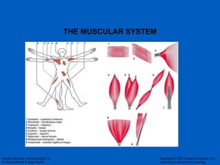
  • 7. Patterns of Arrangements of Fascicles in skeletal Muscles Circular arrangement – the orbicularis oris and orbicularis oculi - muscles around the mouth and eyes, respectively
  • 8. Attachment sites for each skeletal muscle Skeletal muscles span joints and they have at least 2 attachment sites – the ORIGIN and the INSERTION The bone that moves (the movable bone) when the skeletal muscle contracts is known as the INSERTION; and the bone that does not move ( the immovable bone) is the ORIGIN . Hence, when the skeletal muscle contracts, the insertion moves toward the origin. Example: Brachioradialis Origin (O) - lateral supracondylar ridge at distal end of the humerus Insertion(I) – base of styloid process of radius Refer to tables in the lab manual and in the textbook ( pages 329-379)
  • 9. Direct Skeletal Muscle Attachments DIRECT ATTACMENTS - the epimysium of the skeletal Muscle is fused directly to the periosteum
  • 10.
  • 11. Figure 9.2a: Connective tissue sheaths of skeletal muscle, p. 283. (a) Bone Perimysium Endomysium Blood vessel Muscle fiber (cell) Fascicle (wrapped by perimysium) Endomysium (between fibers) Epimysium Tendon INDIRECT ATTACHMENT- the Connective tissue wrappings of the skeletal muscle extends as a tendon or an aponeurosis to anchor the muscle to bone, cartilage or fascia
  • 12. Figure 9.3a-c : Microscopic anatomy of a skeletal muscle fiber , p. 285. Nuclei Fiber Nucleus Light I band Dark A band Sarcolemma Mitochondrion H zone (b) Myofibril (a) (c) Thin (actin) filament Thick (myosin) filament Z disc Z disc
  • 13.
  • 14.
  • 15. Figure 9.5: Relationship of the sarcoplasmic reticulum and T tubules to myofibrils of skeletal muscle, p. 288. Myofibril Myofibrils Triad Tubules of sarcoplasmic reticulum Sarcolemma Sarcolemma Mitochondrion I band I band A band H zone Z disc Z disc Part of a skeletal muscle fiber (cell) T tubule Terminal cisterna of the sarcoplasmic reticulum M line
  • 16.
  • 17. Figure 9.4: Composition of thick and thin filaments, p. 286. (b) (c) (d) (e) (a) Heads Flexible hinge region Tail Myosin head Troponin complex Tropomyosin Actin Thin filament Thick filament Thin filament (actin) Thick filament (myosin) Myosin heads Myosin molecule Portion of a thick filament Portion of a thin filament Longitudinal section of filaments within one sarcomere of a myofibril Transmission electron micrograph of part of a sarcomere
  • 18. Figure 9.11: Role of ionic calcium in the contraction mechanism, p. 294. (a) (b) (c) (d) Actin Actin Tnl TnT Tropomyosin Myosin binding sites Troponin complex TnC Myosin head Myosin binding site Additional calcium ions bind Additional calcium ions bind to TnC Myosin head Actin Overview Troponin Tropomyosin Myosin head Plane of (a) Plane of (d) + Ca 2+
  • 19.
  • 20. Figure 9.3c-e: Microscopic anatomy of a skeletal muscle fiber, p. 285. I band Z disc Z disc I band A band H zone (c) (d) (e) Thin (actin) filament Thick (myosin) filament Thin (actin) filament Elastic (titin) filaments Z disc Z disc M line M line Sarcomere Thick (myosin) filament I band thin filaments only H zone thick filaments only M line thick filaments linked by accessory proteins Outer edge of A band thick and thin filaments overlap
  • 21.
  • 22. Figure 9.6: Sliding filament model of contraction , p. 289. A Z Z I I A Z Z A Z Z H 1 2 3
  • 23.
  • 24. Figure 9.7a: The neuromuscular junction, p. 290. (a) Action potential Axon terminal at neuromuscular junction Sarcolemma of the muscle fiber Nucleus Myelinated axon of motor neuron
  • 25. Figure 9.7: The neuromuscular junction, p. 290. (a) (b) (c) Axon terminal of a motor neuron Junctional folds of the sarcolemma at motor end plate Part of a myofibril Mitochondrion Synaptic cleft T tubule Binding of Ach to receptor opens Na + /K + channel Acetylcholinesterase Synaptic cleft ACh molecules Fusing synaptic vesicle Synaptic vesicle Acetic acid Choline Axon terminal Action potential Axon terminal at neuromuscular junction Sarcolemma of the muscle fiber Nucleus Na + K + Myelinated axon of motor neuron Ca 2+
  • 26. Figure 9.7b : The neuromuscular junction, p. 290. (b) Axon terminal of a motor neuron Junctional folds of the sarcolemma at motor end plate Part of a myofibril Mitochondrion Synaptic cleft T tubule Synaptic vesicle Ca 2+
  • 27.
  • 28. Figure 9.13: A motor unit consists of a motor neuron and all the muscle fibers it innervates , p. 296. (b) (a) Spinal cord Motor neuron cell body Muscle Branching axon to motor unit Muscle fibers Nerve Motor unit 1 Motor unit 2 Muscle fibers Motor neuron axon Axon terminals at neuromuscular junctions
  • 29.
  • 30. Figure 9.10: Excitation-contraction coupling, p. 292. ADP P i Net entry of Na + initiates an action potential which is propagated along the sarcolemma and down the T tubules. T tubule Sarcolemma SR tubules (cut) Synaptic cleft Synaptic vesicle Axon terminal ACh ACh ACh Neurotransmitter released diffuses across the synaptic cleft and attaches to ACh Action potential in T tubule activates voltage-sensitive receptors, which in turn trigger Ca 2+ release from terminal cisternae of SR into cytosol. Calcium ions bind to troponin; troponin changes shape, removing the blocking action of tropomyosin; actin active sites exposed. Contraction; myosin heads alternately attach to actin and detach, pulling the actin filaments toward the center of the sarcomere; release of energy by ATP hydrolysis powers the cycling process. Removal of Ca 2+ by active transport into the SR after the action potential ends. SR Tropomyosin blockage restored, blocking myosin binding sites onactin; contraction ends and muscle fiber relaxes. Ca 2+ Ca 2+ Ca 2+ Ca 2+ Ca 2+ Ca 2+ Ca 2+ Ca 2+ Ca 2+ Ca 2+ 1 2 6 5 4 3
  • 31.
  • 32. Figure 9.12: The cross bridge cycle, p. 295. ATP ADP ADP ATP hydrolysis ADP ATP P i P i Myosin head (high-energy configuration) Myosin head attaches to the actin myofilament, forming a cross bridge. Thin filament As ATP is split into ADP and P i , the myosin head is energized (cocked into the high-energy conformation). Inorganic phosphate (P i ) generated in theprevious contraction cycle is released, initiating the power (working) stroke. The myosin head pivots and bends as it pulls on the actin filament, sliding it toward the M line. Then ADP is released. Myosin head (low-energy configuration) Thick filament A s new ATP attaches to the myosin head, the link between Myosin and actin weakens, and the cross bridge detaches. 1 2 3 4
  • 33.
  • 34. Figure 9.21: Factors influencing force, velocity, and duration of skeletal muscle contraction, p. 304. (a) (b) (c) Increased contractile force Large number of muscle fibers activated Large muscle fibers Asynchronous tetanic contractions Muscle and sarcomere length slightly over 100% of resting length Predominance of fast glycolytic (fatigable) fibers Predominance of slow oxidative (fatigue-resistant) fibers Small load Increased contractile velocity Increased contractile duration
  • 36.
  • 37. Figure 9.20 : Methods of regenerating ATP during muscle activity , p. 303. Creatine Energy source: CP Oxygen use: None Products: 1 ATP per CP, creatine Duration of energy provision: 15s (a) Direct phosphorylation [coupled reaction of creatine phosphate (CP) and ADP] (b) Anaerobic mechanism (glycolysis and lactic acid formation) (c) Aerobic mechanism (aerobic cellular respiration) Energy source: glucose Oxygen use: None Products: 2 ATP per glucose, lactic acid Duration of energy provision: 30–60 s. Energy source: glucose; pyruvic acid; free fatty acids from adipose tissue; amino acids from protein catabolism Oxygen use: Required Products: 38 ATP per glucose, CO 2 , H 2 O Duration of energy provision: Hours O 2 O 2 ATP ATP net gain Glucose (from glycogen breakdown or delivered from blood) Pyruvic acid Glycolysis in cytosol Glucose (from glycogen breakdown or delivered from blood) Pyruvic acid 2 ATP net gain per glucose 38 Lactic acid Released to blood O 2 H 2 O O 2 Fatty acids Amino acids CP ADP Aerobic respiration in mitochondria CO 2
  • 38.
  • 39.
  • 40.
  • 41.
  • 42. Figure 9.25: Innervation of smooth muscle , p. 311. Smooth muscle cell Mucosa Varicosities Autonomic nerve fiber Varicosity Synaptic vesicles Mitochondrion Submucosa Serosa Muscularis externa
  • 43.
  • 44.
  • 45. Figure 9.27: Sequence of events in excitation-contraction coupling of smooth muscle, p. 313. ATP P i P i P i P i ADP Calcium ions (Ca 2+ ) enter the cytosol from the ECF or from the scant SR. Ca 2+ binds to and activates calmodulin. Ca 2+ Ca 2+ Ca 2+ Sarcoplasmic reticulum Plasma membrane Activated calmodulin activates the myosin light chain kinase enzymes. Inactive calmodulin Activated calmodulin Inactive kinase Activated kinase Extracellular fluid Cytoplasm The activated kinase enzyme catalyzes transfer of phosphate to myosin heads, activating the myosin head ATPases. Phosphorylated myosin heads form cross bridges with actin of the thin filaments and shortening occurs. Inactive myosin molecule Activated (phosphorylated) myosin molecule Thin myofilament Thick filament Cross bridge activity ends when phosphate is removed from the myosin heads by phosphorylase enzymes and intracellular Ca 2+ levels fall. 1 2 3 4 5 6
  • 46.
  • 47.

Notes de l'éditeur

  1. Muscle as a tissue
  2. Muscle fibers = skeletal muscle The last bullet was something she mentioned in the lecture Talked about slide 5, 6 SLIDE 4
  3. 3 rd organ system  muscular system Composed mainly of muscles (organs) Skeletal muscle (organ) Composed of a) skeletal muscle tissue – main component b) connective tissue c) blood vessels d) lymphatic vessels e) nerve fibers Arrangement of skeletal muscle fibers inside the skeletal muscle tissue (which are inside the skeletal muscle) Each skeletal muscle fiber (cell) is wrapped in a delicate connective tissue membrane called the ENDOMYSIUM A group of endomysium-covered skeletal muscle fibers forms a FASCICLE  wrapped by CT membrane called the Perimysium A bundle of perimysium-covered fascicles forms skeletal muscle  wrapped in a coarse CT membrane called epimysium Went to slide 9
  4. Blood vessels, nerve fibers, etc to form the organ, Then wrote down bullets on slide 3
  5. Went back to slide 2
  6. Attachment sites for skeletal muscles  attachment may be DIRECT OR INDIRECT  via tendons Most of the skeletal muscle in the body are attached to bones (or cartilage) indirectly 2 advantages to indirect attachment of skeletal muscles: The rope-like tendons can fit into small spaces to reach the bones Skeletal muscles that course over joints will be protected if tendons run directly over the joint sites  skeletal muscles will be damaged at the joint sites as the joints move
  7. Microscopic arrangement of the skeletal muscle fiber  because this explains the mechanism of skeletal muscle contraction Each skeletal muscle (the organ) consists of skeletal muscle fibers that run the entire length of the skeletal muscle Each skeletal muscle fiber  80% of volume occupied by rod-like structures called MYOFIBRILS (run the entire length of skeletal muscle fibers) MYOFIBRILS are composed of smaller filaments called MYOFILAMENTS 2 types of myofilaments thick filament – thicker than the thin filament thin
  8. 2 types of myofilaments thick filament – thicker than the thin filament; composed of structural proteins called myosin structure of a myosin molecule  a rod-like tail that ends in 2 globular heads  MYOSIN HEADS myosin heads have a binding site for ACTIN; also has binding site for enzyme ATPase that hydrolyzes ATP  ADP + Pi; myosin head itself can act as an ATPase THICK FILAMENTS consists of 300 molecules of myosin molecules arranged such that the tails form the core with the heads exposed thin - composed of 3 types of proteins (actin, tropomyosin, troponin) actin – double helical structure that forms the core of the thin filament; contains binding sites for MYOSIN (binding sites on actin bind to the actin binding sites on myosin) tropomyosin – in a relaxed skeletal muscle (not contracted), tropomyosin spirals about the actin blocking myosin binding sites on actin troponin – a three-complex protein that consists of: 1) troponin C  binds calcium ions 2) troponin subunit that binds to tropomosin (TnT) 3) troponin subunit that binds to actin and inhibits actin  inhibitory troponin (TnI)
  9. In a skeletal muscle fiber, the thick and the thin filaments are arranged in an alternating pattern THICK, THIN, THICK, THIN and so on Which explains the straited appearance of skeletal muscles The thin filaments are anchored/stabilized by proteins called the Z disc or Z lines The distance between 2 successive Z lines is referred to as a SARCOMERE  structural unit of skeletal muscle Shortening of sarcomeres results in skeletal muscle contraction ======================== Components of a sarcomere distance between two z-lines z lines anchor the thin filaments in a sarcomere we know the thin and thick filaments alternate in a sarcomere thick filament is referred to as the “A BAND” in a sarcomere the regions of the thin filaments not overlapping with the A band are called the I bands the region of the A band not overlapping with the thin filaments is called the H zone the line bisecting the H zone is called M line  anchors the A band in a sarcomere I band are located toward the Z lines The H zone is located in the central region of the A band
  10. Sliding fillament mechanism of the skeletal muscle contraction Sliding of the thin filament in sarcomeres toward the M line in the H zones causes shortening of sarcomeres as the Z lines are pulled inward Since sarcomeres run the entire length of the myofibrils, the myofibrils also shorten Since the myofibrils run the entire length of skeletal muscle fibers, the fibers also shorten Since skeletal muscle fibers run the entire length of the skeletal muscle, the organ (skeletal muscle) ALSO SHORTENS.
  11. 10-18 started here
  12. Factors that affect the strength of skeletal muscle contraction  force developed by the skeletal muscle Size of the motor units activated Number of motor units activated when we’re looking at recruitment FREQUENCY of skeletal muscle activation  If action potentials are developed at a higher rate such that the skeletal muscle fibers do not have time to sequester calcium ions into the SR  more and more calcium ions remain in the sarcoplasm resulting in stronger and stronger skeletal muscle contraction  this effect is referred to as TEMPORAL (time) or WAVE SUMMATION Slide 35 after slide 34
  13. Drew a graph of strength of skeletal muscle contraction vs. frequency of activation where both increase until a plateau is reached When the skeletal muscle contraction levels off it is called TETANY The response in contraction of a motor unit to a single action potential is what is referred to as a TWITCH
  14. 4) (from slide 33) Length of the sarcomere before contraction begins… The optimum length of sarcomeres = 2.0 microns – 2.25 microns At these lengths  MAXIMUM contraction of the skeletal muscle because  H zone is present and the activated myosin heads can reach/bind to their sites on actin to initiate sliding of the thin filament into the H zone that is available Sarcomere lengths below the optimum (less than 2.0 micron)… The H zone present is decreased  hence, sliding of thin filaments is reduced (impeded) = less sliding of the thin filaments  less shortening of the sarcomeres  less contraction of the skeletal muscle  less force generated Sarcomere lengths above the optimum (more than 2.25 micron) Ample space in the H zone; however there is little or NO overlap between the A band and the thin filaments. Hence, the activated myosin heads can NOT reach and bind to their sites on actin  NO SLIDING of the thin filaments  NO SHORTENING OF SARCOMERES WILL OCCUR  NO CONTRACTION of skeletal muscle  no force generated
  15. Sources of ATP to support skeletal muscle contraction Stored ATP in skeletal muscle fibers  stored ATP can support ONLY 5 seconds of skeletal muscle activity Creatin phosphate (CP) = unique in muscle cells (especially in skeletal muscle fibers) CP  phosphorylates ADP to give ATP (ADP + CP  ATP + Creatine **where creatine kinase is over the arrow as an enzyme) 3) Catabolism (break down) of glucose in the absence of oxygen is referred to as ANAEROBIC CATABOLISM of glucose (Glycolysis) The anaerobic catabolism of glucose (anaerobic respiration) yields 2 molecules of ATP and pyruvic acid (when O2 absent, pyruvic acid  Lactic acid  contributes to MUSCLE FATIGUE  halts skeletal muscle contraction 4) AEROBIC CATABOLISM OF GLUCOSE (with O2)  36 – 38 molecules of ATP; more efficient in generating ATP to support muscle contraction, BUT it involves MORE steps then anaerobic catabolism of glucose
  16. Based on the table in the book s SLOW oxidative fibers also called RED fibers Fast glycolytic fibers are also called WHITE FIBERS Activities the 3 types of skeletal muscle fibers are suited for Slow oxidative fibers  use aerobic respiration to generate ATP hence, they are fatigue-resistant and suited for endurance-type activities A successful marathon runner possesses more of the slow oxidative fibers Fast oxidative fibers are intermediate fibers that are more efficient and fatigue-resistant compared to glycolytic fibers however, they are less efficient when compared to the slow oxidative fibers Hence, you use fast oxidative fibers in medium length activities such as walking, standing Fast glycolytic fibers  largest, use anaerobic respiration. Hence, lactic acid builds up, causing muscle fatigue fatigable so they are suited for intense but short-lived activities such as weight lifting
  17. Matured skeletal muscle grows by HYPERTROPHY  increase in the size of the skeletal muscle fibers (which are actually cells) in the skeletal muscle Skeletal muscle fibers are MULTINUCLEATE cells and therefore, they lose the ability to undergo mitosis which would have resulted in hyperplasia As skeletal muscle grows by hypertrophy, the larger skeletal muscle generates MORE FORCE = “the bigger the skeletal muscle, the greater the force it generates” Skeletal muscle NOT activated or used will undergo muscle atrophy (DECREASE in skeletal muscle size)  decrease the force generated by the skeletal muscle
  18. Usually found lining tracts in the body (tracts = opening to the exterior) No striations
  19. No troponin, z discs absent, no sarcomeres Skeletal muscle fiber (drew T tubule and terminal cisternae) Smooth Muscle cell (shallow dips called caveolae instead of T tubules)
  20. Excitation-contraction coupling of smooth muscle cells Smooth muscle cells may be activated to contract or inhibited from contracting resulting in RELAXATION Smooth muscle cells are innervated by the AUTONOMIC NERVOUS SYSTEM Sympathetic Nervous System Parasympathetic Nervous system Typically, activation of the sympathetic nervous system will result in smooth muscle contraction (exception: in the bronchioles in the lungs, activation of the sympathetic nervous system actually results in relaxation of the smooth muscle  BRONCHODILATION) The autonomic fibers innervating the smooth muscle cells form DIFFUSE JUNCTIONS  wide spaces between the smooth muscle cells and the ends of the autonomic fibers  the ends of the autonomic fibers appear bulbous  referred to as VARICOSITIES. HENCE a diffuse junction forms between the sarcolemma of the smooth muscle cell and a varicosity of an autonomic fiber Activation of the parasympathetic nervous system will result in RELAXATION of the smooth muscle. 3) Unlike skeletal muscle, smooth muscle can be activated to contract or relax by blood-borne chemicals such as hormones 4) Some smooth muscle cells have intrinsic cells that depolarize spontaneously to stimulate smooth muscle contraction  these cells are called PACEMAKER cells Hence, the smooth muscle has the INTRINSIC ability to contract = pacemaker activity THREE WAYS TO CONTRACT SMOOTH MUSCLE (she went to 45)
  21. SEQUENCE OF EVENTS When smooth muscle is activated to contract by the sympathetic nervous system/chemical/pacemaker activity, depolarization of the sarcolemma causes calcium channels in the sarcolemma to open up and these channels will allow calcium ions in the fluid inside the caveolae to enter into the sarcoplasm of the smooth muscle cell. Entry of calcium ions into the sarcolemma triggers more calcium ions to be released from the poorly developed SR in the smooth muscle cells (TRIADS ARE ABSENT) ** because calcium ions must enter from the extracellular fluid, calcium channel blockers reduce or abolish smooth muscle contraction Since troponin is absent in smooth muscle cells, the increased calcium ions bind to a regulatory protein in the sarcoplasm called CALMODULIN ============== 10/27/10 Excitation-Contraction Coupling in smooth muscle cell step 1: Sarcolemma is depolarized by the pacemaker activity or autonomic fiber activation or by chemicals in blood. step 2: Calcium channels open up in the sarcolemma and Ca+ ions enter the cell from the extracellular fluid in the caveolae. Hence, calcium channel blockers can inhibit smooth muscle contraction Ca+ ions are also released from the SR in the smooth muscle cell **calcium ions to support or initiate smooth muscle contraction come from 2 sources a) extracellular fluid b) from the SR step 3: Calcium ions bind to calmodulin to form Calcium-calmodulin complex step 4: calcium calmodulin complex activates a kinase called MYOSIN LIGHT CHAIN kinase (MLCK) step 5: activated MLCK attaches phosphate to the myosin heads  results in activation of the myosin heads step 6: activated myosin heads bind to their sites on actin (in a smooth muscle cell the myosin binding sites on actin are ALWAYS accessible; although tropomysoin is present, it does NOT block myosin binding sites on actin) step 7: the attached activated myosin heads form CROSSBRIDGES and as the phosphate dissociate from the crossbridge, the crossbridge undergoes a change in orientation termed the POWER STROKE which causes the thin filaments to slide downward along the axis of the smooth muscle  results in contraction of the smooth muscle step 8: cross bridge detachment is caused by the enzyme PHOSPHORYLASE To cause complete relaxion of the smooth muscle Phosphorylase to cause crossbridge detachment Calcium ions are pumped (active transport) into the SR and out into the extracellular fluid Turn off the pacemaker activity or turn off the hormone released or stop activation of the autonomic nervous system  sympathetic nervous system **Activation of the autonomic nervous system may cause smooth muscle relaxation or smooth muscle contraction. In contrast, activation of the motor nuerons ALWAYS cause skeletal muscle contraction
  22. 202 material - Cardiac Muscle Striated  have sarcomeres Highly branchedwith lateral contacts called INTERCALATED DISCS  2 parts Desmosomes and Gap Junctions Cardiac muscle is located in the wall of the heart in the middle layer of the heart wall called the myocardium If you look at the excitation-contraction coupling of cardiac muscle, it is similar to smooth muscle 1) Pacemaker activity 2) Autonomic Nervous system 3) Chemicals In addition, calcium ions enter the cardiac cells to trigger release of more Ca+ from the SR in the cardiac muscle cells Cardiac muscle  similar to skeletal muscle structurally, but similar to smooth muscle in terms of FUNCTION