Encephalitis and other brain infection final (autosaved)
•Télécharger en tant que DOCX, PDF•
0 j'aime•831 vues


Recommandé
Contenu connexe
Tendances
Tendances (20)
Similaire à Encephalitis and other brain infection final (autosaved)
Similaire à Encephalitis and other brain infection final (autosaved) (20)
Dernier
Dernier (20)
Encephalitis and other brain infection final (autosaved)
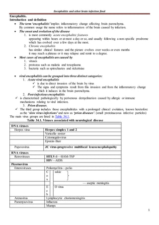
- 1. Encephalitis and other brain infection final 1 Encephalitis. Introduction and definition The term ‘encephalitis’ implies inflammatory change affecting brain parenchyma. By common usage the name refers to inflammation of the brain caused by infection. The onset and evolution of the disease 1. is most commonly acute encephalitic features appearing within hours or at most a day or so, and usually following a non-specific prodrome which has evolved over a few days at the most. 2. Chronic encephalitis has similar clinical features and the picture evolves over weeks or even months it may reach a plateau or it may relapse and remit to a degree. Most cases of encephalitis are caused by 1. viruses 2. protozoa such as malaria and toxoplasma 3. bacteria such as spirochaetes and rickettsiae viral encephalitis can be grouped into three distinct categories: 1. Acute viral encephalitis is due to direct invasion of the brain by virus The signs and symptoms result from this invasion and from the inflammatory change which it induces in the brain parenchyma. 2. Post-infectious encephalitis is characterized pathologically by perivenous demyelination caused by allergic or immune mechanisms relating to viral infection. 3. Prion diseases. The third group includes those encephalitides with a prolonged clinical evolution, known heretofore as the ‘slow virus infections’ and now as ‘prion diseases’. (small proteinaceous infective particles) The main virus groups are listed in Table 34.1. Table 34.1. Viruses associated with neurological disease DNA viruses Herpes virus Herpes simplex 1 and 2 Varicella–zoster Cytomegalovirus Epstein–Barr Papovavirus JC virus-progressive multifocal leucoencephalopathy RNA viruses Retroviruses HTLV-1—HAM-TSP HIV—AIDS Picornovirus Enteroviruses Poliomyelitis—polio C o x sakie — aseptic meningitis E c h O virus Arenavirus Lymphocytic choriomeningitis Paramyxovirus Influenza Mumps
- 2. Encephalitis and other brain infection final 2 Morbillivirus Measles Rhabdovirus Lyssavirus Rabies Bunyavirus California and LaCrosse encephalitis Rift Valley fever Congo–Crimean haemorrhagic fever Retrovirus Orbivirus Colorado tick fever Togavirus Alphavirus Western, Eastern, and Venezuelan equine encephalitis Flavivirus Japanese, Murray Valley, and St Louis encephalitis Central European and Russian spring–summer encephalitis Louping ill Rubivirus Rubella The diagnosis of viral infection of the nervous system Careful attention to historical details meticulous clinical examination Confirmation that a clinical syndrome difficulties 1. demonstration of the presence of the virus or viral antigen in body tissue or fluids 1. the presence of a virus may be due to a co-existing and unrelated infection 2. or a specific antibody response to its presence 2. ↑antibody levels may be persistently raised from a previous infection 3. or the rise may represent an anamnestic generalized immune response to infection 4. There may also be difficulties with inadequate sensitivity of the tests employed 5. problems with cross-reactions that impair specificity 6. and timing the taking of specimens against the progression of the disease Viral identification Virus culture has not been greatly successful in the diagnosis of CNS infections, in large part due to the difficulty of obtaining specimens of neural tissue. It may be possible to cultivate virus from other sources but this is seldom fruitful. Traditionally, the demonstration of a fourfold rise in antibody titre between an acute phase and a convalescent serum sample to a specific virus has been sufficient to confirm the diagnosis. Numerous techniques that employ immunofluorescence and ELISA have greatly increased the sensitivity of diagnosis and the development of monoclonal antibodies has permitted the detection of viral antigen using these techniques. 1. IgM capture ELISA Rapid results can now be obtained 2. Western blotting in which electrophoretic gels use specific antibodies to trap antigen, is very sensitive.
- 3. Encephalitis and other brain infection final 3 Viral nucleic acid can be detected and characterized by sensitive molecular techniques such as Southern blot hybridization and in situ hybridization. 3. The polymerase chain reaction (PCR) is an exquisitely sensitive method to detect nucleic acid, capable of picking out a single viral genome in specimens that contain thousands of cells Clinical features and differential diagnosis of encephalitis However, encephalitis is an unusual consequence of common viral infections, and only a small minority of patients with systemic viral infections develop clinical disease of the CNS. A list of the virus groups that cause encephalitis is found in Table 34.2. Encephalitis may be sporadic or epidemic. Table 34.2. Viral causes of encephalitis Herpes simplex Varicella–zoster Cytomegalovirus – Epstein–Barr virus Human herpes virus 6 -B herpes virus Mumps-Measles→↓by immunization Rabies HIV Arboviruses-JC virus Clinical features I-Common features to all varieties. 1. prodrome of several days non-specific illness with fever, malaise, fatigue, and myalgia. 2. With the onset of encephalitis the patient usually has signs of meningitis as well—headache, fever, and neck stiffness. 3. Encephalitis is implied by signs of mental change disorientation, behavioural and speech disturbance alteration of consciousness which may range from lethargy and drowsiness to deep coma. 4. Epileptic seizures generalized or focal→common 5. focal neurological signs hemiparesis, cerebellar upset, sensory loss ,hallucinations spasticity, speech disturbance and other signs of parietal dysfunction, and memory upset. II_Signs may occur in other organ systems which may point to causal virus e.g skin rashes which may be caused by measles parotitis or orchitis from mumps. Differential diagnosis 1-CNS infection Other non-viral forms of encephalitis have identical features. Meningitis will affect mentation as it develops into meningo-encephalitis. 2-CNS demyelination ADAM more explosive varieties of MS may have an encephalitic onset 3-Brain tumours Intracranial suppuration
- 4. Encephalitis and other brain infection final 4 advanced primary or metastatic brain tumours ↑ICP from whatever cause may lead to coma and to neck stiffness from cerebellar tonsillar herniation. 4-Vascular Intracranial haemorrhage can cause a similar picture, including pyrexia. Cerebral vasculitides a) either primary or secondary to collagen disease, not infrequently present as an encephalopathy b) conditions which give rise to cerebral granulomas such as sarcoidosis may be difficult to differentiate 5-Encephalopathies that complicate metabolic disturbances e.g kidney and liver failure often occur in the context of generalized infection and may be particularly difficult to diagnose Rare encephalopathies such as Wilson's disease must not be forgotten. The history may reveal a characteristic epidemiological pattern the time of year and the disease known to occur in a particular community. Has there been a bite from or exposure to known insect vectors of disease? Enquiry must always be made about recent travel abroad and the activities which were undertaken during that time. Investigation 1-Virology 2-Cerebrospinal fluid this must not be done until it is considered safe to do so and after intracranial space occupation has been excluded by brain imaging Pressure Cells Protein and Glucose Virology ICP may be dangerously ↑↑↑ cerebral oedema may develop rapidly Decision of LP not simple a pleocytosis 10 -2000 or more In the early stages these may be PMNs but in general they are lymphocytic unless there is a necrotizing component, in which case red cells are found. The protein is raised glucose is normal bacteria are not found. Detetection of virus by ELISA DNA probes with hybridization or nucleic acid amplification methods e.g PCR PCR is the quickest and most accurate method by which to diagnose herpes simplex and other encephalitides 3-Electroencephalography The EEG is useful in demonstrating and following up epileptic activity Findings diffuse slow wave activity that becomes slower with increasing severity. There may be focal abnormality reflecting greater structural change on one side of the brain and this seldom has diagnostic significance. In herpes simplex encephalitis Abnormal EEG activity is commonly seen from one temporal lobe and this may spread to the other side as the disease progresses. This may be followed by spike and slow wave activity and PLEDs arising from a temporal lobe This is not pathognomonic. Creutzfeldt–Jakob disease (CJD) and subacute sclerosing panencephalitis (
- 5. Encephalitis and other brain infection final 5 Have a characteristic appearances 4-Imaging brain infection some of the abnormalities are subtle, particularly in the early stages. Advantages of MRI For encephalitis, MR is the modality of choice because it is superior to CT in 1. demonstrating changes of cerebral oedema and white matter disturbance 2. infarction and derangement of the BBB and contrast enhancement 3. structures in the posterior fossa Limitation of MRI there are some limitations to the use of MR with patients who are ill. It is necessary for them to lie still for about 30 min within a tube This is not possible for patients who are ill or confused. Any movement degrades the image →quite long. Not used with patients who are ventilated or has ICP monitoring equipment For these reasons CT is the more commonly used in emergency situations. Also CT is superior at interrogating bone Early changes Later 1. diffuse brain oedema 2. white matter low density changes which may be diffuse or focal as in cases of HS encephalitis with predilection for the temporal lobes 3. sometimes associated with infarction 4. specific changes (not pathognomonic) 1. atrophic changes 2. Imaging will identify areas of necrosis, granulomas abscesses, neoplasm venous thrombosis, and infarction. NB In cases of viral encephalitis, no abnormality may be evident for the first few days of the illness Treatment General measures 1. similar to the management of any unconscious or confused patient care being taken to maintain adequate nutrition, hydration, and ventilation. 2. Epileptic seizures need to be controlled 3. secondary infection must be prevented or treated vigorously if it occurs. 4. raised ICP needs to be controlled to ensure adequate brain perfusion to prevent secondary ischaemia and infarction. ICP monitoring should be considered if the Glasgow Coma Scale falls to 8 or below if there is radiological evidence of significant brain swelling or if there is mass effect. →Hyperventilation and the administration of mannitol →There is no consensus on the use of dexamethasone or other steroids to reduce brain oedema. As no clear evidence that steroids reduce the oedema of CNS infection Most data relate to the use of dexamethasone in the treatment of herpes simplex encephalitis and childhood bacterial meningitis In practice, dexamethasone is given to patients who have raised ICP
- 6. Encephalitis and other brain infection final 6 and who continue to deteriorate. 5. The availability of neuro-intensive care and a multidisciplinary approach →improvement in their management. Antiviral drugs Specific treatment for viral encephalitis is available for only a minority of infections. Acyclovir Related drugs → valacyclovir and famciclovir are under investigation. ↓mortality rate blow 30 5 in cases of HS encephalitis It is also useful for the treatment of varicella/zoster encephalitis. Ganciclovir and foscarnet are of some use in cytomegalovirus infection antiretroviral combination therapy incorporating reverse transcriptase inhibitors (either nucleoside analogues or non- nucleoside drugs) and protease inhibitors Treatment of AIDS Superadded and opportunistic infections should be treated immediately pyrimethamine and sulphadiazine Toxoplasmosis antibiotic combinations Mycobacterium avium complex disease Prognosis Overall the prognosis for recovery from viral encephalitis is good but this depends greatly on the nature of the offending pathogen rabies is universally fatal once established herpes simplex encephalitis can be devastating. The herpes viruses Those viruses which cause disease in man are HSV-1 and -2 varicella–zoster Cytomegalovirus EBV Recently 1. human herpes virus 6 (HHV-6) has been found to produce encephalitis 2. B virus, a herpes virus found in Old World monkeys, has been transmitted to humans by animal bite, and person to person contact has been documented. Herpes simplex -Varicella–zoster Cytomegalovirus - Epstein–Barr virus see paper Human herpes virus 6 →CNS diseases are 1. meningo-encephalitis 2. retinitis, 3. Guillain–Barré polyneuropathy 4. epileptic seizures on the basis of serological studies and PCR studies of CSF causal relationship has not been definitely established found HHV-6 DNA in CSF of 2.2 % of patients with neurological complications of AIDS. Not enough is known to date to make any recommendations about treatment. Herpes B virus human disease has been described in persons bitten by a monkey and is therefore likely to occur only in laboratory workers.
- 7. Encephalitis and other brain infection final 7 It has also been described following person to person contact. Clinical picture The onset is acute with neurological symptoms within 3–5 days and death has occurred within 2 weeks. A localized vesicular eruption occurs around the area of the bite followed by 1. regional lymphadenopathy then malaise, fever, myalgia 2. and neurological signs of myelitis and encephalitis or encephalomyelitis Other viral encephalitides Epidemic encephalitis This form of encephalitis is caused by one of the arboviruses Arbovirus transmitted biologically by haematophagous arthropods such as mosquitoes, ticks, sandflies, and biting midges (Phlebotomus and Culicoides). cause disease in man, such as dengue and yellow fever, where neurological involvement is of minor importance or is caused by haemorrhagic or cardiovascular complications. There are in excess of 20 arboviruses that cause encephalitis in man (Table 34.3). All of them contain RNA and are classified in three families 1. Reoviridae. 2. Bunyaviridae 3. Togaviridae Reovirus Bunyavirus Togavirus Colorado tick fever California- Rift Valley Canyon LaCrosse Jamestown St Louis- Japanese West Nile Murray Valley Far Eastern Kyasanur Forest Louping ill Powassan Eastern equine Western equine Venezuelan equine Togaviridae are symmetrical spherical enveloped virions that range in size from 40 to 90 nm in diameter subdivided by size into larger flaviviruses and smaller alphaviruses. Bunyaviridae are larger and the nucleocapsids have helical symmetry. The Reoviridae contain double stranded nucleic acid and have no envelope. Vectors All arboviral encephalitides are maintained by zoonoses with complex life cycles involving a non- human, vertebrate primary host—usually birds and lower vertebrate and a primary arthropod vector—usually a mosquito or tick. Many arboviruses have different vertebrate hosts and more than one vector. The mechanism of transmission
- 8. Encephalitis and other brain infection final 8 The mosquito or tick bites an infected rodent, primate, or bird, becomes infected, the virus replicates and spreads to the insect brain and salivary gland and within 2 weeks, the insect is infective. It bites a human, injecting virus in the process. This replicates in lymph nodes, spleen and vascular endothelium and viraemia develops. Clinically Most human infections are 1. asymptomatic 2. or lead to a mild flu-like illness of insidious onset with fever, headache, malaise, and myalgia. 3. In the small number who develop encephalitis virus enters the brain, probably by infecting vascular endothelial cells and diffusing through capillaries, spreads rapidly and infects neurones and glia which may die, and an inflammatory response ensues. Humans are infected by accident, and become ‘dead end’ hosts, because the viraemia which results is not of sufficient degree to infect a biting mosquito. Most cases of encephalitis occur in the summer, when arthropods are most active. Climatic factors are important—a late summer can prolong the period for potential infection. St. Louis encephalitis: The most common in the USA Japanese encephalitis: Widely spread in Asia The most frequent cause of viral encephalitis in the world Eastern Equine encephalitis: The most severe with mortality of 50-70% Tick born encephalitis: Widely spread in Central Europe and former Soviet Union Clinical features Features that are common to all, are fever headache, , malaise drowsiness some neck stiffness, and sometimes epileptic seizures. Focal signs may develop less commonly than with other encephalitides. Diagnosis The diagnosis of arbovirus encephalitis (A high degree of suspicion) if there is a history of exposure to insect bites in a geographical area known to harbour the virus. With ease of transcontinental travel recent movements, recreational and occupational activities people who hike, camp or work in forest and scrub exposed to mosquitoes and ticks have a higher exposure to insect bites. 1-Routine blood tests are seldom helpful 2- EEG shows slow wave abnormality in proportion to the degree of impairment of awareness. 3-Imaging with either CT or MR is often normal or may show a tight brain. In some cases of Japanese and St Louis encephalitis MR and CT have demonstrated abnormality in basal ganglia, brainstem, and substantia nigra 4-Serum AB Demonstrating a rise of antibody titre in paired serum samples 5-CSF Showing viral RNA in CSF by one of the PCR techniques
- 9. Encephalitis and other brain infection final 9 or by demonstrating antigen or antibody by ELISA West Nile Virus Seenin Africa, West Asia, Middle East, Eastern Europe and Australia Vector: Culex mosquito Encephalitis or meningioencephalitis Diagnostic clues include: GIT upset Maculopapular rash (up to 5 to 50%) Cranial neuropathies (20% of cases): Most commonly bilateral peripheral facial palsy Optic neuropathy may occur Movement abnormalities: Postural or kinetic tremor Rigidity Myoclonus Lower motor neuron weakness May be isolated or associatedwith encephalitis Polio like flaccid paralysis affect one limb or any combination of limbs Respiratory muscle weakness requiring ventilation may occur Bowel and bladder dysfunction in up to 30% of cases Others May be isolated or associatedwith encephalitis Polio like flaccid paralysis affect one limb or any combination of limbs Respiratory muscle weakness requiring ventilation may occur Bowel and bladder dysfunction in up to 30% of cases Rift Valley Fever Disease is found in Egypt, Sudan, east Africa, Mauritania Clinical picture: Influenza like illness Eye manifestations: Retro-orbital pain Macular retinitis with diminution of vision Meningioencephalits Hepatitis No specific treatment Equine encephalitis St Louis and Japanese encephalitis see paper Tick-borne encephalitis
- 10. Encephalitis and other brain infection final 10 Rabies Rabies is found enzootic almost all of the world except for Australia, Great Britain, Ireland and Japan Reservoir hosts include: Dogs Bats Transmission: Bites Aerosoal exposure rarely reported Virus enters the peripheral nervous systemat site of bite It moves along peripheral nerves to the CNS Once inside the CNS it spreads rapidly Pathology: Little tissue necrosis or cellular loss Negri bodies: intra neuronal inclusions Changes most prominent in brainstem and limbic system Clinical picture Incubation period: Variable from 1-2 months Shorter with head and neck bites Prodromal phase: Headache High grade fever Pain and paresthesia the site of the bite Acute neurological phase: Hydrophobia: Spasms in pharngeal or nuchal muscles lasting from 1-5 minutes Triggered by swallowing attempts or sensory stimuli Seizures Autonomic hyperactivity Behavioral changes: Furious rabies: Agitation and hallucinations predominate May proceede to coma Dumb rabies: Seenin 20% of cases Most commonly associatedwith bat rabies PAresthesia and weakness at site of the bite that proceeds to quadriplegia Investigations Virus can be identified by: Inoculation of mice with patient’s saliva Immune florescence for viral antigens in nuchal biopsy or corneal smears PCR from CSF Serology: Neutralizing antibodies in CSF or serum Diagnostic but not highly sensitive
- 11. Encephalitis and other brain infection final 11 Prevention Pre-exposure prevention: Human deploid cell rabies vaccine Post exposure prevention: Wash wound with soap and water followed by povidone iodine Immediate administration of vaccine Human rabies immune globulin administered as soon as possible Vaccine may cause: GBS,ADEM Management Handling of the animal: Animal can be either killed or kept under observation for 10 days Treatment can be stopped if animal is healthy after period of observation or its brain is negative for rabies No active treatment available Mortality is 100% if patient becomes symptomatic Encephalitis lethargica von Economo's encephalitis The histological and epidemiological features of the disease have pointed to a viral, infectious aetiology but no virus has ever been isolated and that the aetiology of these cases is probably multifactorial. It is probable that the disease has died out, certainly there has been no further epidemic. Clinically the clinical syndrome is defined more by the region of the brain which is affected, Age Most cases occurred in early adult life, and during spring. The onset was acute, sometimes fulminant, with 1. headache, malaise, myalgia 2. delirium, and convulsions. 3. pupillary abnormalities 4. and disturbances of ocular movement were common. Less acute cases would develop a characteristic sleep disturbance with severe lethargy by day from which they could be roused, and insomnia by night. Chronic cases Extrapyramidal manifestations would supervene, including 1. frank parkinsonism with tremor, rigidity, and oculogyric crises. 2. Some would have chorea or myoclonus. Treatment No treatment regime has been shown to be beneficial steroids have benefited two recent cases Measles A highly contagious respiratory borne disease Causes four CNS disorders:
- 12. Encephalitis and other brain infection final 12 Acute encephalitis Post viral encephalitis Measles inclusion body myositis SSPE Acute encephalitis Occurs as a complication of acute measles infection Most likely to occur in children under the age of 2 Other features of acute measles are seen including Fever Conjunctivits Koplik’s spots Maculopapular rash EEG abnormalites are seen in 50% of patients with acute measles in absence of encephalitis Post infectious encephomyelitis Affects children above the age of 2 with normal immunity Pathology (not specific): Autoimmune mediated demyelinating disease Perivascular demyelination, cuffing and gliosis Hemorrhagic changes in severe cases Onset: 2 weeks after rash Main manifestations: Encephalopathy Focal pariesis Ataxia Myoclonus Seizure Rarely, pure myelitis Management: Steroids Plasma pharesis Outcome: Mortality is up to 15% untreated Residual neurological deficits may occur Inclusion body encephalitis A rapidly progressive dementing illness developing 1 to 6 months after measles infection in individuals with impaired cell mediated immunity Pathology: Diffuse inflammatory changes in the brain Eosinophilic inclusion bodies within nuclei of neurons Virus antigen can be detected in brain tissue Clinical picture: Behavioral changes Myoclonus
- 13. Encephalitis and other brain infection final 13 Refractory focal and generalized seizures Altered consciousness Coma and death in a few months Treatment: Mainly supportive Reverse immune suppression of possible Passive immune globulin therapy N.B Measles virus has been suggested, but not proved to play a role in the pathogenesis of Multiple sclerosis Autism Chronic active hepatitis Osteosclerosis Crohn’s disease Subacute sclerosing panencephalitis: see WMD dd Rubella Congenital rubella 80% have CNS involvement Manifestation include Mental retardation Sensorineural hearing loss Motor and posture abnormalities Pigmentary retinopathy Late-onset rubella encephalitis An uncommon progressive rubella panencephalitis that may follow congenital rubella or natural childhood rubella. Onset of neurological deterioration during the second decade of life. Symptoms include : behavioral changes and intellectual decline Ataxia Spasticity Seizures. Differs from SSPE in: Patients are usually older Clinical course is more protracted Patients lack generalized myoclonus or periodic burst-suppression EEG patterns. Serum and CSF anti rubella Abs can be detected MRI: diffuse brain atrophy Mumps Incidence markedly declined after introduction of vaccination Onset:
- 14. Encephalitis and other brain infection final 14 5 days after onset of parotitis In 40-50% meningitis may precede parotitis May occur without salivary Manifestations: Meningeal irritation Disturbed conscious level Complications: Seizures Deafness from labyrinth membrane and sensory transducer damage Myelitis Hydrocephalus following viral replication in choroidal and ependymal cells Influenza Neurological complications include: Myositis Reye syndrome Acute encephalitis: High rate of mortlaity Post infectious encephalopathy Good prognosis Encephalitis lethargica Arena viruses Lymphocytic choriomeningitis virus Aseptic meningitis: the most common Encephalitis: (5-34% ) Ascending or transverse myelitis Bulbar syndromes Parkinsonism Sensorineural hearing loss. Hydrocephalus may arise as a sequelae of ependymitis or ventriculitis Diagnosis: CSF : cell counts of 10-500 WBC Viral culture and serology IgM antibody to LCMV is present in serum and/or CSF during acute meningitic disease Treatment: ribavirin Retroviruses The Retroviridae is a large family of viruses including: HTLV-bovine leukemia group:
- 15. Encephalitis and other brain infection final 15 Human T-cell lymphotropic virus (HTLV) types I and II. Lentivirus group: HIV 1 and 2 DNA Viruses JC virus: PML Enteroviruses Poliomyelitis Infection is by the oro-fecal route 95% of infections are asymptomatic or cause non specific Flu like illness CNS involvement Aseptic meningitis Paralytic illness: less than 1 % of all cases Incubation Period: 7 -14 days Gastroenteritis or flu like symptoms Meningeal syndrome: Fever Headache Meningeal irritation Disturbed conscious level Seizures in infants Paralytic Polio: Starts within days of meningeal irritation Progress over 3-5 days Asymmetric acute flaccid paralysis Myalgia and severe muscle spasms can occur Diaphragm and respiration can be affected Cranial nerve involvement Bulbar form (brainstem involvement) Pure or associatedwith spinal affection High mortality (up to 50%) due to Vasomotor disturbances (hypo or hypertension) Dysphagia Dysphonia Respiratory failure Investigations CSF: Pleocytosis: PMN→Lymphocytosis Proteins: slightly elevated Normal glucose PCR Stool culture: Two stool culture for virus isolation in a qualified laboratory Differential Diagnosis Non paralytic polio: Aseptic meningitis
- 16. Encephalitis and other brain infection final 16 Paralytic polio: Flaccid paralysis with CSF pleocytosis Other viruses: Other enteroviruses Flavi viruses Herpes family myelitis Rabies Carcinomatous meningitis Guillian Barre syndrome Antecedent not concurrent fever More common facial nerve involvement Sensory loss Absent reflexes CSF protein elevated Treatment Notification Mainly supportive with special attention for Immobilization in early stages Respiratory care Pleocarnil used successfully in some cases Rehabilitation in Chronic cases Vaccination Sabin: Live attenuated oral vaccine Advantages: Herd immunity Risk of vaccine related paralytic polio in Infants with unrecognized immune defeciency Immune compromised contacts of vaccinated infants Salk: Inactivated parentral vaccine Used in immune compromised Post polio Syndrome Progressive lower motor neuron weakness 30-40 years after acute polio unrelated to any other neurological or systemic illness Occurs in up to one quarter of cases Etiology: Not fully understood Virus could not be isolated Manifests by: Atrophy Fasciculations EMG: evidence of active denervation in involved muscles Management: Muscle strengthening: avoid fatigue Weight Control
- 17. Encephalitis and other brain infection final 17 Non-viral encephalitis Introduction Encephalitis which is clinically indistinguishable from the viral form of the disease, can be caused by other organisms, bacteria, and parasites. 1. Chlamydial diseases 2. Rickettsial infections 3. Mycoplasma infection 4. Malaria 5. Trypanosomiasis 6. Other parasites 7. Whipple's disease Chlamydial diseases Bacteriology Three species of chlamydiae can cause human disease 1. Chlamydia trachomatis 2. Chlamydia pneumonia 3. Chlamydia psittaci. Chlamydiae Human infection results from inhalation of the organism or by ingestion after handling contaminated plumage. The organism is pathogenic to the lung and infection can range from Asymptomatic to severe and potentially fatal, atypical pneumonia. Liver, spleen, meninges, brain, and heart may also be involved. Clinically The incubation period varies from 4 to 15 days and in some, may be as long as a month. Clinical features may vary widely. mild upper respiratory tract infection which may not be recognized as anything more than a common cold. In others, there is abrupt onset of fever, rigors, and chills, with severe headache, myalgia, and a persistent dry cough. neurological A minority develop meningo-encephalitis which may progress to coma. Rarely, meningo-encephalitis may predominate from the outset Other neurological syndromes including 1. transverse myelitis 2. cranial nerve palsies 3. and cerebellar disturbance Investigations 1. isolation of the organism from respiratory secretions (which is hazardous to laboratory staff) 2. or by the demonstration of antibody rise in acute and convalescent sera. 3. PCR examination of tissue or fluid, and enzyme immunoassays are being developed, but there have been some problems with cross reactions. Treatment of chlamydial infection 1. is with tetracycline, erythromycin, or clarythromycin. 2. Some of the newer quinolones are also effective. For neurological disease parenteral administration and a prolonged course is often necessary because of the risk of relapse. Rickettsial infections
- 18. Encephalitis and other brain infection final 18 Bacteriology On the basis of the clinical syndromes which they produce in humans, and on their antigenicity and growth characteristics, they are divided into three main groups 1. the spotted fever group 2. the typhus group 3. and the scrub typhus group (Table 34.5). 4. Q fever, due to infection with Coxiella burnetii, is contained in a fourth group that includes infections caused by Erlichia species, which can rarely cause an acute meningo-encephalitis. Rickettsiae are inoculated into the human body by the bite of the vector insect, or by scratching of contaminated insect material though skin or mucous membrane. Characteristically, they produce a widespread vasculitis which increases vascular permeability, produces oedema and endothelial cell injury, activates inflammatory and coagulation mechanisms, and may result in widespread organ damage due to thrombosis, haemorrhage, and shock. When this occurs in the brain, meningo-encephalitis results. Q fever is probably acquired through inhalation of the organism from infected animal products. The incubation period, progression, and clinical features of the different syndromes varies, but all generally share the features of 1. high pyrexia, skin rash, headache, and myalgia 2. and if neurological features develop, meningo-encephalitis is evident by the second week. Table 34.5. Human rickettsial diseases Group Organism Vect or Geography Disease Spotted fever R. rickettsii Tick North, Central, South America Rocky Mountain spotted fever Typhus R. prowazekii Lous e Ubiquitous Epidemic typhus R. typhi Flea Ubiquitous Murine typhus Scrub typhus R. tsutsugamushi Mite s Asia, Pacific Australia Scrub typhus Other C. burnettii Non e Ubiquitous Q fever ` Rocky Mountain Spotted fever first described from the Rocky Mountain region of the USA and it soon became evident that the same disease occurred throughout the western hemisphere. Clinically IP is about 1 week and varies in proportion to the bite inoculum. General Most patients can remember and report the insect bite. Fever with rigors, to be followed by a distinctive rash by the 3rd-5th day of the illness Neurological Headache is invariable About 25 %, of patients progress to develop encephalitis followed by stupor and coma if they are not treated appropriately. Diagnosis 1. Blood tests are seldom helpful in the acute stage—rise in serum antibody titre is useful but takes time.
- 19. Encephalitis and other brain infection final 19 2. Immunofluorescence of skin biopsy is said to be specific but not very sensitive and is not widely available. 3. PCR of CSF, blood, and skin biopsy specimens is undergoing evaluation. Treatments The treatments of choice are 1. tetracycline or doxycycline for 10 days to a fortnight, for adults 2. steroids have been used →may effective Mediterranean Spotted fever(Boutonneuse fever) This condition is caused by R. conori The centre of the papule becomes necrotic and dark coloured, the ‘tache noire’, which is characteristic. From there, spread takes place to local lymph nodes then to the general circulation. In 7 days or thereby, symptoms similar to those of Rocky Mountain Spotted fever appear fever, headache, myalgia, arthralgia, lymphadenopathy, and rash. Meningism and encephalopathic features occur in up to 20 % of cases. Epidemic and murine typhus Epidemic and murine typhus are clinically similar. C/P The incubation period is about 2 weeks. The clinical syndrome of the two diseases resembles a severe form of RMSF with the abrupt onset of fever, headache, myalgia and a spreading macular and petechial rash delirium, and encephalopathy. White spots (typhus nodules) may be evident in the retina on fundoscopy. Investigations and treatment are similar to those described above Scrub typhus The clinical features 1. are of fever, general lymphadenopathy 2. and an eschar at the site of the bite. 3. Headache and myalgia are invariable and as many as 10 %develop features of encephalitis. 4. A rash on the trunk, spreading to the limbs, is common. Confirmation of the diagnosis is serological and PCR has been developed and is accurate. Treatment is with tetracycline. Q fever Q fever is caused by Coxiella burnetii, a cocco-bacillus. It infects a large number of animal species, usually asymptomatically, where it localizes to the uterus and mammary glands. Direct or indirect exposure to infected animals may result in clinical disease in humans. Infection usually results from aerosol inhalation of infected material. This may take several forms which include 1. an acute self-limiting febrile illness 2. pneumonia, endocarditis, hepatitis 3. aseptic meningitis, and encephalitis. Neurological complications are in fact quite rare 1. such as hallucinations 2. speech disturbance 3. facial pain in 23 %of patients in one outbreak Patients who are immune compromised may be more at risk of developing disease. Investigations and treatment are similar to those described above. Mycoplasma infection
- 20. Encephalitis and other brain infection final 20 The two major mycoplasma organisms which are associated with neurological disease are 1. Mycoplasma pneumoniae 2. and Mycoplasma hominis. The latter is found in the genitourinary tract and may be a cause of neonatal meningitis. It has been isolated from rare cases of brain abscess. Mycoplasma pneumoniae has been associated with several neurological syndromes. Clinically In the majority who become symptomatic an influenza-like illness creeps on after 2–3 weeks incubation with severe coughing, headache, and myalgia. Neurological complications up to 7 % of hospitalized cases and occur from 3 to 30 days after the upper respiratory infection 1. Aseptic meningitis, encephalitis 2. cerebellar syndrome 3. Guillain–Barré polyneuropathy 4. transverse myelitis 5. and cranial nerve palsies The pathogenic mechanisms are not always clear it seems that direct CNS invasion occurs and in others there may be an immune mediated form of post-infectious encephalitis perhaps with immune complex vasculopathy Diagnosis can be made by the detection of cold haemagglutins in the serum of a patient with neurological complications after an upper respiratory infection. Their absence does not exclude the diagnosis as their presence is fairly non-specific. Increase in serum antibody titres can be confirmatory. PCR of CSF is likely to prove useful Treatment 1. Erythromycin, doxycycline, or tetracycline for 2 weeks is effective treatment for the acute respiratory infection 2. the response of neurological disease is variable and probably depends on the pathogenic mechanism. Prognosis Perhaps 1/3 of patients have persisting neurological signs and severe meningo-encephalitis carries a poor prognosis. Whipple's disease Tropheryma whippelii The method of infection and pathogenesis are not known. Because of the gastrointestinal location of the disease, it is thought to be contracted by ingestion following which it disseminates through the body via lymph and blood. In addition to the gut, the CNS, eyes, heart, lungs, and skin and joints may be involved and the disease may present in any of these systems. Clinical features Whipple's disease is rare. It affects men more than women and occurs at any age, most commonly with onset in the 40s. Clinically Usually there is diarrhoea with malabsorption, weight loss
- 21. Encephalitis and other brain infection final 21 wasting, pyrexia, sometimes of unknown origin, and lymphadenopathy. Onset is insidious and progress is often atypical. The most frequent CNS manifestations 5 and 40 % have neurological manifestations and 5 % the disease may be confined to the CNS 1. Dementia 2. ocular movement disturbance Ophthalmoplegia is of supranuclear type and affects vertical rather than horizontal movement. Internuclear ophthalmoplegia. oculomasticatory myorhythmia and oculo-facial-skeletal myorhythmia are said to be diagnostic 3. movement disorders particularly myoclonus 4. Ataxia - epilepsy -hypothalamic upset 5. meningitis, and focal cerebral signs. 6. Headache is common. Certainly, the triad of 1. Dementia 2. Ophthalmoplegia is highly suggestive of Whipple's disease 3. and myoclonus Diagnosis Demonstration of a positive PCR against T. whippelii in material from affected tissue, including CSF. CT and MR imaging of the brain may show atrophy, mass lesions and contrast enhancement, white matter high signal areas, ring enhancing lesions, and hydrocephalus. Differential diagnosis is wide and includes 1. dementias-encephalopathies, 2. CNS vasculitides, demyelination-granulomatous disease 3. chronic CNS infection. 4. If movement disorder is present with dementia Creutzfeldt–Jakob disease merits exclusion. Treatment 1. The regime currently in favour is a combination of parenteral penicillin and streptomycin for a minimum of 14 days followed by cotrimoxazole orally for up to 2 years 2. or parenteral ceftriaxone for a month followed by 2 years of oral cefixime. This length of treatment is necessary because of the high incidence of relapse. PCR is now recognized to be the best test to monitor progress and it is necessary to check CSF as well as bowel for negative results before discontinuing treatment