X-ray Crystallography & Its Applications in Proteomics
•Télécharger en tant que PPTX, PDF•
135 j'aime•39,145 vues

The Presentation explains about X-ray Crystallography & Its Applications in Proteomics

Recommandé
Contenu connexe
Tendances
Tendances (20)
Similaire à X-ray Crystallography & Its Applications in Proteomics
Similaire à X-ray Crystallography & Its Applications in Proteomics (20)
Dernier
Dernier (20)
X-ray Crystallography & Its Applications in Proteomics
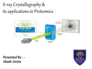
- 1. X-ray Crystallography & its applications in Proteomics Presented By : - Akash Arora
- 2. X-ray crystallography is a technique used for determining the atomic and molecular structure of a crystal, in which the crystalline atoms cause a beam of incident X-rays to diffract into many specific directions. Then they use an X-ray beam to “hit” the crystallized molecule. The electrons surrounding the molecule diffract as the X-rays hit them. This forms a pattern, this type of pattern is called the X-ray diffraction pattern. Introduction
- 3. The English physicist Sir William Henry Bragg pioneered the determination of crustal structure by X-ray diffraction methods X-ray crystallography is a complex field that has been associated with several of science’s major breakthroughs in the 20th century Using X-ray crystal data, Dr. James Watson and Dr. Francis Crick were able to determine the helix structure of DNA in 1953. History
- 4. Why X-ray ?? Why crystal ??
- 5. Why X rays ? An electromagnetic wave of high energy and very short wavelength, which is able to pass through many materials opaque to light.(wavelength 1 angstrom) The wavelength of X-ray photons is on the order of the distance between atomic nuclei in solids (bonds are roughly 1.5-2.5 Å). You can think of it like the waves fit nice between the atoms and "fill" the crystal.
- 6. Why Crystal ? Researchers crystallize an atom or molecule, because the precise position of each atom in a molecule can only be determined if the molecule is crystallized. If the molecule or atom is not in a crystallized form, the X-rays will diffract unpredictably and the data retrieved will be too difficult if not impossible to understand
- 8. X-ray diffraction Diffraction is the slight bending of light as it passes around the edge of an object. X-ray crystallography uses the uniformity of light diffraction of crystals to determine the structure of molecule or atom Then X-ray beam is used to hit the crystallized molecule. The electron surrounding the molecule diffract as the X-rays hit them. This forms a pattern. This type of pattern is known as X-ray diffraction pattern.
- 9. Bragg’s Law There is a definite relationship between the angle at which a beam of X rays must fall on the parallel planes of atoms in a crystal in order that there be strong reflection, the wavelength of the X rays, and the distance between the crystal planes 2d sinƟ = nλ Here d is the spacing between diffracting planes, Ɵ is the incident angle, n is any integer, and λ is the wavelength of the beam.
- 10. When an incident x-ray beam hits a scatterer, scattered x-rays are emitted in all directions. Most of the scattering wave fronts are out of phase interfere destructively. Some sets of wave fronts are in phase and interfere constructively. A crystal is composed of many repeating unit cells in 3-dimensions, and therefore, acts like a 3-dimensional diffraction grating. The constructive interference from a diffracting crystal is observed as a pattern of points on the detector. The relative positions of these points are related mathematically to the crystal’s unit cell dimensions. Destructive Interference Constructive Interference Interference
- 11. X-rays are scattered almost exclusively by the electrons in the atoms, not by the nuclei. The incident electromagnetic wave exerts a force on the electrons. This causes the electrons to oscillate with the same frequency as the incident radiation. The oscillating electrons act as radiation scatters and emit radiation at the same frequency as the incident radiation. Interactions between X-rays and atoms
- 12. The first-and often most difficult-step is to obtain an adequate crystal of the material under study. The crystal should be sufficiently large (typically larger than 0.1 mm in all dimensions), pure in composition and regular in structure, with no significant internal imperfections such as cracks. Researchers crystallize an atom or molecule, because the precise position of each atom in a molecule can only be determined if the molecule is crystallized. Procedure
- 13. The crystal is placed in an intense beam of X-rays, usually of a single wavelength (monochromatic X- rays), producing the regular pattern of reflections. As the crystal is gradually rotated, previous reflections disappear and new ones appear; the intensity of every spot is recorded at every orientation of the crystal. Multiple data sets may have to be collected, with each set covering slightly more than half a full rotation of the crystal and typically containing tens of thousands of reflections.
- 14. In the third step, these data are combined computationally with complementary chemical information to produce and refine a model of the arrangement of atoms within the crystal. The final, refined model of the atomic arrangement-now called a crystal structure is usually stored in a public database. After the diffraction pattern is obtained, the data is then processed by a computer and the structure of the atom or molecule is deduced and visualized.
- 17. Generally a typical x-ray diffraction contain below parts: X-ray source Detector Crystal on the end of mounting needle Liquid nitrogen steam to keep crystal cold Movable mount to rotate crystal Collimator
- 18. X-ray Tube: the source of X Rays Incident-beam optics: condition the X-ray beam before it hits the sample The goniometer: the platform that holds and moves the sample, optics, detector, and/or tube The sample & sample holder Receiving-side optics: condition the X-ray beam after it has encountered the sample Detector: count the number of X Rays scattered by the sample.
- 20. Limitations Small-molecule crystallography typically involves crystals with fewer than 100 atoms in their asymmetric unit; such crystal structures are usually so well resolved that the atoms can be discerned as isolated "blobs." of electron density. By contrast, macromolecular crystallography often involves tens of thousands of atoms in the unit cell. Such crystal structures are generally less well-resolved (more "smeared out"); the atoms and chemical bonds appear as tubes of electron density, rather than as isolated atoms. In general, small molecules are also easier to crystallize than macromolecules; however, X-ray crystallography has proven possible even for viruses with hundreds of thousands of atoms.
- 21. Application of X ray Crystallography XRD is a nondestructive technique To identify crystalline phases and orientation To determine structural properties. To measure thickness of thin films and multi-layers To determine atomic arrangement
- 22. X-ray diffraction is most widely used for the identification of unknown crystalline materials (e.g. minerals, inorganic compounds). Determination of unknown solids is critical to studies in geology, environmental science, material science, engineering and biology. identification of fine-grained minerals such as clays and mixed layer clays that are difficult to determine optically. determination of unit cell dimensions measurement of sample purity
- 23. Applications of X ray Crystallography in proteomics & Conclusion
- 24. A branch of biotechnology concerned with applying the techniques of molecular biology, biochemistry, and genetics to analyzing the structure, function, and interactions of the proteins produced by the genes of a particular cell, tissue, or organism, with organizing the information in databases, and with applications of the data. Proteomics
- 26. Protein Structure Determination Protein structure determination refers to finding the exact orientations and arrangements of different amino acids present in the protein. X ray crystallography helps us to determine the structure of proteins which further helps us to even determine its function.
- 27. Protein Interaction Studies Protein interaction refers to the way in which two or more proteins interact with each other. Studies include the orientation, site of action and the major amino acids (of protein) taking part in a particular reaction ray crystallography helps to determine there Interactions.
- 28. Conformational Studies It is necessary to determine the arrangements as it determines the structure and function of Protein X ray crystallography is an efficient technique to determine it. Conformational studies refers to spatial arrangements of atoms in a molecule that can come about through free rotation of atoms about a chemical bond.
- 29. Enzyme catalysis Determination Enzymes are protein. Determination of structure (specially active site’s) and type of amino acids present in active sites determines catalytic activities, Interaction level of enzymes. X ray crystallography helps us to determine and predict the catalytic efficiency of enzymes.
- 30. X-ray crystallography is essentially a form of very high resolution microscopy. It enables us to visualize protein structures at the atomic level and enhances our understanding of protein function. Specifically we can study how proteins interact with other molecules, how they undergo conformational changes, and how they perform catalysis in the case of enzymes. Armed with this information we can design novel drugs that target a particular protein, or rationally engineer an enzyme for a specific industrial process. Conclusion
- 31. References https://www.jic.ac.uk/staff/david-lawson/xtallog/summary.htm https://en.wikipedia.org/wiki/X-ray_crystallography https://www.youtube.com/watch?v=gLsC4wlrR2A https://www.stolaf.edu/people/hansonr/mo/x-ray.html theconversation.com/explainer-what-is-x-ray-crystallography-22143 Lehninger Principles of Biochemistry Book by Albert L. Lehninger, David L. Nelson, and Michael M. Cox https://en.wikibooks.org/wiki/Structural_Biochemistry