Study the role of estradiol and luteinizing hormones in breast tumors incidence in the women in al najaf governorate- iraq
•
1 j'aime•265 vues

International peer-reviewed academic journals call for papers, http://www.iiste.org

Recommandé
Recommandé
Contenu connexe
Tendances
Tendances (20)
Similaire à Study the role of estradiol and luteinizing hormones in breast tumors incidence in the women in al najaf governorate- iraq
Similaire à Study the role of estradiol and luteinizing hormones in breast tumors incidence in the women in al najaf governorate- iraq (20)
Plus de Alexander Decker
Plus de Alexander Decker (20)
Dernier
Dernier (20)
Study the role of estradiol and luteinizing hormones in breast tumors incidence in the women in al najaf governorate- iraq
- 1. Advances in Life Science and Technology ISSN 2224-7181 (Paper) ISSN 2225-062X (Online) Vol 14, 2013 www.iiste.org Study the role of Estradiol and Luteinizing hormones in breast tumors incidence in the women in Al-Najaf Governorate- Iraq Ali H.Al-Saadi 1 , Ezzate H.Ajeena 2 , Haider K.Zaidan 1 , Mufeed Ewadh*3 1 . Department of Biology, College of Science, University of Babylon- Iraq 2. Department of Biology, College of Science, University of Kufa- Iraq 3. Department of clinical biochemistry,college of medicine ,university of Babylon,Iraq * E-mail ; mewadh@yahoo.com Abstract This study aims to illustration the role of some important risk factors which including age, age at menarche , menopausal status, the levels of estradiol(E2) and luteinizing hormones(LH) in the increasing breast tumors incidence by using 120 women, divided into three groups; control group including forty healthy women, benign group containing forty women with breast benign tumor and malignant group of forty women with malignant breast tumor, this last group subdivided into; malignant presurgery and malignant post surgery groups each one contain twenty women. The results showed that benign breast tumors are more common in women with interval age 31-40 years whereas malignant breast tumors were more common in women with interval age 41-50 years. Early age at menarche wasn’t significantly associated with increasing breast cancer incidence, contrariwise menopausal status which was significantly associated with increasing this disease . The results revealed a significant elevation P≤ 0.05 in the levels of estradiol E2, and Luteinizing hormones in the women with breast cancer before tumor excision and reduction in the levels of these hormones in the women after tumor excision indicating the role of these hormones in the initiation or promotion breast cancer. In conclusion, this study revealed that breast cancer incidence increased in women in forties and fifties of age and increasing the levels of endogenous hormones such as Estradiol, and luteinizing hormones can increased risk of developing breast cancer especially in postmenopausal women. Key word : Brest cancer , Risk factor , Estradiol(E2) , luteinizing hormones 1. Introduction All cancers start as a single cell that loses control of its normal growth and replication processes and they affect many different tissues and types of cells. About 85%of adult cancers which develop from the epithelial cells of the inner and outer lining of the body are called carcinomas (American Institute for Cancer Research,2007). Generally breast cancer is the second most common cancer after lung cancer (WHO,2003).Moreover ,it is the commonest malignancy in females as it constitutes about 3 folds more common than all gynecological malignancies put together, and it has become a major health problem in the developed world (Horner et al.,2008).Breast cancer a heterogeneous disease that encompasses several distinct entities with different biological characteristics and clinical behavior (Carey et al.,2007;Dent et al.,2007). The 2003 world health organization (WHO) classification of breast tumor which includes benign (harmless) tumors and malignant (cancerous )tumors(Peter et al.,2003). Breast cancer is influenced by multiple risk factors which can be classified into four groups (Abulkhair et al., 2010). These groups are: first reproductive factor such as age at menarche, age at menopause, age at first pregnancy and lactation (Veronesi et al., 2005). Second is genetic and family history (Raicevic and Radulovic, 2000). Third is a history of benign proliferative breast diseases (Wang et al., 2004) . Fourth include obesity, diet, al-cohol intake and smoking (International Agency for Research on Cancer, 2002). 2. Materials and methods: 2.1 Sampling of cases. (a) Study group : forty cases of female patients with breast carcinoma were included in this study ,their ages ranging from 25 to 70 years .Twenty cases of these patients were in early detection of breast cancer .The other twenty cases were post-excision tumor patients . (b) Comparative group: forty cases of female patients with benign breast tumor were included in this study, their ages ranging from 15 to 50 years to compare them with malignant and normal breast patients. (c) Control group : forty cases of normal females which were healthy and don’t undergo from any breast diseases or other diseases .these cases were of the same age of malignant group 2.2 Collection of blood samples. Blood samples were collected from healthy control, benign and malignant patients according to the method which used by (Dorgan et al.,2010) 91
- 2. Advances in Life Science and Technology ISSN 2224-7181 (Paper) ISSN 2225-062X (Online) Vol 14, 2013 www.iiste.org 2.3 Measurement of hormones Reagents of estradiol hormone (E2) according to estradiol hormone kit , DRG, Germany. Reagents of luteinizing hormone (LH) according to luteinizing hormone kit , Monobind, USA. 3. Results 3.1 Age The highest percentage of malignant patients in this study was recorded in (41-50 years) with 12 cases (30%) followed by 11 cases (27.5%) were seen (31-40 y), 9 cases (22.5%) were seen in (51-60 y),and 7 cases (17.5%) were seen in (61-70 y),while only one case (2.5%) was noted in (21-30) and there was no cases recorded in ≤20 years .In benign tumor patients the highest percentage was noted in (31-40 y) with 13 cases (32.5%) ,while the lowest percentage in (41-50 y) with 6 cases (15%). The interval age ≤20 years recorded 11 cases (27.5%) which revealed increasing in compared with 10 cases (25%) in (21-30y). While there was no case recorded in age (5160 y),(61-70 y) respectively which reflect the tendency of benign tumor incidence in younger age . As shown in figure (1) 3.2 Age at menarche. Table (1) shows the percentage of women who begin menarche at age more than 12 years in malignant(80%) ,benign(92.5%) breast tumor and control (85%) groups recorded prevalence ,while women of the same groups above who begin menarche at age less than 12 years have little percentage(20%,7.5%,15%)respectively. 3.3 Menopausal status . The results of table (2) showed that the women who are premenopause in malignant group have less percentage 14(35%) in comparing with control 18(45%) and benign groups 37(92.5),while postmenopausal women in malignant group recorded higher percentage 26(65%) than those of control 22(55%)and benign groups 3(7.5%). Benign group showed a significant difference P≤0.05 when being compared with control group, malignant group showed a significant difference P≤0.05 as compared with benign group . 3.4 Estradiol hormone (E2). The results of figure (2) showed a highly significant difference P≤0.000 in the levels of estradiol hormone E2 as compared with the studied groups ,the levels of this hormone in malignant presurgery group(98.30±75.10) was the highest followed by benign group (63.32±31.99) then control group(41.57±30.33) and finally malignant postsurgery group(29.60±20.33).Table (3) explains multiple comparisons in serum estradiol hormone levels among study population .When the comparison was set between control and benign groups ,the result records a significant difference P≤0.017 ,there was highly significant difference P≤0.000 between control and malignant presurgery groups and the results show no a significant difference P≤0.281 between malignant postsurgery and control groups .The results record high significant difference (P≤0.002 and P≤0.003)respectively in comparison between benign versus malignant presurgery and benign versus malignant postsurgery groups. Finally the comparison between malignant presurgery and postsurgery was highly significant P≤0.000. 3.5 Luteinizing hormone . The results of figure (3) proves that a highly significant difference P≤0.001 in the level of luteinizing hormone as compared among studied groups .This table explains that the level of luteinizing hormone when measured in forty healthy women was 1.56±0.80 ,while when being measured in forty benign breast tumor women there was slight increasing 2.87±2.08 ,whereas the level of the same hormone revealed marked increasing 4.78±2.12 as measured in twenty women with breast cancer before surgery then recorded decreasing in it's level 3.64±1.46 in twenty women with breast cancer after surgery. Table (4) shows multiple comparisons among studied groups ,first studied group reveals highly significant difference P≤0.001 as compared with malignant presurgery group while in the same first group there was no a significant difference (P≤0.615, P≤0.247) respectively between control versus benign and control versus malignant postsurgery groups .With regard to second group in which there were two comparisons the first was with significant difference P≤0.000 between benign and malignant presurgery groups. The second comparison was without significant difference P≤0.118 between benign versus malignant postsurgery. Last comparison was between malignant presurgery and malignant postsurgery exhibits no a significant difference P≤0.042. 4. Discussion 4.1 Age In the present study maximum number of women with malignant breast cancer were observed in 41-50 years (12 cases) followed by 51-60 years (11 cases) .The average age was 49.1 years .These results agreed with previous studies in Iraq (Waheda, 1998 ; Madhoor ,2002),(figure 1). Age is the single most important risk factor in breast cancer. Compared with women in their twenties, women are 10 times as likely to develop breast cancer in their thirties ,40 times as likely in their forties ,60 times as likely in their fifties and 90 times as likely after sixties (Forbes, 1997). 92
- 3. Advances in Life Science and Technology ISSN 2224-7181 (Paper) ISSN 2225-062X (Online) Vol 14, 2013 www.iiste.org 4.2 Age at menarche The association between breast cancer risk and the age at menarche is consistent with the hypothesis; breast cancer risk is related to the total extent of breast mitotic activity which driven by estrogen exposure during the luteal phase of menstrual cycle which will determine the probability of tumorigenic somatic events (Pike et al.,1993). The results of this study showed only 20% of women in malignant group and 7.5% women in benign group whose age at menarche began before 12 years while the remaining women in malignant and benign groups their age at menarche began after 12 years, table (1) .Our results supported by (Chang-Claude et al.,2007 ; Barnett et al.,2008) who showed that no association between breast cancer risk and age at menarche. 4.3 Menopausal status The results of the present study show among study population there were 92.5% of women in benign group were premenopause as compared with 35% with same feature in malignant group, table (2) .This result indicates that benign breast diseases were more often found in premenopausal women as (Ernster,1981;Bodian et al.,1993) did . The rate of fibroadenoma (which was included the most important condition in breast benign diseases) rises to 75% for biopsies in women under age twenty years (Greenberg et al.,1998) . On other hand the results of this study revealed that postmenopausal women in malignant group recorded high prevalence (65%) in comparing with benign group which recorded only (7.5%), table (2) , this result was in parallel with(National Breast Cancer Coalition (NBCC),1999) which appeared that breast cancer occurs more often in postmenopausal women about 75% of cases. The increasing in the risk which associated with this menopausal status may be explained possibly due to the increased levels of circulating estradiol hormone and reduced level of sex hormone binding globulin by the conversion of androgens to estradiol in adipose tissues (Calle and Kaaks,2004). 4.4 Estradiol hormone (E2) The present study demonstrated that there was a significant increasing in the levels of serum estradiol hormone in malignant presurgery women, figure (2) and this result was supported by Blair (2010) who explained that the postmenopausal women with elevated plasma or serum estrogen hormone are at increased risk of developing breast cancer. Estrogen can cause cancer by stimulating cell proliferation(promotion) and causing genotoxic damage(initiation), estrogen are highly mitogenic in hormone sensitive tissues such as breast ,prolonged exposure of target tissues and cells to excessive mitogenic stimulation by estrogens has been considered an important etiological factor for induction breast cancer (Hiraku et al.,2001).The result of this study was revealed a significant difference P≤0.017 when comparison made between control and benign groups, table(3) ,these result was supported by Khanna et al. (2012) who found that serum estradiol hormone significantly higher in women with breast benign diseases than in normal control. It has been postulated that both benign and malignant breast tumor have a hormonal origin because of estradiol hormone profound stimulatory influence on breast ductal epithelium and it was thought that it must play a central role in this disease (Wu et al.,1999). 4.5 Luteinizing hormone (LH) . The results show a significant increasing in the levels of serum luteinizing hormone in the women in malignant presurgery group, figure (3) ,this result was confirmed by a study explaining the presence of direct effects of LH on breast cells themselves (Guo et al.,2004) , there was elevation in the levels of serum LH among postmenopausal women with breast cancer as Krishnamoorthy et al.(1994) found. How luteinizing hormone and it's receptors exert their effects on breast cancer cells is unclear ,however, one possibility may explain these effects which refer to that effects take place through luteinizing hormone receptors which present in the ovaries thereby influencing steroid hormone production , another possibility is that effects happen through luteinizing hormone receptors present in malignant tumor cells as some studies have detected these receptors in both normal and neoplastic breast tissues and in breast cancer cell lines (Kuijper et al.,2009). As luteinizing hormone (LH) and luteinizing hormone receptors (LHR) are both involved in estradiol hormone synthesis , functionally important polymorphisms in these genes could alter the levels of estrogen exposure and thereby contribute to breast cancer risk determination (Powell et al.,2003). References Abulkhair,O.A.;AlTahan,F.M.;Young,S.E.;Musaad,S.M.andJazieh,M.A.(2010). The first national public breast cancer screening program in Saudi Arabia. J.Annals of Saudia medicine.,30(5):350-357 American Institute for Cancer Research and World Cancer Research Fund.(2007).Food,Nutrition,Physical activity ,and the Prevention of Cancer :a Global Perspective. Washington DC: AICR. Barnett,G.C.; Shah,M.; Redman,K.; Easton,D.F.; Ponder,B.A.; Pharoah,P.D.(2008). Risk factors for the incidence of breast cancer: do they affect survival from the disease? J. Clin. Oncol.,26:3310–6. 93
- 4. Advances in Life Science and Technology ISSN 2224-7181 (Paper) ISSN 2225-062X (Online) Vol 14, 2013 www.iiste.org Blair,I.A.(2010).Analysis of estrogen in serum and plasma from postmenopausal women :past present and future .Steroid., 75(4-5):297-306. Bodian, C. A., Perzin, K. H., Lattes, R., Hoffmann, P. and Abernathy, T. G. (1993). Prognostic significance of benign proliferative breast disease. Cancer., 71: 3896–3907 Carey,L.A.; Dees,E.C.; Sawyer,L.(2007). The triple negative paradox: primary tumor chemosensitivity of breast cancer subtypes. Clin. Cancer Res., 13:2329–2334. Chang-Claude,J.;Andrieu,N.;Rookus,M.;Brohet,R.;Antoniou,A.C.(2007). Age at Menarche and Menopause and Breast Cancer Risk in the International BRCA1/2 Carrier Cohort Study. Cancer Epidemiol Biomarkers Prev., 16(4):740–746. Dent,R.; Trudeau,M.; Pritchard,K.I. (2007). Triple-negativebreast cancer: clinical features and patterns of recurrence. Clin.Cancer Res., 13:4429–4434. Dorgan,F.J.;Stanczyk,Z.F.;Kahle,L.L.;and Brinton,A.L.(2010). Prospective case-control study of premenopausal serum estradiol and testosterone levels and breast cancer risk. J.Breast cancer research.,12:98. Ernester,V.L. (1981). The epidemiology of benign breast disease. Epidemiol. Rev., 3, 184. Forbes, J.F.(1997). The incidence of breast cancer: the global burden, public health considerations. Seminars in Oncology., 24(1, suppl 1):S1-20–S1-35. Guo,S.; Russo,I.H.; Lareef,M.H. & Russo,J. (2004). Effect of human chorionic gonadotropin in the gene expression profile of MCF-7 cells. International J. of Oncol., 24 399–407. Hiraku,Y.; Yamashita,N.; Nishiguchi,M. and Kawanishi,S.(2001). Catechol estrogens induce oxidative DNA damage and estradiol enhances cell proliferation. International J. of Cancer., 92(3):333-337. Horner,M.J.; Ries,L.A.G.; Krapcho,M.;Neyman,N.; Aminou,R.;Howlader,N. (2008). SEER Cancer Statistics Review. National Cancer Institute.Bethesda,MD.p 5. International Agency for Research on Cancer.(2002). Weight Control and Physical Activity. Lyon, France Kaaks,R.; Rinaldi,S.; Key,T.J.; et al.(2005). Postmenopausal serum androgens, oestrogens and breast cancer risk: The European prospective investigation into cancer and nutrition. Endocr. Relat. Cancer., 12: 10711082. Khanna,S.;Singh,S.;Khanna,H.D.;Kumar,M. and Gupta,S.K.(2012). Evaluation of estradiol levels ,lipid profile ,estrogen receptor status and its correlation with histological variant in benign breast diseases . World J. of path., 1:10-13. Krishnamoorthy,L.;Zachariah,E.;Ramaswamy,P.G.; and Anantha,N.(1994). Circulating peptide and steroid hormone levels in patients with breast cancer .Indian J. of Clin.Biochem.vol.9(1):37-39. Kuijper,M.T.; Ritstier,R.K.;Verhoef-Post,M.; Piersma,D.; et al.(2009). LH receptor gene expression is essentially absent in breast tumor tissue:implications for treatment. Mol. Cell Endocrinol.,302(1):58-64. Madhoor,B.M.(2002).Immunological Study on Patients with Breast Cancer. Msc. thesis College of medicine .University Al-Mustansiriyah. National Breast Cancer Coalition (1999). 15 percent increased appropriation for breast cancer research at the national institutes of health .Washington D.C Peter Devilee; Fattaneh,A.; Tavassoli. (2003). World Health Organization: Tumours of the Breast and Female Genital Organs. Oxford . Pike,M.C.;Spicer,D.V.;Dahmoush,L.; and Press,M.F.(1993). Estrogens, proliferation and breast cancer risk .Epidemiol.Rev.,15:17-35. progestron,normal Raicevic,L.; and Radulovic,S.(2000).Breast cancer susceptibility gene 1- BRCA1. .,8(1):21-3. 94 breast cell J.Archive of Oncology
- 5. Advances in Life Science and Technology ISSN 2224-7181 (Paper) ISSN 2225-062X (Online) Vol 14, 2013 www.iiste.org Veronesi,U.; Boyle,P.; Goldhirsch,A. (2005). Breast cancer .Lancet .,365:41-1727. Waheda, N. Elia.(1998).A study of certain immunological parameters in women a fflicted with breast cancer in Iraq. Ph D , College of Science. University of Baghdad. Wang,J.; Costantino,J.P.; Tan-Chiu,E.; Wickerham,D.L.; Paik,S. and Wolmark, N.(2004). Lower-category benign breast disease and the risk of invasive breast cancer. J. Natl. Cancer Inst.,96:616-20. World Health Organization International Agency for Research on Cancer (WHO) .(2003). World Cancer Report .Retrived on 2002-3,p.335. Wu,A.H.; Pike,M.C.; and Stram,D.O.(1999). Meta-analysis dietary fat intake, serum estrogen levels and the risk of breast cancer. J.Natl. Cancer Inst., 91:529 – 534. Table (1) Distribution of control, benign and malignant breast tumor groups according to age at menarche Control group Benign group Malignant group Age at χ2 % % % menarche(year) No. No. No. ≤12 6 15 3 7.5 8 20 ≥ 12 34 85 37 92.5 32 80 Total 40 100 40 100.0 40 100.0 2.583 Table (2) Distribution of control, benign and malignant breast tumor groups according to menopausal status . Control group Benign group Malignant group Menopausal status χ2 No . % No . % No. % Premenopause 18 45 37 92.5 14 35 Postmenopause 22 55 3 7.5 26 65 Total 40 100 40 100.0 40 100.0 a= significant differences when comparing benign with control group at P<0.05 significant differences when comparing malignant with benign group at P<0.05 95 30.638 a c c=
- 6. Advances in Life Science and Technology ISSN 2224-7181 (Paper) ISSN 2225-062X (Online) Vol 14, 2013 www.iiste.org Table (3) Result of multiple comparisons of estradiol hormone among control, benign and malignant (pre and postsurgery) groups Studied group Control Groups P. value Sig . Significant at <0.05 0.000 Highly significant at <0.05 Malignant postsurgery 0.281 Non significant at<0.05 Malignant presurgery 0.002 Highly significant at<0.05 Malignant postsurgery Malignant presurgery 0.017 Malignant presurgery Benign Benign 0.003 Highly significant at <0.05 Malignant postsurgery 0.000 Highly significant at <0.05 Table (4) Result of multiple comparisons of luteinizing hormone among control, benign and malignant (pre and postsurgery)groups Studied group Groups P. value Sig . Control Non significant at<0.05 0.001 Highly significant at<0.05 Malignant postsurgery 0.247 Non significant at<0.05 Malignant presurgery 0.000 Highly significant at<0.05 Malignant postsurgery Malignant presurgery 0.615 Malignant presurgery Benign Benign 0.118 Non significant at<0.05 Malignant postsurgery 0.042 Significant at<0.05 96
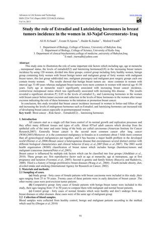
- 7. Advances in Life Science and Technology ISSN 2224-7181 (Paper) ISSN 2225-062X (Online) Vol 14, 2013 www.iiste.org Figure (1): Distribution of age of malignant and benign breast cancer patients . * Estradiol hormone (E2) pg/ml 120 98.3 100 80 63.32 60 41.57 40 25.57 20 0 control group benign group malignant malignant presurgery group postsurgery group Cases groups *= Significant difference at P≤0.05 Figure (2): Serum estradiol hormone levels (E2) (pg/ml) in control, benign and malignant (pre and postsurgery) groups . * Luteinizing hormone (mIU/ml) 6 4.78 5 3.64 4 2.87 3 2 1.56 1 0 control group benign group malignant presurgery group malignant postsurgery group Cases groups *= Significant difference at P≤0.05 Figure (3): Serum luteinizing hormone levels (µIU/ml) in control, benign and malignant (pre and postsurgery)groups. 97
- 8. This academic article was published by The International Institute for Science, Technology and Education (IISTE). The IISTE is a pioneer in the Open Access Publishing service based in the U.S. and Europe. The aim of the institute is Accelerating Global Knowledge Sharing. More information about the publisher can be found in the IISTE’s homepage: http://www.iiste.org CALL FOR JOURNAL PAPERS The IISTE is currently hosting more than 30 peer-reviewed academic journals and collaborating with academic institutions around the world. There’s no deadline for submission. Prospective authors of IISTE journals can find the submission instruction on the following page: http://www.iiste.org/journals/ The IISTE editorial team promises to the review and publish all the qualified submissions in a fast manner. All the journals articles are available online to the readers all over the world without financial, legal, or technical barriers other than those inseparable from gaining access to the internet itself. Printed version of the journals is also available upon request of readers and authors. MORE RESOURCES Book publication information: http://www.iiste.org/book/ Recent conferences: http://www.iiste.org/conference/ IISTE Knowledge Sharing Partners EBSCO, Index Copernicus, Ulrich's Periodicals Directory, JournalTOCS, PKP Open Archives Harvester, Bielefeld Academic Search Engine, Elektronische Zeitschriftenbibliothek EZB, Open J-Gate, OCLC WorldCat, Universe Digtial Library , NewJour, Google Scholar