Radiation Protection Course For Orthopedic Specialists: Lecture 3 of 4: Basics Radiation Protection
•
2 j'aime•398 vues

Radiation Protection Course For Orthopedic Specialists: Lecture 3 of 4: Basics Radiation Protection

Recommandé
Contenu connexe
Tendances
Tendances (20)
Similaire à Radiation Protection Course For Orthopedic Specialists: Lecture 3 of 4: Basics Radiation Protection
Similaire à Radiation Protection Course For Orthopedic Specialists: Lecture 3 of 4: Basics Radiation Protection (20)
Plus de Amin Amin
Plus de Amin Amin (20)
Dernier
Dernier (20)
Radiation Protection Course For Orthopedic Specialists: Lecture 3 of 4: Basics Radiation Protection
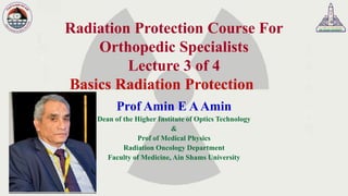
- 1. Radiation Protection Course For Orthopedic Specialists Lecture 3 of 4 Basics Radiation Protection Prof Amin E AAmin Dean of the Higher Institute of Optics Technology & Prof of Medical Physics Radiation Oncology Department Faculty of Medicine, Ain Shams University
- 3. Dealing With Ionizing Radiation It should be emphasised that ionizing radiation needs to be treated with CARErather than FEAR.
- 4. Aspects of the Problem • There are four main aspects of the problem to be considered. –Firstly, radiological procedures should be based on a demonstrated medical need. –Secondly, when radiological procedures are required, it is essential that patients be protected from excessive radiation during the exposure.
- 5. Aspects of the Problem –Thirdly, it is necessary that personnel in radiology departments be protected from excessive exposure to radiation in the course of their work. –Finally, personnel in the vicinity of radiology facilities and the general public require adequate protection.
- 7. Specific Effects Of Radiation Radiological protection aims at avoiding deterministic effects by setting dose limits below their thresholds. Stochastic effects are believed to occur, albeit with low frequency, even at the lowest doses and therefore have been taken into account at all doses.
- 8. Aims Of Radiation Protection • Deterministic effects ❖RP aims to ELIMINATE them. • Stochastic effects ❖RP aims to REDUCE them.
- 9. The Aim Of Radiological Protection The primary aim of radiological protection is to provide an appropriate standard of protection for man without unduly limiting the beneficial practices giving rise to radiation exposure.
- 10. Sources Of Radiation Background ❖Natural sources of radiation ❖Artificial sources of radiation
- 11. Natural Sources Of Radiation Background ❖Cosmic rays ❖Terrestrial radiation ❖Radionuclides in the body ❖Radon gas and its decay products
- 12. Let’s Compare Backgrounds • Sea level - 30 mrem/year from cosmic radiation • 10,000 ft. altitude - 140 mrem/year from cosmic radiation
- 13. Terrestrial Radiation ❖ Terrestrial radiation comes from radioactivity emitting from Primordial radio nuclides - these are radio nuclides left over from when the earth was created. ❖ Common radionuclides created during formation of earth: ❖ Radioactive Potassium (K-40) found in bananas, throughout the human body, in plant fertilizer and anywhere else stable potassium exists. ❖ Radioactive Rubidium (Rb-87) is found in brazil nuts among other things.
- 14. Terrestrial Radiation • Greatest contributor is 226Ra (Radium) with significant levels also from 238U, 232Th, and 40K. –Igneous rock contains the highest concentration followed by sedimentary, sandstone and limestone. –Fly ash from coal burning plants contains more radiation than that of nuclear or oil-fired plants. ++
- 15. Another Look at Sources
- 16. Artificial Sources Of Radiation Background Two artificial sources of radiation to which every body is exposed; ❖Fall-out from nuclear explosions. ❖Radioactive waste, including discharges from nuclear establishments.
- 17. Artificial Sources Of Radiation Background Two artificial sources of radiation to which not all the population are exposed; ❖Radiation used for medical purposes. ❖Occupational exposure to radiation.
- 19. Relative Contribution Of Different Sources Of Background Radiation Radon 32% Terestri al 19% Body 17% Cosmic Rays 14% Medical 12% Thoron 5% Artificial 1% Average radiation exposure from all sources: 2.8 mSv/year
- 20. Annual Dose To The General Population From Natural And Man-made Sources Radiation Source Effective Dose Equivalent (mrem/year) Percentage of Total Natural Cosmic Cosmogenic Terrestrial Inhaled (due to radon) In the Body Subtotal 27 1 28 200 39 295 8% - 8% 55% 11% 82% Man-made Medical X-rays Nuclear Medicine Consumer Products Others Subtotal 39 14 10 <1 64 11% 4% 3% - 18% Rounded Total 360 100%
- 21. System Of Radiation Protection “System of RP” is the name given by the ICRP to the application of the 3 basic principles of RP (no part should be taken in isolation): ❖ Justification ❖ Optimization ❖ Limitation
- 22. The framework of radiation protection (I) • Justification of a practice • Optimization of protection • Application of individual limits
- 23. Principles of Protection in Practices ❖Justification of a practise - no practice should be adopted unless it produces sufficient benefit to the exposed individuals or to society to offset the radiation detriment it causes. ❖Optimization of protection - the magnitude of individual doses, the number of people exposed, and the likelihood of incurring exposures should be kept as low as reasonably achievable, economic and social factors being taken into account. ❖Individual dose limits - exposure should be restricted so, that exposure of any individual from authorized source does not exceed any relevant dose limit.
- 24. Practices Vs Intervention Human activities which increases the overall exposure to radiation are called practices. Other human activities which can decrease the overall exposure by influencing the existing causes of exposure are called intervention.
- 25. The Framework Of Radiation Protection II In diagnostic radiology the radiation sources (X-rays) are deliberately used and are under control. Such situations are called by the International Commission on radiation Protection (ICRP) “practices”. The basic components of the system of protection for “practices” can be summarized as follows: No practice involving exposures to radiation should be adopted unless it produces at least sufficient benefit to the exposed individuals or to society to offset the radiation detriment it causes (this is called “justification of a practice”).
- 26. The Framework Of Radiation Protection III In relation to any particular source of radiation within a practice (e.g. X-rays in radiodiagnostic), all reasonable steps should be taken to adjust the protection so as to maximize the net benefit, economic and social factors being taken into account (this is called “optimization of protection”). A limit should be applied to the dose (other than from medical exposures) received by any individual as the result of all practices to which he is exposed (this is called “application of individual dose limits”).
- 27. Justification Justification means that any dose exposure MUST have a benefit to exposed individuals or to society. Thus, if the exposure has no benefit it is not justified.
- 28. Justification Benefit of the radiation exposure must outweigh the risk of exposure vs
- 29. Justification • No practice involving exposures to radiation should be adopted unless it produces sufficient benefit to the exposed individuals or to society to offset the radiation detriment it causes. • Justification of exposures is primarily the responsibility of the medical professional i.e. the Radiologist. • The expected clinical benefit associated with each type of procedure should have been demonstrated to be sufficient to offset the radiation detriment.
- 30. Optimization ❖ Optimization means that minimum risk and maximum benefits should be achieved, economic and social factors being taken into account. ❖ Optimization includes the ALARA criterion: doses should be “As Low As Reasonably Achievable”, economic and social factors being taken into account. BENEFIT RISK
- 31. Optimisation • For every exposure, operators must ensure that doses arising from the exposure are kept as low as reasonably practicable and consistent with the intended diagnostic purpose. • THIS IS OPTIMISATION
- 32. ALARA ALARA (As Low As Reasonably Achievable) refers to the continual application of the optimization principle in the day-to-day practice.
- 33. Limitation ❖ Doses should not exceed specific values, called “individual dose limits”. ❖ These dose limits are established in order to keep away from the “maximum risk level” so that no individual is exposed to a radiation risk that is judged to be unacceptable in any normal circumstance.
- 34. Limitation • Limits are set such that deterministic effects never happen • Limits are set such that chances of stochastic effects are minimised
- 35. Types Of Exposure There are three types of radiation exposure; Occupational exposure; Which is the exposure incurred at work, and principally as a result of work. Medical exposure; Which is principally the exposure of persons as part of their diagnosis or treatment. Public exposure; Which includes all other exposures (i.e. exposure incurred by members of the public from authorized radiation sources, excluding any occupational and medical exposure and exposure from natural background radiation). Their justification (for those of non natural origin) is the general benefit brought by the use of ionizing radiation in Medicine or Industry.
- 36. Legal Dose Limits - Patients • For examinations directly associated with illness – there are no dose limits.
- 37. Legal Dose Limits – Radiation Workers • Radiation workers are those exposed to radiation as part of their occupation • No benefit – only risk
- 38. Legal Dose Limits • Receive high levels of radiation exposure • Very unlikely for dental • Require annual health check • Compulsory dose monitoring • For classified worker – Whole body 20 mSv per year effective dose (18 years old and above) – Lens of eye 150 mSv per year equivalent dose – Skin 500 mSv per year equivalent dose – Extremities (hands and feet etc) 500 mSv per year equivalent dose
- 39. Radiation Dose Limits ❖ Old Radiation Dose Limits ❖ 50 milliSieverts per year (mSv/y) for occupational exposure ❖ 5 mSv/y for the general public ❖ New Radiation Dose Limits ❖ 20 mSv/y for occupational exposure (5 year average) with a maximum of 50 mSv in any one year ❖ 1 mSv/y for the general public
- 40. Radiation Dose Limits • 20 mSv/y for occupational exposure • 1 mSv/y for the general public • No limit for medical exposure
- 41. Dose limits (public) (*) In special circumstances, an effective dose of up to 5 mSv in a single year provided that the average dose over five consecutive years does not exceed 1 mSv per year. Public dose limitApplication 1 mSv in a year (*)Effective dose Annual equivalent dose in: 15 mSvThe lens of the eye 50 mSvThe skin
- 42. Dose Limits (Occupational Exposure) The occupational exposure of any worker should be controlled so that the following limits be not exceeded: Occupational dose limitApplication 20 mSv per year, averaged over defined periods of 5 years 50 mSv in any single yearEffective dose Annual equivalent dose in: 150 mSvThe lens of the eye 500 mSvThe skin 500 mSvThe hands and feet
- 43. Individual Dose Limits Public exposure • an effective dose limit for a member of the public (e.g. office worker in room next door to x-ray) is 1 mSv in any single year • an effective dose of 5 mSv in a single year in special circumstances provided that the average dose over 5 consecutive years does not exceed 1 mSv per year; • an equivalent dose to the lens of eye of 15 mSv in a year • an equivalent dose to the extremities or the skin of 50 mSv in a year
- 44. Individual Dose Limits Occupational exposure • an effective dose of 50mSv in any single year; • an effective dose of 100 mSv in 5 consecutive years; (an effective dose of 20 mSv per year averaged over 5 consecutive years); • an equivalent dose to the lens of eye of 150 mSv in a year • an equivalent dose to the extremities or the skin of 500 mSv in a year
- 45. Recommended Dose Limits In Planned Exposure Situations (ICRP 103) PublicOccupationalType of limit 1 mSv in a year20 mSv per yearEffective dose 15 mSv 50 mSv 150 mSv 500 mSv 500 mSv Lens of the eye Skin Hands and feet
- 46. Optimisation – Staff Dose Investigation Level (DIL) ❖ Dose Investigation Level ▪ 1.2 mSv per year ▪ Or 0.1 mSv per month ❖ This is a level of dose that should trigger an investigation in conjunction with your RPA, and ensures that you do not receive anywhere close to the legal limit.
- 47. Investigational Levels Level 2Level 1 3.75 mSv1.25 mSvWhole body 55.00 mSv17.50 mSvDistal Extremities
- 48. Pregnant Workers A female worker should, in becoming aware that she is pregnant, notify the employer in order that her working conditions may be modified if necessary.
- 49. Pregnant workers The notification of pregnancy should not be considered a reason to exclude a female worker from work; however, the employer of a female worker who has notified pregnancy should adapt the working conditions in respect of occupational exposure so as to ensure that the embryo or fetus is afforded the same broad level of protection as required for members of the public.
- 50. The Occupational Exposure Of Women ❖ The basis for the control of the occupational exposure of women who are not pregnant is the same as that for men and the ICRP recommends no special occupational dose limit for women in general. ❖ Once pregnancy has been declared, the conceptus should be protected by applying a supplementary equivalent dose limit at the surface of the woman’s abdomen (lower trunk) of 2 mSv for the remainder of the pregnancy.
- 51. Medical Exposure ❖ Medical radiation is the largest radiation source other than natural background ❖ Medical radiation dose accounts for about 95% of doses from “man-made” sources ❖ There are about 2 billion diagnostic x-ray examinations, 32 million nuclear medicine procedures and 5.5 million radiation therapy treatments annually
- 52. Medical Exposure Exposure incurred by : • patients as part of their own medical or dental diagnosis or treatment; • persons (other than occupationally exposed), voluntarily helping in the support and comfort of patients; • volunteers in a program of biomedical research involving their exposure.
- 53. Medical Exposure • Medical exposure is different from most other uses of radiation. Too little or too much dose are bad in both diagnosis and therapy • The task is to provide enough dose for the task but not much more
- 54. Dose limitation for comforters and visitors of patients (I) The dose limits should not apply to comforters of patients, i.e., to individuals exposed while voluntarily helping (other than in their employment or occupation) in the care, support and comfort of patients undergoing medical diagnosis or treatment, or to visitors of such patients.
- 55. ➢ However, the dose of any such comforter or visitor of patients should be constrained so that it is unlikely that his or her dose will exceed 5 mSv during the period of a patient's diagnostic examination or treatment. ➢ The dose to children visiting patients who have ingested radioactive materials should be similarly constrained to less than 1 mSv. Dose Limitation For Comforters And Visitors Of Patients (II)
- 56. Some cases are not considered for dose limits, although they may increase the effective dose: ❖Natural background radiation ❖Origin: cosmic radiation and natural radioactive elements in the environment (2-3 mSv/year) ❖Radiation received as consequence of medical exposure ❖It may represent an increment of dose > than natural radiation, but it is not taken into consideration for dose limits. Not Considered For Dose Limits
- 57. Code of Practice ➢ Detailed procedures for patient, worker and visitor safety. ➢ Identification of all "Controlled Areas", displaying schematically the location of all external radiation devices.
- 58. Code of Practice ➢ Radiation dose information to patients for all routine diagnostic examinations, as determinate by direct measurement. ➢ Establishment of a quality control program to maintain optimum standards for quality and safety.
- 59. Controlled Areas • This is an area where it is necessary to follow special procedures to restrict exposure to ionizing radiation, or • An area where any person is likely to receive 1/10th of the dose limit or more.
- 60. Controlled Areas • High radiation areas and each radiation area where the possibility presents of approaching 10% of the occupational dose limit shall be treated as controlled areas.
- 61. Controlled Areas The specific requirements are: 1. The area must be secured when it is not occupied by responsible personnel. 2. The area must be posted with proper signs indicating the radiation zone(s) and the sources which is present. 3. Personnel monitoring must be provided where appropriate, as determined by the Radiation Safety Office. 4. Surveys must be performed to maintain surveillance on the hazards which might be present, and records kept. 5. Personnel must receive written instructions as to the hazards present in the area.
- 63. Dose monitoring • Film badges • Thermoluminescent dosemeters (TLD) – Badge – Extremities • Ionisation chambers
- 65. Who Should Wear Radiation Dosimeters Or Badges? • Those “likely” to exceed 10% of their annual limit are required • Minors & Declared Pregnant Workers*
- 66. Monitoring is conducted at either monthly or quarterly intervals depending upon the radiation level associated with the work environment. The radiation monitor shall be worn under the protective garment in such a manner as to record the maximum incident exposure. Personnel Monitoring
- 67. Film Badges Copper Tin Image filter Open window Al2O3 strip
- 68. Film Badge Advantages ➢ Inexpensive ➢ Easy to handle ➢ Reasonably accurate ➢ Provides a permanent dose record ➢ Measure type and energy of radiation ➢ Simple and robust
- 69. Film Badge Disadvantages ❖ No immediate indication of exposure ❖ Processing can lead to errors ❖ Prone to filter loss
- 70. TLD • Similar use as for film badges • They absorb radiation and release this as light when heated – Advantages: • Re-usable • Easy to read out – Disadvantages: • Read out is destructive • Limited info on type of radiation
- 71. Personal Dose Monitoring Extremity TLD “ring badge” • Wear facing source • Wear under gloves • Exchange every 3 months
- 72. Area Survey • Area Surveys are used to measure: – radiation level – contamination level
- 73. • Area Surveys should be conducted: – Daily whenever radionuclides are used – Monthly even if no experiments • Keep record of all surveys conducted Area Survey
- 74. Area Survey
- 75. • A complete radiation survey shall be performed for each X-ray installation, at the time of acceptance and after any important change or repair to confirm the adequacy of structural shielding required, added shielding, or its accessories. Area Survey
- 76. Procedures To Reduce Dose To X-ray Personnel
- 77. X-ray Tube Primary Beam Scattered Radiation Staff Patient The Real Risk To Staff
- 78. Protection From External Radiation External hazards arise from ❖radioactive sources ❖machines producing radiation eg x-rays Protection Methods fall under three headings 1) Time limit the exposure time 2) Distance use inverse square law 3) Shielding attenuate the beam
- 79. Basic Principles of Dose Reduction •Time •Distance •Shielding
- 80. Reduction of External Dose ❖Minimize the time spent near the radiation source ❖Maximize the distance away from the source ❖Make use of available shielding
- 81. Time An ALARA principle is to reduce the time in a radiation field 100 200 300 mrem 100 mrem/hr 1 hour 2 hours 3 hours
- 82. 20 Time Dose is proportional to the time exposed It is wise to spend no more time than necessary near radiation sources
- 84. Methods For Minimizing Time I • Pre-plan and discuss the task thoroughly prior to entering the area. • Use only the number of workers actually required to do the job. • Have all necessary tools before entering the area. • Use mock ups and practice runs. • Take the most direct route to the job site.
- 85. • Never loiter in an area controlled for radiological purposes. • Work efficiently but swiftly. • Do the job right the first time. • Perform as much work outside the area as possible. Methods For Minimizing Time II
- 87. Inverse Square Law For a point source, the Radiation Dose decreases as the square of the distance from the source. Dose a 1/d2
- 88. In air, x-rays obey the Inverse Square Law. I∞1/d2 •Double distance = 1/4 dose •Triple distance = 1/9 th dose.
- 89. Distance Another ALARA principle is to maximize the distance from source 0 1 2 3 4 ft. ft. ft. ft. ft. 100 25 11 6 mrem/hr Inverse square 1 1/4 1/9 1/16
- 90. Distance •Double distance = 1/4 dose •Triple distance = 1/9 th dose.
- 91. 22 Distance •It is recommended that an individual remains as far away as possible from the radiation source . •Procedures and radiation areas should be designed such that only minimum exposure takes place to individuals doing the procedures or staying in or near the radiation areas.
- 92. Methods For Maintaining Distance From Sources Of Radiation I • The worker should stay as far away as possible from the source of radiation. • For point sources, the dose rate follows the inverse square law. If you double the distance, the dose rate falls to 1/4. If you triple the distance, the dose rate falls to 1/9.
- 93. • Be familiar with radiological conditions in the area. • During work delays, move to lower dose rate areas. • Use remote handling devices when possible. Methods For Maintaining Distance From Sources Of Radiation II
- 94. Consequence • Distance is very efficient for radiation protection as the dose falls off in square • Examples: – long tweezers for handling of sources – big rooms for imaging equipment
- 96. Shielding • Materials “absorb” radiation • Proper shielding = Less Radiation Exposure • Plexiglass vs. Lead
- 97. Shielding
- 98. Shielding
- 101. Shielding For Various Types of Radiation Alpha −− ++ Beta Gamma and X-rays Neutron Paper Plastic Lead Concrete n g
- 102. Shielding •Various high atomic number (Z) materials that absorb radiations can be used to provide radiation protection •The ranges of alpha and b particles are short in matter the containers themselves act as shields for these radiations –Alpha can be stopped by a piece of paper –Beta low molecular weight element Al or glass can stop its effect. (Whay don’t we use lead for shielding of beta radiation?) •Gama radiations are highly penetrating absorbing material must be used for shielding of g-emitting sources –Lead is most commonly used for this purpose.
- 103. Proper Uses Of Shielding Shielding reduces the amount of Radiation dose to the worker. Different materials shield a worker from the different types of radiation.
- 104. Proper Uses Of Shielding It should be remembered that the placement of shielding may actually increase the total dose (e.g., man-hours involved in placement, Bremsstrahlung, etc.).
- 106. X-Ray Tube Shielding • The shielding of the housing must be such that, at each rating specified by the manufacturer for that tube, the leakage radiation, measured at a distance of one metre in any direction from the focal spot of the x-ray tube, does not exceed 0.1% of the exposure rate at the same distance along the central axis of the useful beam.
- 107. Protective Clothing 1. Protective body aprons 2. Gonad shields 3. Protective gloves
- 108. Protective Body Aprons • Protective body aprons used for radiographic or fluoroscopic examinations with peak x-ray tube potentials of up to 150 kVp must provide attenuation equivalent to at least 0.5 mm of lead.
- 109. Gonad Shields • Contact-type gonad shields used for routine diagnostic radiology must have a lead equivalent thickness of at least 0.25 mm and should have a lead equivalent thickness of 0.5 mm at 150 kVp. Contact- type gonad shields must be of sufficient size and shape to exclude the gonads completely from primary beam irradiation.
- 110. Protective Gloves • Protective gloves used in fluoroscopy must provide attenuation equivalent to at least 0.25 mm of lead at 150 kVp.
- 111. Eye Shielding Wear safety glasses/goggles to protect the eyes from beta radiation, when applicable.