
Recommandé
Contenu connexe
Tendances
Tendances (20)
Similaire à Gastrulation
Similaire à Gastrulation (20)
Plus de Amani Riyadh
Plus de Amani Riyadh (20)
Dernier
Dernier (20)
Gastrulation
- 2. Embryo development stages األمشاجتكوين(Gamete formation) اإلخصاب(Fertilization) الالقحة تكوين(الزيجوت()Zygote) التفلج(Cleavage)وتكويناملفلجةالبالستولةأو التبطين(Gastrulation)تكوينالجاسترولة ي التعض(Organogensis)االعضاءتكوين
- 3. Gastrulation and the three-layer formation The terms "gastrula" and "gastrulation" were coined by Ernst Haeckel, in his 1872 work "Biology of Calcareous Sponges".
- 4. Characters of the gastrulation stage The gastrula phase is characterized by the following: 1. It is the first stages of cellular differentiation. 2. Embryonic cells begin to follow in specific layers through morphogenetic movements. 3. The three embryonic layers (external Ectoderm, medium Mesoderm and Endoderm internal layer). 4. Cellular divisions lose synchronization between them. 5. The metabolism changes within the cells and the oxidation process becomes the dominant. 6. Nucleus at this stage has more impact than before in the process of control of development and shows chromosomes or paternal genes and its effect.
- 5. Gastrulation Gastrulation rearranges the cells of a blastula into a three-layered embryo, called a gastrula, which has a primitive gut or called archenteron. The three layers produced by gastrulation are called embryonic germ layers 1. The ectoderm forms the outer layer 2. The endoderm lines the digestive tract, and lungs 3. The mesoderm partly fills the space between the endoderm and ectoderm
- 6. • The blastula consists of numerous cells, the positions of which were established during cleavage. • During gastrulation, these cells are given new positions and new neighbors, and the multilayered body plan of the organism is established. • The cells that will form the endodermal and mesodermal organs are brought to the inside of the embryo, while the cells that will form the skin and nervous system are spread over its outside surface. • Thus, the three germ layers-outer ectoderm, inner endoderm, and interstitial mesoderm-are first produced during gastrulation. • In addition, the stage is set for the interactions of these newly positioned tissues. Gastrulation
- 7. The three layers (Endoderm, Mesoderm, and Ectoderm) are formed and organized in their proper locations during gastrulation. This fate map diagram of a Xenopus blastula shows: cells whose fate is to become ectoderm in blue and green,(skin and neural tube) Cells whose fate is to become mesoderm in red,(muscles, bones heart, blood, kidney …) Cells whose fate is to become endoderm in yellow.(digestive system, liver lunges…) Notice that the cells that will become endoderm are NOT internal, yet. It will become internal during gastrulation stage. Gastrulation
- 9. • Gastrulation involves some combination of several types of movements. • These movements involve the entire embryo, and cell migrations in one part of the gastrulating embryo must be intimately coordinated with other movements that are taking place simultaneously. • Although patterns of gastrulation vary enormously throughout the animal kingdom, there are only a few basic types of cell movements. Type of cell movement during gastrulation
- 10. 1. Invagination: The in folding of a region of cells.(e.g= sea urchin gastrula) 2. Involution: The inward movement of an expanding outer layer so that it spreads over the internal surface of the remaining external cells.(e.g frog gastrula). 3. Ingression: The migration of individual cells from the surface layer into the interior of the embryo. The cells become mesenchymal (i,e" they separate from one another) and migrate independently.(e.g Drsophlia and sea urchin mesoderm) 4. Delamination: The splitting of one cellular sheet into two more or less parallel sheets. (chick embryo gastrolation) 5. Epiboly, The movement of epithelial sheets (usually of ectodermal cells) that spread as a unit (rather than individually) to enclose the deeper layers of the embryo.(e.g formation of ectoderm in frog, sea urchin and tunicates embryos). Type of cell movement during gastrulation
- 12. The frog blastula is many cell layers with blastocoel at the end of cleavage stage. The blastula start to form the dorsal lip were the cell in the animal pole start to move from the outside to the inside via the dorsal lip of Blastopore. Cells of the dorsal lip originate in the gray crescent area (in the fertilized ova) and invaginate to the inside of the embryo. Cells continue to move from the embryo surface to the inside of the embryo by involution. These invagenated cells become the endoderm and mesoderm. The blastocoel is replaced by a new cavity inside the gastrula stage called archenteron which will become the cavity of elementary canal later. The surface of the embryo is now ectoderm, the innermost layer is endoderm, and the middle layer is mesoderm. The blastopore encircles a yolk plug when gastrulation is completed. Witch will become the posterior area of the embryo. Gastrulation in the frog
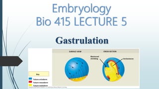
- 13. •Gastrulation begins on dorsal side Below the equator, in region of grey crescent. • Cells invaginate to form a slender blastopore. •Dorsal lip of blastopore will become important organizing region of embryo (Spemann organizer) •Cells become elongated as they contact the inner surface (Bottle cells). Future ectoderm Key Future endoderm Future mesoderm SURFACE VIEW Animal pole Vegetal pole Early gastrula Blastopore Blastocoel Dorsal lip of blasto- pore CROSS SECTION Dorsal lip of blastopore Bottle cell Cell Movements in Amphibian Gastrulation
- 14. • A small group of cells change shape, narrowing at the exterior edge of the blastula. • This change in cell shape, called apical constriction, creates a local invagination, which pushes more interior cells upwards and begins to roll a sheet of cells towards the interior. • The constricted cells are called bottle cells, due to their shape (like an upside-down bottle in these images). How does the blastopore lip form?
- 15. Gastrulation in the frog
- 16. Future ectoderm Key Future endoderm Future mesoderm SURFACE VIEW Blastocoel shrinking CROSS SECTION Archenteron Next steps: •Involution of the cells at the marginal zone (outer sheet spreads over inner sheet). Cells from Animal pole undergo epiboly Converge at the blastopore, When reach blastopore, travel inward Bottle cells continue to migrate, form leading edge of archenteron (primitive gut) . Cell Movements in Amphibian Gastrulation
- 17. Once the padded or gastrophila is formed until the Pericardial plug begins to decrease as a result of the crawl of the animal pole cells around it from each direction, then the lips eventually become a small longitudinal mischief known as the primitive line has two fine holes that close the abdomen and the dorsal remains open for a while and then close later. After the process of lining, the fetus transforms from a hollow ball with a single layer, into a three-layer outer body known as ectoderm, internal known as endoderm, and cells that migrate from the outside to the inside are the middle class, which is known as the Mesoderm. From these three embryonic layers, all organs of the fetus arise later. Gastrulation in Amphibian
- 18. SURFACE VIEW Blastopore Late gastrula Blastopore Blastocoel remnant Yolk plug CROSS SECTION Ectoderm Mesoderm Endoderm Archenteron Future ectoderm Key Future endoderm Future mesoderm •Mass of yolk left by surrounding blastopore = yolk. • plug Mass of yolk left by surrounding blastopore = yolk plug. •Cells from the dorsal lip (the first cells that migrated inward) become prechordal plate (will form head mesoderm). •Next cells that involute form chordamesoderm (will become notochord) Important for patterning the nervous system. Gastrulation in Amphibian
- 19. Future ectoderm Key Future endoderm Future mesoderm SURFACE VIEW Animal pole Vegetal pole Early gastrula Blastopore Blastocoel Dorsal lip of blasto- pore CROSS SECTION Dorsal lip of blastopore Late gastrula Blastocoel shrinking Archenteron Blastocoel remnant Archenteron Blastopore Blastopore Yolk plug Ectoderm Mesoderm Endoderm Gastrulation in Amphibian
- 20. What causes gastrulation? What factors are responsible for all of the complex and orchestrated movements that occur during gastrulation? There are at least 2 hypotheses and they are not mutually exclusive. 1. Cellular behaviors observed in each gastrula region are caused by local accumulations of gene products that are already present in the fertilized oocyte. 2. One region of the blastula could be determined to organize the behavior of all other regions. The dorsal lip of the blastopore will become the spemann organizer that appears to be particularly important in regulating the fate of other areas. 2 genes, goosecoid and noggin from the embryonic organizer area seem to cause the cell invagination in the blastopore area to mediate this effect.
- 21. How does the blastopore lip form?
- 22. Gastrulation in the birds The epiblast and the hypoblast: • By the time a hen has laid an egg, the blastoderm contains some 20,000 cells. • At this time, most of the cells of the area pellucida remain at the surface, forming an "upper layer,“ called the epiblast. • While other area pellucida cells have delaminated and migrated individually into the sub germinal cavity to form hypoblast islands. • The resulting two-layered blastoderm (epiblast and hypoblast) is joined at the marginal zone of the area opaca, and the space between the layers forms a blastocoel.
- 23. • The avian embryo comes entirely from the epiblast. • The hypoblast does not contribute any cells to the developing embryo. • Rather, the hypoblast cells form portions of the external membranes, especially the yolk sac and the stalk linking the yolk mass to the endodermal digestive tube. • Hypoblast cells also provide chemical signals that specify the migration of epiblast cells. • The three germ layers of the embryo proper (plus the amnion) are formed solely from the epiblast. Gastrulation in the birds
- 24. The primitive streak • The major structural characteristic of avian, reptilian and mammalian gastrulation is the primitive streak. • The primitive streak becomes the blastopore lips of amniote embryos. • The primitive streak first arises from a local thickening of the epiblast at the posterior edge of the area pellucida, called Koller's sickle, and the epiblast above it. Gastrulation in the birds occur via
- 25. The Formation of primitive streak
- 26. The Formation of primitive streak 1. The streak is First visible as cells accumulate in the middle layer, Followed by a thickening of the epiblast at the posterior marginal zone. 2. This thickening is initiated by an increase in tile height (thickness) of the cells forming the center of the primitive streak. 3. The presumptive streak cells around them become globular and motile, and they appear to digest away the extracellular matrix underlying them. 4. This process allows these cells to undergo intercalation and convergent extension. 5. Convergent extension is responsible for the progression of the streak doubling in streak length is accompanied by a concomitant halving of its width. 6. As cells converge to form the primitive streak, a depression forms within the streak. This depression is called the primitive groove, and it serves as an opening through which migrating cells pass into the deep layers of the embryo.
- 28. • As soon as the primitive streak has formed, epiblast cells begin to migrate through it and into the blastocoel. • The streak thus has a continually changing cell population. Cells migrating through the anterior end pass down into the blastocoel and migrate anteriorly, forming the endoderm, head mesoderm, and notochord. • cells passing through the more posterior portions of the primitive streak give rise to the majority of mesodermal tissues. Formation Of Endoderm And Mesoderm
- 29. A cross-section through the embryo allows us to observe the three germ layers that form during gastrulation: ectoderm, mesoderm, and endoderm.
- 30. Mammalian Gastrulation • Birds and mammals are both descendants of reptilian species (albeit different reptilian species). • It is not surprising, that mammalian development parallels that of reptiles and birds. • the gastrulation movements of reptilian and avian embryos, which evolved as an adaptation to yolky eggs, are retained in the mammalian embryo even in the absence of large amounts of yolk.
- 31. Images of human embryos during gastrulation,13 - 19 days post ovulation. Notice the primitive streak, which is analogous to the blastopore of Xenopus.
- 33. Schematic diagram showing The derivation of tissues in human and rhesus monkey embryos