1. Mix&Go is a novel surface chemistry that uses metal polymers to activate surfaces for protein binding. It forms thin films below 5 nm that allow proteins to bind as mono-layers while maintaining stability and function.
2. Experiments showed that Mix&Go could activate a variety of surfaces for protein binding, including glass, polystyrene, and gold. It formed stable mono-layers of proteins like streptavidin and antibodies.
3. Mix&Go performed better than conventional methods in assays, requiring less capture protein to achieve similar signals. This suggests it forms cleaner mono-layers versus stacked multi-layers with other methods. Mix&Go could enable new biosensor designs requiring stringent protein-surface interfaces.
Simple methods for forming protein monolayers lab on-a-chip 2013
1. Simple Methods for Forming Protein Monolayers
Chung E, Cooper SJ, Ohse BT, Gao Y, Jennins D, Koudijs MM, Yang L, Ling T, Vukovic P, Wong A, Maeji,NJ
Anteo Diagnostics Ltd, Brisbane, Australia; www.anteodx.com
Results, Cont.
Fig.1. The polymeric metal ions of Mix&Go
chelate by avidity to available electron donating
groups on the synthetic substrate surface.
Since not all the chelation potential is used to
bind Mix&Go to the substrate, the remaining
chelation points of Mix&Go are available for
protein binding.
Therefore, Mix&Go can be considered a
“molecular glue”.
Mix&Go solutions comprise less than 0.5% cationic polymers in aqueous solution. The polymers are
<5,000D and like poly-Lysine (another cationic polymer) can form very thin films in the 1 nm range.
However, unlike poly-Lysine which depends on electrostatic interactions with carboxylic acid and other
acidic residues for binding to surfaces, Mix&Go depends on the coordination forces of the metal ions.
These coordination forces are essentially irreversible due to the multi-valent nature of metal polymers.
In addition, Mix&Go can be applied to many other surfaces, e.g. polystyrene (PS) which cannot form
stable ion pairs with poly-Lysine. But, like poly-Lysine coated surfaces which have residual amino
groups for subsequent binding with other molecules, Mix&Go surfaces have residual chelation potential
that can be utilised to bind proteins.
Previously, we have shown on a number of different nano- and micron-sized latex-type particles that
Mix&Go has the following advantages compared to standard benchmark methods:
Rapid and stable surface activation
Significant savings in antibody and particle use for equivalent or better performance
Increased sensitivity and dynamic range
Excellent reproducibility and scalability
Using two types of Mix&Go with different polymeric structures (designated N10 and C10), the objective
of our work was to demonstrate coating of proteins and other materials onto surfaces commonly used
in the fabrication of biosensors.
Methods
To activate surfaces for protein binding, the substrates in this study were immersed in Mix&Go C10 or
N10 solutions from 10 min up to 1 hr at room temperature. The substrates were subsequently washed
with deionised water and are ready for immediate use. Alternatively, activated Mix&Go surfaces can be
stored after drying.
Prior studies have shown that Mix&Go activated microparticles are stable for over 2 years. Preliminary
studies on Mix&Go glass slides and Mix&Go PS plates indicate that these coated substrates have
comparable stability.
Results
Comparison of assays performed in 96- vs. 384-well plates shows increased advantages with
miniaturization (Fig. 5). The advantages of Mix&Go are especially relevant in miniaturized devices and
lab-on-a-chip applications where a need to form clean boundaries between proteins and their
underlying surfaces exists.
14 Days @ 37°C
20,000
20,000
15,000
15,000
15,000
10,000
10,000
10,000
5,000
5,000
5,000
0
0
0
200 μg/mL
50 μg/mL
200 μg/mL
10 μg/mL
50 μg/mL
10 μg/mL
200 μg/mL
50 μg/mL
10 μg/mL
Fig. 2.
Biotinylated
mouse IgG was spotted
at 10, 50 and 200 g/ml
using a contact printer,
detected with goat antimouse Alexaflour 647,
and
read
using
ArrayWoRx scanner.
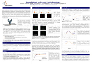
In order to investigate whether streptavidin slides had stability issues, one of a few commercially available
streptavidin slides was purchased to better understand streptavidin stability issues. These commercially
available slides are 3D surfaces having covalently coated streptavidin. Initial studies indicated
comparable performance between Mix&Go and commercially available streptavidin slides but on blocking
with excess Biotin, there was still comparable signal on the commercially available product while there
was no signal with Mix&Go slides. This data suggest that streptavidin binding to the commercially
available slides was non-specific, whereas binding to the Mix&Go slide was specific.
Streptavidin Mix&Go Slides
Commercial Streptavidin Slides
No Blocker
No Blocker
12.5 25 40 100 200
12.5 25 40 100 200
g/ml biotin-RPE
Fig. 3. Biotin-RPE was printed at 5 different
concentrations (12.5, 25, 40, 100, and 200
g/ml)
onto
commercially
available
streptavidin and streptavidin Mix&Go slides.
Subsequently, the slides were blocked with
biotin prior to printing. No Biotin-RPE binds
to the streptavidin Mix&Go slides whereas
significant – non-specific – binding can be
observed on the commercially available
streptavidin slides
g/ml biotin-RPE
Blocked with Biotin
Blocked with Biotin
12.5 25 40 100 200
12.5 25 40 100 200
g/ml biotin-RPE
g/ml biotin-RPE
It is common to use 3D surfaces to protect and maintain protein conformation. These studies show that
proteins as mono-layers can be just as or even more stable when Mix&Go coated surfaces are used.
3.000
3.000
Mix&Go
Mix&Go
2.500
2.500
Passive
2.000
2.000
1.500
1.500
1.000
1.000
0.500
0.500
0.000
Passive
0.000
0
1,500
3,000 4,500 6,000 7,500
Ag Concentration (pg/mL)
0
9,000 10,500
1,500
3,000
4,500
6,000
7,500
Ag Concentration (pg/mL)
9,000
10,500
Fig 5. With decreasing
surface area for capture
antibody
binding
the
relative performance of
Mix&Go surfaces over
conventional
passive
binding increases.
Mix&Go on Gold Surfaces
Silica oxide, titanium oxide, aluminium oxide, iron oxide and nearly all other metal oxides have residual
oxygen species on their surfaces which allows for direct chelation of Mix&Go to form an activated
surface for protein binding. However, inert gold surfaces were not considered an ideal surface for
Mix&Go. To test Mix&Go coating of inert gold and to see if Mix&Go affects electromagnetic waves, e.g.
in Localised Surface Plasmon Resonance (LSPR), Mix&Go was coated onto plain gold colloids of 100
nm. A λ-max shift of 4nm from 571nm to 575nm was observed (see Fig. 6). This λ-max shift may
reflect the poly-dispersity of the gold colloids and clearly indicates that activation of gold colloids is
easily achieved without serious aggregation or clumping of the colloids.
0.8
Fig. 6. 100 nm gold colloids (Sigma) were
coated with Mix&Go to form activated surfaces
for protein binding. The coating resulted in a λmax shift of 4nm. The shape of the curve
indicates improved dispersity of these colloids in
suspension after Mix&Go activation.
0.7
Mix&Go
0.6
Passive
0.5
0.4
0.3
0.1
0
400
450
500
550
600
650
700
750
800
Wavelength (nm)
Mix&Go on Polystyrene (PS) Surfaces
Greiner low binding 96-well microtitre plates (Cat. No. 655101) were activated with Mix&Go solution,
washed with deionised water, and dried to give Mix&Go activated plates. These plates were compared
to passive binding on Nunc Maxisorp plates in a capture antibody titration experiment. GM-CSF capture
antibodies at 7 concentrations (0.125, 0.25, 0.5, 1, 2, 4 and 8 g/ml) were coated onto these microtitre
plates (100 l), blocked with 1%BSA in PBS and tested with a standard antigen concentration of 5000
pg/ml. Detection was with biotinylated antibodies followed by streptavidin-HRP and TMB.
As shown in Fig. 4, maximum assay performance plateaus quickly when 0.25 g/ml or higher
concentrations of capture antibody are used to coat Mix&Go activated plates. In contrast, Nunc
Maxisorp plates, which bind antibodies by passive absorption, show a steady increase in signal that
suggests more antibodies are being coated onto the surface. While an increase in assay signal is
desirable, the data suggests antibody stacking to form a sterically hindered antibody layer. In contrast,
the Mix&Go data strongly indicates antibody mono-layer formation on the PS surface. In addition to a
cleaner boundary layer between antibody and underlying PS surface, Mix&Go better preserves antibody
function. This effect is more pronounced on smaller surface areas where each antibody is more
relevant.
Mix&Go on Glass Microarray Slides
OD (450-620nm)
2
It is well-known that conformational stability of proteins is often poor on such “2D” glass slides.
Streptavidin is reputed to be an especially difficult protein for microarray slides. However, streptavidin on
Mix&Go activated slides showed no significant stability issues. Streptavidin slides stored at 14 days at
both RT and at 37°C performed well compared to freshly made streptavidin slides (Fig. 2).
Mix&Go 384-well Greiner vs. Nunc Maxisorp
0.2
2.5
Schott Nexterion Glass B slides were activated with Mix&Go solution, washed with deionised water, and
dried to give Mix&Go activated Schott slides. Using a 16-well gasket (ProPlate, Grace Bio-Labs),
streptavidin (Prozyme) was coated onto these slides using 50 g/ml streptavidin in MES buffer (pH5.2)
for 1 hr. The slides were subsequently blocked with 5% BSA.
Mix&Go 96-well Greiner vs. Nunc Maxisorp
OD (450-620nm)
14 Days @ 25°C
Freshly Coated
OD Normalized
Miniaturization and/or label-free detection methods create a need for stringent uniformity at the interface
between a synthetic surface and biomolecules, e.g. antibodies and other capture agents. However, direct
immobilization of antibodies to synthetic surfaces, such as silicon wafers, ceramics, or plastics damages
proteins and therefore a compromise needs to be made in most cases between having an antibody monolayer and maintaining its stability and function.
Mix&Go™, a novel chelation-based surface chemistry, was developed to enable protein binding onto most
surfaces used in point-of-care devices. Mix&Go’s metal polymers bind any surfaces having electron
donating potential to form a thin, stable, and activated surface. Each chelation point alone binds only
weakly but multiple chelation points together enable gentle yet strong protein binding. Mix&Go represents
a “one-size-fits-all” surface chemistry approach in situations where maximum antibody performance within
mono-layers is critical.
Results, Cont.
1.5
1
Mix&Go Greiner
0.5
Passive Nunc Maxisorp
0
0
1
2
3
4
5
cAb (mg/mL)
6
7
8
9
Fig 4. GM-CSF capture antibody titration
on Mix&Go activated Greiner plates was
compared to Nunc Maxisorp plates.
These Greiner plates cost approx. 10% of
Nunc plates and used far less capture
antibody to achieve a comparable signal
in a sandwich assay. Mix&Go hit a
plateau at just over 0.25 g/ml indicating
a monolayer. In contrast, Nunc plates
continued to creep slowly upwards
suggesting that antibodies were stacking
on top of others.
0.8
0.7
0.6
Mix&Go + IgG
OD Normalized
Introduction
0.5
Passive + IgG
0.4
0.3
Fig 7. On binding antibody/BSA there is a
further shift in λ-max of 9nm giving a total shift
of 13nm from the bare gold colloid. The
dispersity of these protein coupled colloids did
not change dramatically but the same procedure
on non-Mix&Go colloids led to severe clumping
of the colloids.
0.2
0.1
0
400
450
500
550
600
650
700
750
800
Wavelength (nm)
Co-addition of antibody/BSA protein mixture to Mix&Go activated gold colloids (total 10ug protein per
400 ul gold solution) lead to a further λ-max shift of 9nm to 584nm (Fig 7). These preliminary results
show that Mix&Go activation is not detrimental to label-free detection methods such as LSPR. More
interestingly, the activation procedure did not lead to serious clumping of the colloids, a very common
problem in modifying nanoparticles of all types.
Conclusion
We have used Mix&Go, an aqueous metal polymer solution, to create protein binding surfaces on
materials commonly used in biosensors whether they are nanoparticles or planer. Coating surfaces
with Mix&Go and binding proteins to these surfaces is an easy and fast process and consistent with
manufacturing of disposable consumables. Importantly, Mix&Go forms a very thin film in the low nm
region but still helps maintain protein functionality in a mono-layer. To date, Mix&Go has been shown
not to be detrimental to the application of label-free detection strategies such as LSPR. Mix&Go will
prove to be a easy-to-use, affordable and universal solution in biochip development and manufacture.