Ashley Otter - Research Poster - 15th Medical Biodefense Conference, Munich
•
1 like•268 views

Our data showing the use of our Anthrax bacteriophages entitled "The Enemy of My Enemy if My Friend: Anthrax Specific Bacteriophages." that was presented at the 15th Medical Biodefense Conference in Munich (April 26th - 29th 2016)
Recommended

Recommended
More Related Content
What's hot
What's hot (12)
Viewers also liked
Viewers also liked (12)
Similar to Ashley Otter - Research Poster - 15th Medical Biodefense Conference, Munich
Similar to Ashley Otter - Research Poster - 15th Medical Biodefense Conference, Munich (20)
Recently uploaded
Recently uploaded (20)
Ashley Otter - Research Poster - 15th Medical Biodefense Conference, Munich
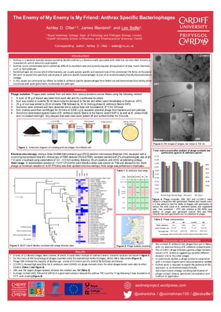
- 1. RW AB1 LC1H911 3B6 Plaque appearance Clear Turbid Clear Clear Capsid Diameter (nm) 62 ±6 70 ±5 77 ±6 123 ±6 Tail Length (nm) 197 ±20 344 ±32 406 ±47 449 ±40 Classification SiphoviridaeSiphoviridaeSiphoviridaeMyoviridae The Enemy of My Enemy is My Friend: Anthrax Specific Bacteriophages Ashley D. Otter1,2, James Blaxland2 and Les Baillie2. 1 Royal Veterinary College, Dept. of Pathology and Pathogen Biology, London. 2 Cardiff University School of Pharmacy and Pharmaceutical Sciences, Cardiff. Corresponding author: Ashley D. Otter – aotter@rvc.ac.uk aednetproject.wordpress.com Introduction • Anthrax is a bacterial zoonotic disease caused by Bacillus anthracis, a disease usually associated with cattle but can also infect humans, a characteristic which terrorists could exploit. • Anthrax spore contaminated land is extremely difficult to decontaminate and currently requires the application of toxic, harsh chemicals such as formaldehyde. • Bacteriophages are viruses which infect bacteria, are usually species specific and represent one of the commonest life forms on the planet • We wish to exploit this specificity and employ B. anthracis specific bacteriophages as part of an environmentally friendly decontamination strategy. • In this poster we summarize our efforts to isolate B. anthracis specific bacteriophages from Welsh soil and demonstrate their ability, when combined with spore germinants, to eliminate the pathogen. Discussion and Conclusion • We isolated B. anthracis lytic phages from soil in Wales with no previous history of B. anthracis contamination • The LC1H911 phage infected a gamma phage resistant variant of B. anthracis suggesting a possible different receptor site to the other phages • In preliminary studies, a phage cocktail in conjunction with a mixture of germinants reduced bacterial numbers • Further work is required to exploit the potential of this approach as an environmentally friendly decontamination strategy including optimisation of phage cocktail mixture, germinant concentration and method of application. Methods Phage isolation: Phages were isolated from soil taken from various locations across Wales using the following method: 1. A total of 25 g of topsoil was taken from each site and it’s coordinates recorded 2. Soil was stored in a sterile 50 ml falcon tube for transport to the lab and either used immediately or frozen at -20°C. 3. 25 g of soil was added to 25 ml ofsterile TSB followed by 10 ml mid-log phase B. anthracis Sterne 34F2. 4. Samples were vortexed and then decanted into a culture flask and incubated at 37°Cfor 24 hours 5. Soil slurries were then centrifuged for 20 mins at 5,000 x g to separate potential phage from bacteria and soil particles. 6. Solutions were tested against lawns of B. anthracis strains Sterne & the Sterne variantSdT12 as well as B. cereus 4342 and incubated overnight. Any plaques that were seen were picked off and purified further for 3 rounds. Electron microscopy: Using a Zeiss SIGMA field emission gun (FEG) electron microscope (Brighton Uni), equipped with a scanning transmission electron microscopy (STEM) detector (FEG-STEM), samples stained with 2%phosphotungstic acid at pH 7.4 were visualised using parameters of 3.2 – 3.5 mm working distance, 20 µm aperture and 20 kV accelerating voltage. Host range: A standardized solution of ~1x10⁸ CFU/ml for each Bacillus strain was spread on TSA and allowed to dry. Each phage (a minimum solution of 1x10⁸ PFU/ml) was then spotted onto the overlays. Host range was performed in triplicates. @asherichia / @cornishman100 / @lesbaillie1 Results • A total of 12 Bacillus phages were isolated, of which, 4 could infect multiple B. anthracis strains. Isolation locations are found in Figure 5. • On the basis of EM morphology all phages classified under the Siphoviridae family of phages, whilst 3B6 is Myoviridae(Figure 2). • Phage 3B6 infected the majority of Bacillus spp - a total of 47 strains out of a total of 58 (full data not shown). • LC1H911 showed high specificity for B. anthracis, even LSU463, a Ɣ phage resistant strain. No other phages tested were able to infect LSU463 (Table 1 and Figure 3). • 3B6 was the largest phages isolated, whereas the smallest was RW (Table 2). • A phage cocktail (AB1, RW and LC1H911) in a germinant solution reduced the anthrax TVC count by ~2 log following 5 days incubation at 37°C with shaking (Figure 4) Soil from sample site 25g soil Bacteria and phage separated Vortex + 25 ml TSB + 10 ml B. anthracis Centrifuge 20 mins. 5,000 x g 37°C 18 –24 hours Filter Phage solution Spot test Overlay showing phage plaques Figure 1: Schematic diagram of isolating anthrax phages from Welsh soil Table 2: Phage characteristics. Table 1: B. anthracis host range. LC1H911 AB1 3B6Gamma RW 100 nm Figure 2: EM images of phages, bar shown is 100 nm. Figure 4: Phage ’cocktails’ (RW, AB1 and LC1H911) were tested in conjunction with germinants Alanine and Inosine over a 5 day period to test the ability of phages with germinants to reduce the total count of B. anthracis spores and vegetative cells. Test conditions – 37ºC with shaking at 250 RPM, one addition of phage and germinant mixture. BHI and PBS controls had both germinants but no presence of phage. Total viablecountsafter addition of phage cocktails and germinants against B. anthracis spores Anthrax cluster Figure 3: MLST treeof Bacillus combined with phage infection data. Figure 5: Phage isolation locations.