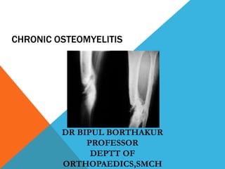
2. INTRODUCTION
Chronic osteomyelitis is an infection of bone and marrow of more than six weeks
duration characterised by recurrent attacks of inflammation with discharging
sinuses and presence of infected dead bone(sequestra).
Causes: pyogenic bacteria, mycobacteria, or fungus but the term usually indicate
pyogenic infection.
5. An acute becomes
chronic due to one of the
following reasons:
1. Improper drainage of
the pus in acute
osteomyelitis
2. Formation of an
undrained non
collapsable cavity in
the bone
6. 3. Presence of sequestra
4. Presence of foreign
bodies in case of
osteomyelitis following
open injuries
9. PATHOGENESIS
Infection at the bone locus
creates an increase of
intramedullary pressure as a
result of inflammatory
exudate stripping the
periosteum; this leads to
vascular thrombosis, followed
by bone necrosis and the
10. Usually, necrosis of the
large segments of bone
leads to sequestrum
formation. These
sequestra with infected
material are surrounded
by sclerotic bone that is
relatively avascular.
11. The haversian canals are
blocked with scar tissue, and
the bone is surrounded by
thickened periosteum and
scarred muscle
Antibiotics cannot penetrate
these relatively avascular
tissues and are hence
ineffective in clearing the
12. New bone formation occurs at
the same time (involucrum)
around the dead bone
Multiple openings appear in
this involucrum, (cloacae)
through which exudates and
debris from the sequestrum
pass via the sinuses.
14. Sequestrum:
It is a piece of dead bone
separated from healthy
bone. The area of dead
bone gets demarcated by
granulation tissue and
gradually separates and
forms a loose piece of
sequestrum.
15. Types of sequestrum-
1) corraliform- in pyogenic
infections
2) ivory sequestrum –
syphillis
3) feathery sequestrum- in
tuberculosis of long
bones
16. 4) sand sequestrum – in
vertebral tuberculosis
5) black sequestrum – in
fungal infections
6) ring sequestrum – in pin
tract infections and
amputation stumps
17. CLINICAL FEATURES
Presentation-
Unlike acute
osteomyelitis, chronic
osteomyelitis causes
no acute
constitutional
symptoms
The presenting
features may be those
of a long-standing,
discharging sinus or
chronic bone pain that
persists despite
treatment.
18. Patients may also
present with acute
exacerbations and
usually have a
history of acute
osteomyelitis,
sometimes dating
back to childhood.
Some times it may
present with a
pathological
19. On examination
drainage of pus
and small
sequestra through
the skin sinus is
found which is the
hall mark of
chronic
osteomyelitis.
23. Laboratory blood
studies are nonspecific
and gives no indication
of severity of infection.
ESR and CRP are
elevated in most
patients but WBC is
elevated only in 35%
24. DIAGNOSIS CONT..
X ray usually
shows bone
resorption with
thickening and
Sclerosis of the
surrounding
bone. A cavity
may appear as
an osteolytic
area.
28. DIAGNOSIS CONT..
M.R.I – More useful
for soft tissue
evaluation. It also
shows areas of bony
edema. It reveal an
area of high signal
intensity
surrounding the
active disease (rim
sign). Sinus tracts
also appears as
areas of increased
29. DIAGNOSIS CONT..
Isotope bone
scanning –
technetium 99m bone
scans which shows
increased uptake in
areas of increased
blood flow or
osteoblastic activity
but tend to lack
specificity. It has a
30. Gallium scan – show
increased uptake in areas of
leukocyte or bacteria
accumulation. A normal
gallium scan virtually
excludes osteomyelitis and
can be useful as follow up
examination.
31. Indium 111-labled leucocyte
scan- Specially useful to
differentiate chronic
osteomyelitis from reactive
bone disease or neuropathic
arthropathy.
Biopsy with culture
sensitivity – It is the gold
standard for establishing
diagnosis.
34. TREATMENT CONT.
Aims are –
1) adequate debridement
2) appropriate
reconstruction of bone and
soft tissue defect
3) appropriate antibiotic
therapy
35. SURGICAL TREATMENT
Sequestrectomy and
curettage – The
infected area of bone
is exposed and all
sinus tracks
completely excised.
The indurated
periosteum is incised
and elevated on both
sides. Drill is used to
outline a cortical
window at the
36. SURGICAL TREATMENT CONT
Remove all sequestra,
purulent material,
scarred and necrotic
tissue. If sclerotic
bone seals off a cavity
within medullary
canal, open it on both
directions to allow
blood vessels to grow
37. SURGICAL TREATMENT CONT
After removing
all suspicious
matter excise the
over hanging
edges of bone
and avoid leaving
are dead space.
38. SURGICAL TREATMENT CONT.
PAPINEAU technique-
this procedure is based
on following principles-
1) Granulation tissue
markedly resist
infection.
2) Autogenous
cancellous bone grafts
are rapidly
revascularized and are
resistant to infection.
39. SURGICAL TREATMENT CONT.
The operation is divided
into three stages-
a)excision of infected
tissue with or without
stabilization using an
external fixator or
intramedullary rod.
Dressing continued till
healthy appearing
granulation tissue is
40. SURGICAL TREATMENT CONT.
b)Cancellous bone
grafting in concentric
and overlapping layers
c)Wound coverage- in
some cases spontanous
epithelialization results,
otherwise skin grafts,
myocutaneous flaps or
muscle pedicle flaps can
be used.
41. SURGICAL TREATMENT CONT.
POLYMETHYLMETHAC
RYLATE(PMMA)
antibiotic bead chains-
the rationale for this
treatment is to deliver
high level of antibiotics
locally in
concentrations that
exceed the mic. The
antibiotic is leached
from beads into the
postoperative wound
hematoma and
secretion.
42. SURGICAL TREATMENT CONT.
Before the beads
are implanted the
infected and dead
tissue should be
debribed. the
beads are
implanted in the
bony defect.
43. SURGICAL TREATMENT CONT.
Aminoglycosides are
most commonly used,
but cephalosporins
and vancomycin also
used. Short term (10
days), long term (6
weeks), or permanent
implantation is
possible. The limb
should be
appropriately
44. SURGICAL TREATMENT CONT
Biodegradable Antibiotic
Delivery systems – Various
biodegradable antibiotic
delivery systems have been
evaluated. The main
advantage to these is that a
second procedure is not
required to remove the
implant.
45. SURGICAL TREATMENT CONT
Furthermore, some of these
biodegradable substrates
contain calcium, which can be
used in new bone formation. As
these beads resorb they are
slowly replaced by new bone
and soft tissue, and this process
may decrease the need for
further reconstructive or
coverage procedures.
46. SURGICAL TREATMENT CONT.
Soft Tissue Transfer – Soft
tissue transfers to fill dead
space left behind after
extensive debridement may
range from a localized muscle
flap on a vascular pedicle to
microvascular free tissue
transfer.
47. SURGICAL TREATMENT CONT.
The transfer of vascularised
muscle tissue improves the
local biological environment
by bringing in a blood
supply that is important in
the host’s defence
mechanisms, as well as for
antibiotic delivery and
osseous and soft tissue
48. SURGICAL TREATMENT CONT.
Most commonly a local muscle
flap is used in the treatment
of chronic osteomyelities of
the tibia. The gastrocnemius
muscle is used for defects
about the proximal third, and
soleus muscle is used for
defects at middle third.
49. SURGICAL TREATMENT CONT.
Ilizarov Technique – the
Ilizarov technique has been
helpful in the treatment of
chronic osteomyelitis and
infected non unions. This
technique allows radical
resection of the infected
bone.
50.
51. SURGICAL TREATMENT CONT.
A corticotomy is performed
through normal bone proximal
and distal to the area of
disease. The bone is
transported until union is
achieved.
Disadvantages include the time
required to achieve a solid
union and the high incidence of
52. SURGICAL TREATMENT CONT.
Hyperbaric Oxygen Therapy –
Hyperbaric oxygen therapy
has not proved to be reliably
effective. The use of
hyperbaric oxygen can be
recommended only as an
adjuvant to more traditional
methods of treatment.
55. BRODIE ABSCESS
A Brodie abscess is a
localized form of
subacute
osteomyelitis that
occurs most often
in the long bones of
the lower
extremities of
young adults.
Before physeal
closure, the
56. In adults the
metaphyseal-
epiphyseal area is
involved.
Intermittent pain
of long duration is
the presenting
complaint, along
with local
tenderness over
the affected area.
57. On plain roentgenograms a
Brodie abscess generally
appears as a lytic lesion with a
rim of sclerotic bone but can
have a markedly varied
appearance
Careful evaluation of plain films is
mandatory because a Brodie
abscess can be easily mistaken
for a variety of neoplasm
The lesion is thought to be
caused by organisms of low
virulence. S. aureus is cultured
in 50% of patients, in 20%
culture is negative.
58. This condition often
requires an open biopsy
with curettage to make
the diagnosis. The
wound should be closed
loosely over a drain.
59. GARRE’S OSTEOMYELITIS
Sclerosing Osteomyelitis of Garre –
Sclerosing osteoyemilitis is a
chronic from of disease in which
the bone is thickened and
distended but abscesses and
sequestra are absent
The disease affects children and
young adults
Its cause is unknown, but it is
thought to be an infection caused
by a low-grade, possibly anaerobic
60. Patients report intermittent
pain of moderate intensity
and usually of long duration.
Swelling and tenderness over
the affected bone may be
found
Roentgenograms show an
expanded bone with
generalized sclerosis
The ESR usually is slightly
elevated
Biopsy shows only chronic,
low-grade, nonspecific
61. A secondary lesion at a distant
site can occur years after
onset. No treatment has been
predictably helpful, but
fenestration of the sclerotic
bone and antibiotics are
advisable
The condition must be
distinguished from osteoid
62.
63. SALMONELLA OSTEOMYELITIS
Subacute type of
osteomyelitis usually
occurring in the ulna, ribs,
and vertebrae
Occurs some months or
years after attack of typhoid
or paratyphoid fever
Commonly associated with
sickle cell anaemia
64. Presents as an abscess
within the diaphysis of
bone
Blood widal tests may be
positive
Biopsy and culture
sensitivity done to
establish diagnosis.
Surgery is always required.
65. FUNGAL (MYCOTIC) INFECTION
Mycotic osteomyelitis is
the general term used to
describe a group of
diseases caused by
fungal infections of bone
There are two main
organisms- Actinomyces
and Maduramyces.
66. FUNGAL (MYCOTIC) INFECTION
Actinomycosis from cattle, occurs in
man in the soft tissues like mouth,
appendix, caecum and lung. Bone
affected secondarily, mandible most
commonly. The infection may spread
from lung to thoracic spine and from
caecum to pelvis. Multiple abscesses
result with the typical amorphous
yellow granules or sulphur granules
formed of fungal colonies.
67. FUNGAL (MYCOTIC) INFECTION CONT.
The affected bone has a
moth-eaten appearance. In
the spine the condition is
distinguished from
tuberculosis by sparing of
intervertebral discs and
absence of vertebral
collapse and kyphosis. The
heads of ribs and
transverse processes are
68. FUNGAL (MYCOTIC) INFECTION CONT.
Treatment is classically with the
penicillins, addition of
streptomycin or tetracycline
may be necessary. Antibiotic
should be continued for 6
month at least. Surgical
excision of the affected bone is
required for treatment of
69. MADURA FOOT:
~ first described by
Gill in 1832 from
madurai. The
organism usually
enter through a cut
in the foot, from
there they spread
through
subcutaneus tissue
and tendon sheaths.
Bones infected by
direct invasion
Patient may present
at early stage with
tender
70. ~ as the condition
forms tumour like
mass it was called
mycetoma
~ Swelling
gradually spreads
and blister forms
which ultimately
involves the whole
foot. X ray shows
multiple bony
cavities or
progressive bone
destruction.
71. ~ It later bursts
and forms
multiple
discharging
sinuses
~ pus contains
black granules
which are fungal
colonies from
which organism
can be isolated.
72. ~ Treatment :
1.penicillin or
dapsone orally
may be
effective but
usually
unsatisfactory,
i.v.
amphotericin B
is advocated
which is fairly
toxic.
74. TUBERCULOUS OSTEOMYELITIS
Tuberculous dactylitis
~ occurs in children and young
adults
~ infection starts in the shaft of the
phalanx and causes erosion and
gradual destruction of the bone
~ subperiosteal new bone
formation and thickening of bone
this phenomenon is peculiar to
the tuberculous infection in the
75. ~The surrounding
soft tissue also swell
up and cold abscess
often forms and
bursts to form
chronic sinuses
~ the patient
presents with a
painful spindle
shaped swelling of
finger which is called
spinosa ventosa
76. SYPHILITIC OSTEOMYELITIS
Syphilitic affections of bone occur in
the inherited and acquired forms of
the disease and in the latter they are
more serious in the tertiary stage.
They differ from tuberculus
affections in that the shaft is more
frequently involved while the joints
escape
The causative organism is Treponema
pallidum
The tibia, femur and humerus and the
cranial bones are most common
sites of syphilitic osteomyelitis.
77. SYPHILITIC OSTEOMYELITIS CONT.
Manifestations –
1. Pain – this may vary from slight
dull ache to most excruciating
pains. There are no local
abnormalitis on clinical exam. and
a diagnosis of neuralgia is often
made.
2. Periostitis – frequently occurs and
affects multiple long bones.
3. The periosteal node – the
characteristic lesion is a localized
swelling of shaft which usually
involves a portion of the
circumference, and may surround
78. 1. Diffuse osteoperiostitis – this
is a chronic inflammation
affecting the whole bone or
the greater portion of it inside
the periosteal envelope.
X-ray shows double outline
which is very characteristic. A
second sheath of compact
bone surrounds the original
compact layer but an
intervening space exists
which may be filled with
granulation tissue.
79. 2. Syphilitic osteochondritis –
children with inherited
syphilis show an irregularity
of the epiphyseal line. This
irregularity is due to
transformation of cartilage
into bone. There is
thickening of epiphysis and
pain on passive movement.
80. 3. Gummatous
Osteomyelitis - Gumma
can occur on surface of
the bone or within it. The
surface gumma
resembles an ordinary
periosteal nod except that
its speedily softens at its
centre. A gumma is in the
interior of long bone is a
serious condition as it is
mistaken for a malignant
81. 4. Syphilitic dactylitis – the
importance of syphilis of the
phalanges lies in the fact that it
may be mistaken for
tuberculosis. But there is little
tendency to break down and
ulcerate as in tuberculosis. The
condition is usually painless.
Antibiotics are usually
ineffective. Pathological
fracture from break down of
gamma needs stabilization.