SlideShare une entreprise Scribd logo
1  sur  1
Télécharger pour lire hors ligne
Myungsik Yoo, Christopher Park, Jennifer Moore, Melitta Schachner
W. M. Keck Center for Collaborative Neuroscience, Department of Cell Biology and Neurosciences, Rutgers University, Piscataway, NJ
Non-viral single doxycycline inducible system for human neural stem cell-derived
embryonic stem cells
1. Abstract
2. Introduction
3. Methodology
- Plasmid vector construction
The vector used for the initial construct was a pd2EGFP (Clontech) back bone,
subcloned with Chicken actin promoters (CAG) driving GFP and transactivator,
oppositely, and seven repeat tetracycline response elements (7XTRE) in between
the two CAG promotors.
- Predifferentiation of hNSC-ESC
We followed a slightly modified adherent monolayer differentiation protocol from
human embryonic stem cells, first published by Austin Smith’s lab (Nat, 2007)
- Transfection protocol with Amaxa to generate pTet-off GFP-hNSC-ESC
The transfection of proliferated pre-differentiated cells was done using the Amaxa
96 well transfection kit (Lonza, Amaxa)
- Clonal stable selection after transfection: N2a, hNSC-ESC
After 7 and 14 days, selection of N2a and hNSC-ESC clonal cells, respectively,
were picked into a new dish by G418 (200 mg/ml).
- Cell transplantation into cyclosporin immunosuppressed spinal cord
Cell transplantation (105 cells/ml) was performed immediately after opening of
spinal cord. 1 µL of the cells were transplanted at the center of spinal cord with
the injection lasting for 7 minutes.
Embryonic stem cell derivatives are potential avenues of therapy to cure
irreversible neuronal cell damage (Abdel-Salam 2011). In order to more efficiently
use embryonic stem cell derivatives, we hypothesized using an inducible
paradigm. Initially, the inducible system was developed in eukaryotic cells and
organisms using antibiotic and hormone regulating systems for clinical and basic
research, these were started in 1995 using chrimeric transactivator (tTA) fused
bacterial Tet repressor with viral protein 16 (VP-16) (Gossen et al. 1995).
Advanced binary tetR-mediated inducible gene regulation system, applicable to
eukaryotic cells had better regulation than the initial system by providing less
leaking and less toxicity (Yao et al. 1998). More recently, the lentiviral vectors were
developed for gene delivery to primary cells and non-immortalized cells (Cockrell
et al. 2007). However two major issues arised; the lenti-viral system could not
package the big-sized target cDNA well because of its limited size packaging and
the virus DNA sequences in the plasmid vector are silenced by the virus protection
mechanism in the cells (Suzuki et al. 2006). Thus, we have developed a novel
non-viral single doxycycline inducible system, which works efficiently in
immortalized neuroblastoma cells (N2a) by doxycycline and predifferentiated
hNSC-ESC. The clonal stable human cell line pTet-off GFP-hNSC-ESC also works
efficiently In vivo in the cyclosporin immunosuppressed spinal cord.
5. Conclusion
7. References
8. Funding
We have successfully constructed a non-viral single inducible system, which
efficiently regulates in immortalized neuroblastoma cells (N2a) and
predifferentiated hNSC-ESC. In addition, we found the system’s capability to
function in vivo testing, using GFP-hNSC cells line in the spinal cord.
These results suggest that the pTet-off GFP system can be delivered in a non-
viral manner and can be used to control the expression of doxycycline inducible
genes in stem cells in vitro and in vivo.
6. Discussion
New Jersey Commission for Spinal Cord Research
-Abdel-Salam OM. (2011). Stem cell therapy for Alzheimer's disease. CNS Neurol. Disord.
Drug. Targets 10: 459-485.
-Cockrell AS, Kafri T. (2007). Gene delivery by lentivirus vectors. Mol. Biotechnol. 36: 184-
204.
-Gossen M, Freundlieb S, Bender G, Muller G, Hillen W, Bujard H. (1995). Transcriptional
activation by tetracyclines in mammalian cells. Science 268: 1766-1769.
-Nat R, Nilbratt M, Narkilahti S, Winblad B, Hovatta O, Nordberg A. (2007). Neurogenic
neuroepithelial and radial glial cells generated from six human embryonic stem cell lines
in serum-free suspension and adherent cultures. Glia 55: 385-399.
-Suzuki M, Kasai K, Saeki Y. (2006). Plasmid DNA sequences present in conventional
herpes simplex virus amplicon vectors cause rapid transgene silencing by forming
inactive chromatin. J. Virol. 80: 3293-3300.
-Yao F, Svensjo T, Winkler T, Lu M, Eriksson C, Eriksson E. (1998). Tetracycline repressor,
tetR, rather than the tetR-mammalian cell transcription factor fusion derivatives,
regulates inducible gene expression in mammalian cells. Hum. Gene. Ther. 9: 1939-1950.
Figure 2. Plasmid vector map of non-viral single Tet-off GFP
system for doxycycline-dependant transgene regulation. The
vector, initially, used for the construct was a pd2EGFP (Clontech)
back bone, which was subcloned using multi cloning sites. The
genes, Chicken actin promoter (CAG), highly active in human
neural stem cells, which drives the GFP gene, and transactivator
gene, were separately constructed and subcloned to the final
vector oppositely. The PCR product of seven repeat tetracycline
response elements was subcloned between the two CAG
promotors. The benefits of this system are the use of a non-viral
sequence, single plasmid system driving two separate strong CAG
promoters, multiple tetracycline response elements, and a strong
repressor Kruppel-associated box fused with a tetracycline
repressor to enhance inducible.
A B C
A B
C
100
50
D
%cellviability
0 0.5 2
mg/ml dox
Figure 7. Doxycycline was non-toxic and did not affect GFP
expression in the non-inducible human neural GFP stem cell
line. Non-inducible human stable cell line generated after
transfected pCAG-GFP as the same protocol with inducible human
cell line. Cells without doxycycline (A), cells with 0.5 mg/ml
doxycycline (B), and cells with 2 mg/ml doxycycline (C). Cells
exposed with doxycycline (B and C) showed no difference in cell
number and viability to the cells lacking exposure to doxycycline
(D). Scale bars, 100 µm.
Figure 6. GFP off, time-dependently regulated using pTet-off GFP stable
clonal cell line human neural-stem cells (hNSC), by doxycycline.
Illustrated in A,B, and C, the cells exposed to doxycycline resulted in the
decrease of GFP. D, E, F illustrate cells in the absence of doxycycline,
resulting in the increase of GFP. Cells without doxycycline after 4 days (A),
cells with doxycycline 1 mg/ml after 4 days (B), and cells with doxycycline 1
mg/ml after 8 days (C). Scale bars, 100 µm
Figure 5. Pre-differentiated human H9 embryonic stem cells express
neural and neuronal markers. A) A phase contrast image of the cells
including rosette forming units. B, C, and D, illustrate immunofluorescence
staining of A2B5, Nestin, and doublecortin (DCX). Bar graph of cells positive
with immunoreactivity, most cells being positive for neural stem cell marker
nestin, about 20% of cells positive for glial progenitor marker A2B5, 15% of
cells positive for early neuronal progenitor marker DCX, however astrocyte
marker GFAP and embryonic stem cell marker Oct4 were not detected (H).
Scale bars, 100 µm.
Figure 4. GFP on and off, time-dependently regulated using
pTet-off GFP stable cell line N2a-#12 by doxycycline. Illustrated
in A,B, and C, the cells exposed to doxycycline resulted in the
decrease of GFP. D, E, F, and G illustrates cells in the absence of
doxycycline, resulting in the increase of green fluorescent protein.
Cells without doxycycline after 1 day (A), cells with doxycycline 1
mg/ml after 3 days (B), and cells with doxycycline 1 mg/ml after 6
days (C). Cells without doxycycline after 1 day (D), cells without
doxycycline after 3 days (E), cells without doxycycline after 6 days
(F), and cells without doxycycline after 9 days (G). Bar graph of
GFP on and off (H). Scale bars, 100 µm.
Figure 3. Generation of single clonal Tet-off GFP N2a stable cell lines.
After transfection of the Tet-off-GFP plasmid, the cell lines were selected by
G418 (200 mg/ml) for 7 days. Individual clonal cell lines were picked and
separately passaged. Cell lines N2a-#5 (A,D), N2a-#12 (B,E), N2a-#13 (C,F),
were tested without doxycycline (A, B,C) and with 1 mg/ml doxycycline (D, E,
F), for 5 days. The cell line N2a-#12 was the best cell line regulated by
doxycycline. The bar graph (G) marks the percentage of GFP present for
each cell line with and without doxycycline. Asterisks indicate significant
differences between the groups **p<0.01 assessed by one-way ANOVA
followed by Tukey’s post-hoc analysis. Error bars represent SEM. Scale bars,
100 µm.
Figure 1. Schematic representation of the mechanism for ptet-off-GFP
system using doxycycline. This system uses the single plasmid
doxycycline Tet-off system in which seven repeated tetracycline response
elements are located between two chicken actin promoters which drive
expression of GFP and the tetracycline reverse transactivator oppositely. (a)
In the absence of doxycycline, the tetracycline response elements are latent
to the two Chicken actin promotors, which drive expression of the green
fluorescence protein gene and the transactivator. (b) In the presence of
doxycycline, TREs recruit transactivators fused with a strong repressor
Kruppel-associated box, which leads to the silencing of two gene expression
through the inactivation of CAG promotors.
Figure 8. Non-viral single Tet-off hNSCs-GFP regulated by doxycycline, in vivo. The spinal cords were
transplanted with 10^5 cells in cyclosporin-immunosuppressed mice, and 7 days after transplantion the mice were
perfused and analyzed. The mice which were not fed doxycycline in drinking water, showed similar levels of GFP
and red quantum dot expression after 7 days (A). The mice which were fed doxycycline in drinking water, showed a
decrease of GFP expression while the quantum dot showed no significant change, after 7 days (B). The bar graph
(C), illustrates the comparison of the mice fed with doxycycline and the mice fed without doxycycline. In 7 days the
cells expressing GFP were reduced by about 61% and in 10 days the cells expressing GFP were reduced by about
56%. Asterisks indicate significant differences between the groups *p<0.05 as assessed by one-way ANOVA
followed by Tukey’s post-hoc analysis. Error bars represent SEM. Arrows (in A-B) indicate the injury site. Scale
bars, 0.3 mm
4. Results
The conventional approach for the doxycycline inducible system typically utilizes a
lentiviral delivery system to target the cell of interest. However, a significant
disadvantage of lentiviral delivery is the difficulty faced when trying to efficiently
package viral constructs that are larger than 10kb. We have successfully
developed a novel non-viral system that can express genes in response to
doxycycline in vivo and in vitro. After clonally isolating a stable mouse
neuroblastoma cell line (N2a) and the H9 human embryonic stem cell line (hNSC-
ESC), we were able to demonstrate that 0.5 mg/ml doxycyline completely
eliminated GFP expression after 3 days in N2a cells and 8 days in hNSC-ESC.
GFP expression was restored 9 days after doxycycline removal in N2a cells and
10 days after removal in hNSC-ESC. To test the ability of these cells to respond to
doxycycline in vivo, we transplanted 105 hNSC-ESCs labeled with quantum dots
(red; 655 nm) into the non-lesioned spinal cord. When the mice were subjected to
doxycycline, sacrificed and analyzed for the ratio of quantum dot expressing cells
to GFP expressing cells, we found that GFP expression was reduced by 61% and
56% in the doxycycline treated group. These results suggest that the pTet-off GFP
system can be delivered in a non-viral manner and can be used to control the
expression of doxycycline inducible genes in stem cells in vitro and in vivo.
The non-viral inducible single system used here was capable of efficiently
regulating (over 98%) in vitro conditions for immortalized neuroblastoma cells
(N2a) and pre-differentiated hNSC-ESC. However, in vivo results for hNSC-ESC
showed an estimate of 68% regulation.
The position of the tetracycline response elements (TREs) proves to be significant
for expression. The position of TREs must be placed at 5’ head in order to function
properly, if placed at 3’ end then expression will be inhibited, regardless of the
presence of doxycycline.

Contenu connexe

Tendances

Olivia_Creasey_Science2012_Poster
Olivia_Creasey_Science2012_PosterOlivia_Creasey_Science2012_Poster
Olivia_Creasey_Science2012_PosterOlivia Creasey
 
RNA-Seq To Identify Novel Markers For Research on Neural Tissue Differentiation
RNA-Seq To Identify Novel Markers For Research on Neural Tissue DifferentiationRNA-Seq To Identify Novel Markers For Research on Neural Tissue Differentiation
RNA-Seq To Identify Novel Markers For Research on Neural Tissue DifferentiationThermo Fisher Scientific
 
NIH Summer 2015 Poster_GabrielleDotson
NIH Summer 2015 Poster_GabrielleDotsonNIH Summer 2015 Poster_GabrielleDotson
NIH Summer 2015 Poster_GabrielleDotsonGabrielle Dotson
 
64 andrew fire - 6506559 - genetic inhibition by double-stranded rna
64   andrew fire - 6506559 - genetic inhibition by double-stranded rna64   andrew fire - 6506559 - genetic inhibition by double-stranded rna
64 andrew fire - 6506559 - genetic inhibition by double-stranded rnaMello_Patent_Registry
 
SHSARP paper final
SHSARP paper finalSHSARP paper final
SHSARP paper finalKaylee Racs
 
Targeted and Unbiased Screen for Genetic Suppressors of the Legionella pneumo...
Targeted and Unbiased Screen for Genetic Suppressors of the Legionella pneumo...Targeted and Unbiased Screen for Genetic Suppressors of the Legionella pneumo...
Targeted and Unbiased Screen for Genetic Suppressors of the Legionella pneumo...Chetan Hebbale
 
Global Gene Expression Profiles from Breast Tumor Samples using the Ion Ampli...
Global Gene Expression Profiles from Breast Tumor Samples using the Ion Ampli...Global Gene Expression Profiles from Breast Tumor Samples using the Ion Ampli...
Global Gene Expression Profiles from Breast Tumor Samples using the Ion Ampli...Thermo Fisher Scientific
 
OS18 - 8.a.2 Rational Design of Attenuated FMDV Vaccines by elevation of –Cpg...
OS18 - 8.a.2 Rational Design of Attenuated FMDV Vaccines by elevation of –Cpg...OS18 - 8.a.2 Rational Design of Attenuated FMDV Vaccines by elevation of –Cpg...
OS18 - 8.a.2 Rational Design of Attenuated FMDV Vaccines by elevation of –Cpg...EuFMD
 
Comparative analysis of gene regulation in mouse rat and human
Comparative analysis of gene regulation in mouse rat and humanComparative analysis of gene regulation in mouse rat and human
Comparative analysis of gene regulation in mouse rat and humanconstantina mylona
 
BrandeisBiologyREUResearchPoster_final_final
BrandeisBiologyREUResearchPoster_final_finalBrandeisBiologyREUResearchPoster_final_final
BrandeisBiologyREUResearchPoster_final_finalVictor Suarez
 
20140824 Abnormalities in human pluripotent cells due to reprogramming mechan...
20140824 Abnormalities in human pluripotent cells due to reprogramming mechan...20140824 Abnormalities in human pluripotent cells due to reprogramming mechan...
20140824 Abnormalities in human pluripotent cells due to reprogramming mechan...Alim Polat
 
Akhtar et al. Stem Cell Reports (2015)
Akhtar et al. Stem Cell Reports (2015)Akhtar et al. Stem Cell Reports (2015)
Akhtar et al. Stem Cell Reports (2015)Aslam Akhtar, MS
 
CRISPR cas9 mediated TERT disruption in cancer cells
CRISPR cas9 mediated TERT disruption in cancer cells CRISPR cas9 mediated TERT disruption in cancer cells
CRISPR cas9 mediated TERT disruption in cancer cells ChiLerFam
 

Tendances (20)

Olivia_Creasey_Science2012_Poster
Olivia_Creasey_Science2012_PosterOlivia_Creasey_Science2012_Poster
Olivia_Creasey_Science2012_Poster
 
RNA-Seq To Identify Novel Markers For Research on Neural Tissue Differentiation
RNA-Seq To Identify Novel Markers For Research on Neural Tissue DifferentiationRNA-Seq To Identify Novel Markers For Research on Neural Tissue Differentiation
RNA-Seq To Identify Novel Markers For Research on Neural Tissue Differentiation
 
ISMB2010_Poster
ISMB2010_PosterISMB2010_Poster
ISMB2010_Poster
 
Mekuria's poster
Mekuria's posterMekuria's poster
Mekuria's poster
 
PNAS 1992 FBH.PDF
PNAS 1992 FBH.PDFPNAS 1992 FBH.PDF
PNAS 1992 FBH.PDF
 
NIH Summer 2015 Poster_GabrielleDotson
NIH Summer 2015 Poster_GabrielleDotsonNIH Summer 2015 Poster_GabrielleDotson
NIH Summer 2015 Poster_GabrielleDotson
 
64 andrew fire - 6506559 - genetic inhibition by double-stranded rna
64   andrew fire - 6506559 - genetic inhibition by double-stranded rna64   andrew fire - 6506559 - genetic inhibition by double-stranded rna
64 andrew fire - 6506559 - genetic inhibition by double-stranded rna
 
Final UROP poster 2013
Final UROP poster 2013Final UROP poster 2013
Final UROP poster 2013
 
SHSARP paper final
SHSARP paper finalSHSARP paper final
SHSARP paper final
 
Targeted and Unbiased Screen for Genetic Suppressors of the Legionella pneumo...
Targeted and Unbiased Screen for Genetic Suppressors of the Legionella pneumo...Targeted and Unbiased Screen for Genetic Suppressors of the Legionella pneumo...
Targeted and Unbiased Screen for Genetic Suppressors of the Legionella pneumo...
 
Global Gene Expression Profiles from Breast Tumor Samples using the Ion Ampli...
Global Gene Expression Profiles from Breast Tumor Samples using the Ion Ampli...Global Gene Expression Profiles from Breast Tumor Samples using the Ion Ampli...
Global Gene Expression Profiles from Breast Tumor Samples using the Ion Ampli...
 
OS18 - 8.a.2 Rational Design of Attenuated FMDV Vaccines by elevation of –Cpg...
OS18 - 8.a.2 Rational Design of Attenuated FMDV Vaccines by elevation of –Cpg...OS18 - 8.a.2 Rational Design of Attenuated FMDV Vaccines by elevation of –Cpg...
OS18 - 8.a.2 Rational Design of Attenuated FMDV Vaccines by elevation of –Cpg...
 
Comparative analysis of gene regulation in mouse rat and human
Comparative analysis of gene regulation in mouse rat and humanComparative analysis of gene regulation in mouse rat and human
Comparative analysis of gene regulation in mouse rat and human
 
BrandeisBiologyREUResearchPoster_final_final
BrandeisBiologyREUResearchPoster_final_finalBrandeisBiologyREUResearchPoster_final_final
BrandeisBiologyREUResearchPoster_final_final
 
cronier 2007
cronier 2007cronier 2007
cronier 2007
 
Diaz-Arrastia, Ramon
Diaz-Arrastia, RamonDiaz-Arrastia, Ramon
Diaz-Arrastia, Ramon
 
20140824 Abnormalities in human pluripotent cells due to reprogramming mechan...
20140824 Abnormalities in human pluripotent cells due to reprogramming mechan...20140824 Abnormalities in human pluripotent cells due to reprogramming mechan...
20140824 Abnormalities in human pluripotent cells due to reprogramming mechan...
 
2017 BDSRA Berg CLN3
2017 BDSRA Berg CLN32017 BDSRA Berg CLN3
2017 BDSRA Berg CLN3
 
Akhtar et al. Stem Cell Reports (2015)
Akhtar et al. Stem Cell Reports (2015)Akhtar et al. Stem Cell Reports (2015)
Akhtar et al. Stem Cell Reports (2015)
 
CRISPR cas9 mediated TERT disruption in cancer cells
CRISPR cas9 mediated TERT disruption in cancer cells CRISPR cas9 mediated TERT disruption in cancer cells
CRISPR cas9 mediated TERT disruption in cancer cells
 

Similaire à NJ Stem Cell Symposium 2011 Abstract

A putative mesenchymal stem cells population isolated from adult human testes
A putative mesenchymal stem cells population isolated from adult human testesA putative mesenchymal stem cells population isolated from adult human testes
A putative mesenchymal stem cells population isolated from adult human testes◂ Justin (M) Gaines ▸
 
Pells et al [2015] PLoS ONE 10[7] e0131102
Pells et al [2015] PLoS ONE 10[7] e0131102Pells et al [2015] PLoS ONE 10[7] e0131102
Pells et al [2015] PLoS ONE 10[7] e0131102Steve Pells
 
20614 ftp
20614 ftp20614 ftp
20614 ftpsofiles
 
Gene expression profiling in apoptotic mcf 7 cells infected with newcastle di...
Gene expression profiling in apoptotic mcf 7 cells infected with newcastle di...Gene expression profiling in apoptotic mcf 7 cells infected with newcastle di...
Gene expression profiling in apoptotic mcf 7 cells infected with newcastle di...Mohamed Khalid Ali Xundhur
 
Discovering my research interest by eric garson sheffield university 2017
Discovering my research interest by eric garson   sheffield university 2017Discovering my research interest by eric garson   sheffield university 2017
Discovering my research interest by eric garson sheffield university 2017Eric Garson
 
UTF-8''Final Assessing post-synaptic partners of Dentate Granule Cells in a M...
UTF-8''Final Assessing post-synaptic partners of Dentate Granule Cells in a M...UTF-8''Final Assessing post-synaptic partners of Dentate Granule Cells in a M...
UTF-8''Final Assessing post-synaptic partners of Dentate Granule Cells in a M...Grant Pizzo
 
FINALDRAFT_JAKE_LIZZIE_POSTER_2014_2
FINALDRAFT_JAKE_LIZZIE_POSTER_2014_2FINALDRAFT_JAKE_LIZZIE_POSTER_2014_2
FINALDRAFT_JAKE_LIZZIE_POSTER_2014_2Lizzie Paquette
 
ISSCR-2014-poster
ISSCR-2014-posterISSCR-2014-poster
ISSCR-2014-posterAlicia Lee
 

Similaire à NJ Stem Cell Symposium 2011 Abstract (20)

A putative mesenchymal stem cells population isolated from adult human testes
A putative mesenchymal stem cells population isolated from adult human testesA putative mesenchymal stem cells population isolated from adult human testes
A putative mesenchymal stem cells population isolated from adult human testes
 
Pells et al [2015] PLoS ONE 10[7] e0131102
Pells et al [2015] PLoS ONE 10[7] e0131102Pells et al [2015] PLoS ONE 10[7] e0131102
Pells et al [2015] PLoS ONE 10[7] e0131102
 
14825.full
14825.full14825.full
14825.full
 
Fall project
Fall projectFall project
Fall project
 
Research Article Presentation.pptx
Research Article Presentation.pptxResearch Article Presentation.pptx
Research Article Presentation.pptx
 
Grindberg - PNAS
Grindberg - PNASGrindberg - PNAS
Grindberg - PNAS
 
20614 ftp
20614 ftp20614 ftp
20614 ftp
 
Medicina
Medicina Medicina
Medicina
 
Gene expression profiling in apoptotic mcf 7 cells infected with newcastle di...
Gene expression profiling in apoptotic mcf 7 cells infected with newcastle di...Gene expression profiling in apoptotic mcf 7 cells infected with newcastle di...
Gene expression profiling in apoptotic mcf 7 cells infected with newcastle di...
 
Gene expression profiling in apoptotic mcf 7 cells infected with newcastle di...
Gene expression profiling in apoptotic mcf 7 cells infected with newcastle di...Gene expression profiling in apoptotic mcf 7 cells infected with newcastle di...
Gene expression profiling in apoptotic mcf 7 cells infected with newcastle di...
 
Oncogene1.PDF
Oncogene1.PDFOncogene1.PDF
Oncogene1.PDF
 
Discovering my research interest by eric garson sheffield university 2017
Discovering my research interest by eric garson   sheffield university 2017Discovering my research interest by eric garson   sheffield university 2017
Discovering my research interest by eric garson sheffield university 2017
 
Cancer Research 2
Cancer Research 2Cancer Research 2
Cancer Research 2
 
Ferretti et al., BBI 2016
Ferretti et al., BBI 2016Ferretti et al., BBI 2016
Ferretti et al., BBI 2016
 
PlOSone paper
PlOSone paperPlOSone paper
PlOSone paper
 
UTF-8''Final Assessing post-synaptic partners of Dentate Granule Cells in a M...
UTF-8''Final Assessing post-synaptic partners of Dentate Granule Cells in a M...UTF-8''Final Assessing post-synaptic partners of Dentate Granule Cells in a M...
UTF-8''Final Assessing post-synaptic partners of Dentate Granule Cells in a M...
 
FINALDRAFT_JAKE_LIZZIE_POSTER_2014_2
FINALDRAFT_JAKE_LIZZIE_POSTER_2014_2FINALDRAFT_JAKE_LIZZIE_POSTER_2014_2
FINALDRAFT_JAKE_LIZZIE_POSTER_2014_2
 
Zeine et al. J. Neuroimmunology 1993
Zeine et al. J. Neuroimmunology 1993Zeine et al. J. Neuroimmunology 1993
Zeine et al. J. Neuroimmunology 1993
 
1584-09
1584-091584-09
1584-09
 
ISSCR-2014-poster
ISSCR-2014-posterISSCR-2014-poster
ISSCR-2014-poster
 

NJ Stem Cell Symposium 2011 Abstract

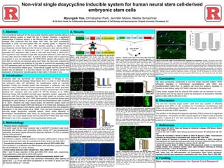
  • 1. Myungsik Yoo, Christopher Park, Jennifer Moore, Melitta Schachner W. M. Keck Center for Collaborative Neuroscience, Department of Cell Biology and Neurosciences, Rutgers University, Piscataway, NJ Non-viral single doxycycline inducible system for human neural stem cell-derived embryonic stem cells 1. Abstract 2. Introduction 3. Methodology - Plasmid vector construction The vector used for the initial construct was a pd2EGFP (Clontech) back bone, subcloned with Chicken actin promoters (CAG) driving GFP and transactivator, oppositely, and seven repeat tetracycline response elements (7XTRE) in between the two CAG promotors. - Predifferentiation of hNSC-ESC We followed a slightly modified adherent monolayer differentiation protocol from human embryonic stem cells, first published by Austin Smith’s lab (Nat, 2007) - Transfection protocol with Amaxa to generate pTet-off GFP-hNSC-ESC The transfection of proliferated pre-differentiated cells was done using the Amaxa 96 well transfection kit (Lonza, Amaxa) - Clonal stable selection after transfection: N2a, hNSC-ESC After 7 and 14 days, selection of N2a and hNSC-ESC clonal cells, respectively, were picked into a new dish by G418 (200 mg/ml). - Cell transplantation into cyclosporin immunosuppressed spinal cord Cell transplantation (105 cells/ml) was performed immediately after opening of spinal cord. 1 µL of the cells were transplanted at the center of spinal cord with the injection lasting for 7 minutes. Embryonic stem cell derivatives are potential avenues of therapy to cure irreversible neuronal cell damage (Abdel-Salam 2011). In order to more efficiently use embryonic stem cell derivatives, we hypothesized using an inducible paradigm. Initially, the inducible system was developed in eukaryotic cells and organisms using antibiotic and hormone regulating systems for clinical and basic research, these were started in 1995 using chrimeric transactivator (tTA) fused bacterial Tet repressor with viral protein 16 (VP-16) (Gossen et al. 1995). Advanced binary tetR-mediated inducible gene regulation system, applicable to eukaryotic cells had better regulation than the initial system by providing less leaking and less toxicity (Yao et al. 1998). More recently, the lentiviral vectors were developed for gene delivery to primary cells and non-immortalized cells (Cockrell et al. 2007). However two major issues arised; the lenti-viral system could not package the big-sized target cDNA well because of its limited size packaging and the virus DNA sequences in the plasmid vector are silenced by the virus protection mechanism in the cells (Suzuki et al. 2006). Thus, we have developed a novel non-viral single doxycycline inducible system, which works efficiently in immortalized neuroblastoma cells (N2a) by doxycycline and predifferentiated hNSC-ESC. The clonal stable human cell line pTet-off GFP-hNSC-ESC also works efficiently In vivo in the cyclosporin immunosuppressed spinal cord. 5. Conclusion 7. References 8. Funding We have successfully constructed a non-viral single inducible system, which efficiently regulates in immortalized neuroblastoma cells (N2a) and predifferentiated hNSC-ESC. In addition, we found the system’s capability to function in vivo testing, using GFP-hNSC cells line in the spinal cord. These results suggest that the pTet-off GFP system can be delivered in a non- viral manner and can be used to control the expression of doxycycline inducible genes in stem cells in vitro and in vivo. 6. Discussion New Jersey Commission for Spinal Cord Research -Abdel-Salam OM. (2011). Stem cell therapy for Alzheimer's disease. CNS Neurol. Disord. Drug. Targets 10: 459-485. -Cockrell AS, Kafri T. (2007). Gene delivery by lentivirus vectors. Mol. Biotechnol. 36: 184- 204. -Gossen M, Freundlieb S, Bender G, Muller G, Hillen W, Bujard H. (1995). Transcriptional activation by tetracyclines in mammalian cells. Science 268: 1766-1769. -Nat R, Nilbratt M, Narkilahti S, Winblad B, Hovatta O, Nordberg A. (2007). Neurogenic neuroepithelial and radial glial cells generated from six human embryonic stem cell lines in serum-free suspension and adherent cultures. Glia 55: 385-399. -Suzuki M, Kasai K, Saeki Y. (2006). Plasmid DNA sequences present in conventional herpes simplex virus amplicon vectors cause rapid transgene silencing by forming inactive chromatin. J. Virol. 80: 3293-3300. -Yao F, Svensjo T, Winkler T, Lu M, Eriksson C, Eriksson E. (1998). Tetracycline repressor, tetR, rather than the tetR-mammalian cell transcription factor fusion derivatives, regulates inducible gene expression in mammalian cells. Hum. Gene. Ther. 9: 1939-1950. Figure 2. Plasmid vector map of non-viral single Tet-off GFP system for doxycycline-dependant transgene regulation. The vector, initially, used for the construct was a pd2EGFP (Clontech) back bone, which was subcloned using multi cloning sites. The genes, Chicken actin promoter (CAG), highly active in human neural stem cells, which drives the GFP gene, and transactivator gene, were separately constructed and subcloned to the final vector oppositely. The PCR product of seven repeat tetracycline response elements was subcloned between the two CAG promotors. The benefits of this system are the use of a non-viral sequence, single plasmid system driving two separate strong CAG promoters, multiple tetracycline response elements, and a strong repressor Kruppel-associated box fused with a tetracycline repressor to enhance inducible. A B C A B C 100 50 D %cellviability 0 0.5 2 mg/ml dox Figure 7. Doxycycline was non-toxic and did not affect GFP expression in the non-inducible human neural GFP stem cell line. Non-inducible human stable cell line generated after transfected pCAG-GFP as the same protocol with inducible human cell line. Cells without doxycycline (A), cells with 0.5 mg/ml doxycycline (B), and cells with 2 mg/ml doxycycline (C). Cells exposed with doxycycline (B and C) showed no difference in cell number and viability to the cells lacking exposure to doxycycline (D). Scale bars, 100 µm. Figure 6. GFP off, time-dependently regulated using pTet-off GFP stable clonal cell line human neural-stem cells (hNSC), by doxycycline. Illustrated in A,B, and C, the cells exposed to doxycycline resulted in the decrease of GFP. D, E, F illustrate cells in the absence of doxycycline, resulting in the increase of GFP. Cells without doxycycline after 4 days (A), cells with doxycycline 1 mg/ml after 4 days (B), and cells with doxycycline 1 mg/ml after 8 days (C). Scale bars, 100 µm Figure 5. Pre-differentiated human H9 embryonic stem cells express neural and neuronal markers. A) A phase contrast image of the cells including rosette forming units. B, C, and D, illustrate immunofluorescence staining of A2B5, Nestin, and doublecortin (DCX). Bar graph of cells positive with immunoreactivity, most cells being positive for neural stem cell marker nestin, about 20% of cells positive for glial progenitor marker A2B5, 15% of cells positive for early neuronal progenitor marker DCX, however astrocyte marker GFAP and embryonic stem cell marker Oct4 were not detected (H). Scale bars, 100 µm. Figure 4. GFP on and off, time-dependently regulated using pTet-off GFP stable cell line N2a-#12 by doxycycline. Illustrated in A,B, and C, the cells exposed to doxycycline resulted in the decrease of GFP. D, E, F, and G illustrates cells in the absence of doxycycline, resulting in the increase of green fluorescent protein. Cells without doxycycline after 1 day (A), cells with doxycycline 1 mg/ml after 3 days (B), and cells with doxycycline 1 mg/ml after 6 days (C). Cells without doxycycline after 1 day (D), cells without doxycycline after 3 days (E), cells without doxycycline after 6 days (F), and cells without doxycycline after 9 days (G). Bar graph of GFP on and off (H). Scale bars, 100 µm. Figure 3. Generation of single clonal Tet-off GFP N2a stable cell lines. After transfection of the Tet-off-GFP plasmid, the cell lines were selected by G418 (200 mg/ml) for 7 days. Individual clonal cell lines were picked and separately passaged. Cell lines N2a-#5 (A,D), N2a-#12 (B,E), N2a-#13 (C,F), were tested without doxycycline (A, B,C) and with 1 mg/ml doxycycline (D, E, F), for 5 days. The cell line N2a-#12 was the best cell line regulated by doxycycline. The bar graph (G) marks the percentage of GFP present for each cell line with and without doxycycline. Asterisks indicate significant differences between the groups **p<0.01 assessed by one-way ANOVA followed by Tukey’s post-hoc analysis. Error bars represent SEM. Scale bars, 100 µm. Figure 1. Schematic representation of the mechanism for ptet-off-GFP system using doxycycline. This system uses the single plasmid doxycycline Tet-off system in which seven repeated tetracycline response elements are located between two chicken actin promoters which drive expression of GFP and the tetracycline reverse transactivator oppositely. (a) In the absence of doxycycline, the tetracycline response elements are latent to the two Chicken actin promotors, which drive expression of the green fluorescence protein gene and the transactivator. (b) In the presence of doxycycline, TREs recruit transactivators fused with a strong repressor Kruppel-associated box, which leads to the silencing of two gene expression through the inactivation of CAG promotors. Figure 8. Non-viral single Tet-off hNSCs-GFP regulated by doxycycline, in vivo. The spinal cords were transplanted with 10^5 cells in cyclosporin-immunosuppressed mice, and 7 days after transplantion the mice were perfused and analyzed. The mice which were not fed doxycycline in drinking water, showed similar levels of GFP and red quantum dot expression after 7 days (A). The mice which were fed doxycycline in drinking water, showed a decrease of GFP expression while the quantum dot showed no significant change, after 7 days (B). The bar graph (C), illustrates the comparison of the mice fed with doxycycline and the mice fed without doxycycline. In 7 days the cells expressing GFP were reduced by about 61% and in 10 days the cells expressing GFP were reduced by about 56%. Asterisks indicate significant differences between the groups *p<0.05 as assessed by one-way ANOVA followed by Tukey’s post-hoc analysis. Error bars represent SEM. Arrows (in A-B) indicate the injury site. Scale bars, 0.3 mm 4. Results The conventional approach for the doxycycline inducible system typically utilizes a lentiviral delivery system to target the cell of interest. However, a significant disadvantage of lentiviral delivery is the difficulty faced when trying to efficiently package viral constructs that are larger than 10kb. We have successfully developed a novel non-viral system that can express genes in response to doxycycline in vivo and in vitro. After clonally isolating a stable mouse neuroblastoma cell line (N2a) and the H9 human embryonic stem cell line (hNSC- ESC), we were able to demonstrate that 0.5 mg/ml doxycyline completely eliminated GFP expression after 3 days in N2a cells and 8 days in hNSC-ESC. GFP expression was restored 9 days after doxycycline removal in N2a cells and 10 days after removal in hNSC-ESC. To test the ability of these cells to respond to doxycycline in vivo, we transplanted 105 hNSC-ESCs labeled with quantum dots (red; 655 nm) into the non-lesioned spinal cord. When the mice were subjected to doxycycline, sacrificed and analyzed for the ratio of quantum dot expressing cells to GFP expressing cells, we found that GFP expression was reduced by 61% and 56% in the doxycycline treated group. These results suggest that the pTet-off GFP system can be delivered in a non-viral manner and can be used to control the expression of doxycycline inducible genes in stem cells in vitro and in vivo. The non-viral inducible single system used here was capable of efficiently regulating (over 98%) in vitro conditions for immortalized neuroblastoma cells (N2a) and pre-differentiated hNSC-ESC. However, in vivo results for hNSC-ESC showed an estimate of 68% regulation. The position of the tetracycline response elements (TREs) proves to be significant for expression. The position of TREs must be placed at 5’ head in order to function properly, if placed at 3’ end then expression will be inhibited, regardless of the presence of doxycycline.