
Recommandé
Contenu connexe
Tendances
Tendances (20)
En vedette
En vedette (20)
Similaire à Cardiovascular System
Similaire à Cardiovascular System (20)
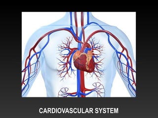
Cardiovascular System
- 2. CARDIOVASCULAR SYSTEM The cardiovascular system transports substances such as oxygen and nutrients between tissues and organs. It also helps to transport and eliminate waste products. The heart, blood vessels and blood form a sophisticated network that transports these materials around the body. They are carried by the blood through the blood vessels and are kept in motion by the pumping action of the heart.
- 3. The cells in the body need oxygen and it is the blood that brings it from the lungs to the various tissues and organs. When breathing, oxygen passes across the alveolar – capillary (respiratory) membrane and is picked up by the blood. Newly oxygenated blood travels along the pulmonary circuit to the heart, where it is pumped to other parts of the body via the systemic circuit. Once the blood reaches these other tissues, the oxygen it contains is released and exchanged for carbon dioxide. Deoxygenated blood is returned to the heart where it is pumped back to the lungs to drop off carbon dioxide and pick up oxygen, completing the cycle.
- 4. THE HEART This small muscle is able to pump approximately 5 to 6 litres of blood per minute, even when you are at rest. It pumps more than 7,000 litres of blood a day through an estimated 100,000km of blood vessels. Snugly enclosed within the mediastinum, the medial cavity of the thorax, the heart extends obliquely from the second rib to the fifth intercostal space. The human heart is a hollow, muscular pump that is divided into four chambers. The two top chambers are called atria, and the two lower chambers the ventricles. The chambers perform different functions. The atria collect the blood that enters the heart and push it to the ventricles, while the ventricles push blood out of the heart and into the arteries to go to the rest of the body. The two atria are separated by an interatrial septum while the interventricular septum divides the two ventricles.
- 5. BLOOD FLOW THROUGH THE HEART In order to move blood through your heart, your heart chambers undergo alternating periods of relaxation (diastole) and contraction (systole) allowing the chambers to fill up with, and pump blood, respectively. The regular contraction and relaxation of the heart is controlled by its electrical system. The heart must beat regularly to deliver oxygen to the body’s cells to keep it functioning properly. The right atrium of the heart receives deoxygenated blood from two major veins, the superior vena cava and the inferior vena cava, as well as a smaller coronary sinus that drains blood from the heart wall. When this chamber contracts blood moves out of the right atrium and into the right ventricle through the tricuspid valve. Once the right ventricle is sufficiently filled with blood this chamber contracts; pumping blood via the pulmonary arteries into the pulmonary circuit of the cardiovascular system. Newly oxygenated blood enters the left atrium of the heart through the pulmonary veins. Once this chamber is filled with blood the left atrial wall contracts; pushing blood into the left ventricle through the bicuspid valve. After the left ventricle has filled with blood this chamber contracts; forcing blood out of the ventricle and into the aorta. From the aorta, blood travels through the systemic circuit of the blood vessels, bringing oxygen to tissue cells throughout the body.
- 6. THE HEART VALVES The heart valves act as gates that allow blood to pass between heart chambers or from heart chambers to their associated blood vessels. They include the tricuspid, pulmonary, bicuspid (or mitral) and aortic valves. The tricuspid valve is located between the atrium and ventricle on the right side of the heart. When this valve is open, blood passes from the right atrium into the right ventricle. The tricuspid valve prevents the reverse of blood flow back into the atrium by closing during ventricular contraction. With the tricuspid valve closed, the only outlet for blood in the right ventricle is through the pulmonary trunk. The pulmonary trunk splits into the left and right pulmonary arteries which connect to the left and right lungs, respectively. The entrance to the pulmonary trunk is guarded by the pulmonary valve. The pulmonary valve is made up of three leaves that open when the right ventricle contracts and close when this chamber relaxes, allowing blood to flow from the right ventricle into the pulmonary arteries but not the reverse. The bicuspid or mitral valve regulates the flow of blood from the left atrium to the left ventricle. Like the tricuspid valve, the bicuspid valve closes during ventricular contraction. The bicuspid valve is composed of two leaves. The aortic valve consists of three leaves found at the entrance to the aorta. This valve lets blood out of the left ventricle as it contracts and blocks the pathway of blood from the aorta back into the left ventricle when this chamber relaxes.
- 7. BLOOD The blood transports nearly everything that must be transported within the body. It exits the heart via arteries which branch repeatedly until they become tiny capillaries. By diffusing across the capillary walls, oxygen and nutrients leave the blood and enter the body tissues, and carbon dioxide and wastes move from the tissues to the bloodstream. Blood is a sticky, opaque fluid with a characteristic metallic taste and is heavier, thicker and about five times more viscous than water. Depending on the amount of oxygen it is carrying, the colour of blood varies from scarlet (oxygen rich) to a dark red (oxygen poor). Its temperature is about 380 C, always slightly higher than body temperature. Blood is pumped through the body at a speed of about 30cm/second, with a complete circulation time of 20 seconds. The blood volume of an average-sized male is 5-6 litres; an average-sized female has 4-5 litres. Blood constitutes about 8% of the total body weight.
- 8. FUNCTIONS OF THE BLOOD • distribution - blood transports oxygen from the lungs to the cells of the body and carbon dioxide from the cells to the lungs; • it also carries nutrients from the gastrointestinal tract to the cells; heat and waste products away from the cells, and hormones from endocrine glands to other body cells; • regulation - blood regulates pH through buffers. It also adjusts body temperature through the heat-absorbing and coolant properties of its water content and its variable rate of flow through the skin where excess heat can be lost to the environment. Blood also influences the water content of cells, principally through dissolved ions and proteins; and • protection - the clotting mechanism protects against blood loss and certain phagocytic white blood cells or specialised plasma proteins such as antibodies protect against foreign microbes and toxins.
- 9. COMPOSITION OF BLOOD Blood is a liquid connective tissue composed of: • 45% of its volume is composed of formed elements (cells and cell fragments); and •55% is plasma (liquid containing dissolved substances). The formed elements constituting 45% of the volume consist of: •erythrocytes (red blood cells); •leucocytes (white blood cells); and •thrombocytes (platelets).
- 10. ERYTHROCYTES (RED BLOOD CELLS) Red blood cells make up over 99% of your blood cells. Blood gets its red colour from the protein haemoglobin within the red blood cells. Haemoglobin enables red blood cells to transport oxygen around the body. Production of erythrocytes takes place in red bone marrow. Erythrocytes are completely dedicated to their job of respiratory gas exchange (oxygen and carbon dioxide transport). Most oxygen carried in blood is bound to haemoglobin. The haemoglobin in erythrocytes combines easily and reversibly with oxygen and with carbon dioxide and transports them through blood vessels. When haemoglobin combines with oxygen, it forms a bright red compound called oxyhaemoglobin; when oxygen is released, the compound darkens and becomes deoxyhaemoglobin.
- 11. LEUCOCYTES (WHITE BLOOD CELLS) Leucocytes also develop in red bone marrow but are far less numerous than erythrocytes. Leucocytes are crucial to our defence against disease. They form a mobile army that helps protect the body from damage by bacteria, viruses, parasites, toxins and tumour cells. They combat these by engulfing them in a process called phagocytosis (eating).
- 12. THROMBOCYTES (PLATELETS) Platelets are fragments of cells that are less than half the size of red blood cells. Platelets help repair blood vessels by adhering to damaged walls and help to trigger blood coagulation (or clotting) which prevents bleeding and blood escaping from the blood vessel.
- 13. PLASMA Plasma is a straw-coloured liquid consisting of 91.5% water, containing a variety of dissolved substances. These include plasma proteins, nutrients, gases, electrolytes, hormones, enzymes and waste products.
- 14. BLOOD VESSELS The blood vessels form a closed system of tubes that carry blood away from the heart, transport it to the tissues of the body then return it to the heart. The three main types of blood vessels are arteries capillaries and veins.
- 15. ARTERIES Arteries are the blood vessels that carry blood away from the heart to the tissues of the body. They have two major properties, elasticity and contractibility. As the ventricles contract, blood is pushed into the arteries which expand to accommodate it. As the ventricles relax, the elastic recoil of the artery walls forces the blood onwards.
- 16. ARTERIOLES These are very small (almost microscopic) arteries that deliver blood to the capillaries. Arterioles plat a key role in regulating blood flow from the arteries into the capillaries. During vasoconstrictions, blood flow to the capillaries is restricted; during vasodilation, the flow is significantly increased.
- 17. CAPILLARIES Capillaries are the smallest blood vessels that connect from the arterioles to the venules. Their very thin walls enable the capillaries to be the site of exchange of nutrients and other substances (such as oxygen and carbon dioxide) between the blood and tissue calls of the body.
- 18. VENULES • When several capillaries unite. They form small veins called venules. Venules collect blood from capillaries and drain it into veins. Venules are similar in structure to arterioles; their walls are thinner near the capillaries and thicker as they progress toward the heart.
- 19. VEINS Veins carry blood towards the heart. By the time blood leaves the capillaries and moves into veins, it has lost a great deal of pressure. A simple way to remember: arteries and Arterioles carry blood away from the heart, while veins and venules bring it back to visit the heart.
- 20. HEART DISEASES Cardiovascular disease (CVD) is a collective term for diseases of the heart and blood vessels. The term commonly includes diseases such as coronary heart disease, heart failure, cardiomyopathy, congenital heart disease, peripheral vascular disease and stroke