2. SLO-Specific Learning Objectives
• Parts of male urethra
• Features of prostatic, membranous
and spongy urethra.
• Sphincters of urethra
• Clinical Anatomy
3. • DEFINITION-
• It is a common tubular passage for the elimination
of urine and semen.
• EXTENT-
• From the internal urethral orifice at the apex of
the trigone of the bladder to the external urethral
orifice to the tip of glans penis.
5. MEASUREMENTS-
• LENGTH-
• Total length- 20 cms
• Prostatic part- 3 cm
• Membranous part-
• Anterior wall- 2cm
• Posterior wall-1.25 cm
• Spongy part- 15 cm
• BREADTH-
• Average breadth-6mm
• Minimum breadth-(at external orifice)-4mm
• Membranous urethra-breadth- 5mm
• Maximum breadth-(at prostatic urethra)-8mm
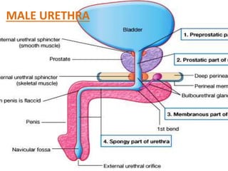
6. DESCRIPTION OF THE PARTS-
• PROSTATIC PART-
• It runs vertically downwards
from the base to the apex, at
the junction of anterior 1/3rd
and posterior 2/3rd of the gland.
It is the widest and most
dilatable part of the urethra.
• It is 3cm long, fusiform in shape
with crescentic shape with
convexity infront.
• In empty conditions anterior
and posterior walls come in
contact with each other.
7. FEATURES IN THE POSTERIOR WALL
• Urethral crest-It is a median longitudinal mucous
fold which gradually increases in height reaches
maximum as it descends; height is maximum
about 3mm in the middle. The crest is due to the
insertion of the trigonal muscle of bell.
• Colliculus seminalis-
• It is rounded elevation at the middle of the crest
and shows three openings- prostatic utricle in
the middle and ejaculatory duct on each side of
the utricle.
• Prostatic utricle-
• It is a mucous-cul-de-sac, about 6mm in length,
extends upwards and backwards from colliculus
to the median lobe of prostate. It is developed
from the paramesonephric ducts and
corresponds with development of vagina in
females; hence called as male vagina or known
as masculinus vaginus.
8. • Ejaculatory duct-
• It is 2 cm long and formed
by the union of vas
deferens and seminal
vesicle. It opens on each
side of the prostatic utricle.
• Prostatic sinuses-
• These are two mucosal
gutters on each side of the
crest and receive the
openings of the prostatic
glands
9. • MEMBRANOUS PART-
• It is the 5mm in diameter
and narrowest part of
urethra.
• It runs from the prostate
upto the bulb of penis.
• It pierces the perineal
membrane.
• On cross section it is
stellate shaped. It is
surrounded by the
sphinctre urethrae muscle.
10. • SPONGY URETHRA-
• It lies in the corpus spongiosum
penis and is 15 cm long.
• It passes through the bulb, body
and glans penis and terminates
at the external urethral orifice
close to the tip of glans penis.
• It shows two dilatations-
• Intrabulbar fossa- within the
bulb of penis. It is 3cm long.
• It receives the ducts of
bulbourethral glands.
• Terminal fossa (navicular fossa)
in the glans penis. It is 1.25 cm
long.
• Between these two dilatations
the urethra is uniform in calibre
of about 6mm.
11. • FEATURES OF SPONGY
URETHRA-
• Urethral glands-(Littre’s
gland)-
• These are simple tubular
glands which open in the
entire spongy part except the
terminal fossa.
• Urethral lacunae-
• These are pits like mucosal
recesses which project from
the entire urethra except the
terminal fossa.
• One lacuna is larger known as
lacuna magna whose mouth
is guarded by a mucosal fold
called as valvule of Guerin.
12. SPHINCTRES OF URETHRA-
• There are two sphinctres-
• Internal sphinctre or sphinctre vesicae-
• It is involuntary and surrounds the internal
urethral orifice.
• External urethral sphinctre-
• It is voluntary and is derived from sphinctre
urethrae muscle which surrounds
membranous urethra.
13. • BLOOD SUPPLY-AND-VENOUS DRAINAGE-
• By the branches of internal pudendal, inferior vesical, middle
rectal and urethral branch of the artery to the bulb of penis.
Veins correspond to the arteries.
• LYMPHATIC DRAINAGE-
• Prostatic part and membranous part- into internal and
external iliac nodes.
• Spongy part- into deep inguinal and into external iliac nodes.
• NERVE SUPPLY-
• Sympathetic nerves- from the L1 and L2 segments of spinal
cord; through the superior hypogastric plexus.
• Parasympathetic nerves- from the S2, S3 and S4 segments of
spinal cord through the pelvic spalnchnic nerves.
• Somatic nerves-for the terminal part through the pudendal
nerve.
14. DEVELOPMENT
• Prostatic part- above the colliculus seminalis from
the mesoderm of mesonephric ducts; and below
the colliculus from the endoderm of pelvic part of
urogenital sinus.
• Membranous part- from the endoderm of pelvic
part of urogenital sinus.
• Spongy urethra- upto the glans penis-from the
endoderm of phallic part of urogenital sinus and
the part within the glans penis from the ectoderm
on the undersurface of the glans penis.
15. APPLIED ANATOMY-
• DEVELOPMENTAL ANOMALIES
• Hypospadis-
• It is the condition where the
urethra opens, instead of tip of
glans penis, anywhere on the
undersurface of the penis.
16. • Epispadis-
• In this condition the
urethra opens on the
dorsal surface of the
penis close to the
anterior abdominal
wall.
17. • Ectopia vesicae-
• In this condition the
anterior abdominal wall
below umbilicus and
the anterior wall of the
bladder are deficient.
• Thus the interior of the
bladder is exposed
showing the trigone of
bladder.
18. • Extravasation of urine in males-
• Male urethra may be ruptured either
by instrumentation when a metal
catheter or a sound is inserted into the
bladder, or by an accidental fall on the
perineum.
• Usually membranous urethra is
ruptured in the superficial perineal
pouch.
• The urine is extravasated in the
superficial perineal pouch passes
forwards deep to dartos muscle of
scrotum, then to the fascia of penis
which leads to the oedematous
swelling of these structures.
• On further collection the urine may
appear in the anterior abdominal wall
deep to the fascia of Scarpa
(membranous layer of superficial
fascia) and extends upto the axilla, but
cannot appear infront of the thigh due
to the fusion of scarpas fascia with the
fascia lata.