
Recommandé
Recommandé
Contenu connexe
Tendances
Tendances (20)
En vedette
En vedette (20)
Similaire à Implant Maintenance and Disease Prevention
Similaire à Implant Maintenance and Disease Prevention (20)
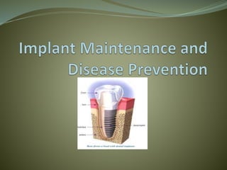
Implant Maintenance and Disease Prevention
- 2. Implant Checklist Health history, pharmacology, and dental health Assess Patient Self Care Evaluate Clinically and Radiographically Review and Document
- 3. What Do We See? Tissue consistency, form and color Inflammation Bleeding Exudate Bone level (radiographic) Occlusion and parafunctional habits Mobility Patient hygiene adequacy
- 5. Signs of Peri-implant Mucositis Red, shiny tissue Sensitivity Presents of plaque and calculus Bleeding Involves soft tissue only
- 7. Tissue Appearance In The Presence Of Peri-implantitis Red, purple, cyanotic Glossy, fibrotic Enlarged, cratered Bleeding Exudate
- 8. Probing Considerations Peri-mucosal seal Penetration of junctional epithelium Closer to proximal bone than around a natural tooth Difficult to get the probe parallel to the long axis of the implant Six sites are usually not attainable Anatomical situation is different Pocket Contamination Potential for damage
- 9. Probing Parameters Gentle Pressure In the 1st year all sites; evaluate color, form, consistency, BOP, depth After 1 year of stable probing use radiographs to check M/D Compare
- 10. Radiographs •Monitor M/D Bone •Diagnostically Acceptable •Slight change from baseline •Capture most apical bone •Vertical BW offers the least degree of distortion
- 11. Implant Maintenance Regular hygiene protocol (3-6 month intervals) Scaling only in the presence of plaque or calculus Polish with a fine paste only Yearly radiograph of implant(s)
- 12. Patient Education Review Homecare •Flossing Implants •Floss Threaders •Interdental brushes •Tapered end tuft brush •Antimicrobial Rinse
- 13. Auxiliary Aids