http://www.permadontics.com Dr. Berger and Dr. Aires are leading researchers in Dental implant technologies. Often lecturing and writing research papers for the industry and other doctors.
2. 2 Volume 31, Number 1, 2016
Aires/Berger
These guides were initially produced by dental labo-
ratories on gypsum dies. However, recent advances
in CAD/CAM technology utilizing stereolithographic
manufacturing of the guides have improved the
production and shown a higher predictability of
outcomes with better planning and accurate im-
plant placement.8,9
There is some evidence that placing the defini-
tive abutment on the day of implant insertion can
improve the chances of success.2 Further, patients
with a failing dentition usually wish to get teeth that
are functioning the same day of surgery. Patients re-
port higher satisfaction during osseointegration of
implants if they receive the prosthesis immediately
compared to delayed loading protocols.10
Implants can be placed in a tilted manner in con-
trast to the upright position of natural teeth. Tilt-
ing implants allows the use of longer implants and
is enabled by multiunit abutments. Furthermore,
it spares areas such as nerves, sinuses, or vessels
and allows a wider anterior-posterior spread to sup-
port the prosthesis, thereby reducing the extent of
cantilevers, which can fracture and are unfavorable
for loading implants.11 In addition, tilting can help
avoid major bone grafting. There is sufficient evi-
dence that tilting does not adversely affect implant
success.5
Tilted implants and immediate placement of im-
plants to replace failing dentitions are demanding
procedures. Surgical templates facilitate placement
of implants in the planned position and can be creat-
ed with planning software using computed tomog-
raphy (CT) or cone beam computed tomography
(CBCT) data. Another option would be to plan and
manufacture the guides on 3D stereolithographic
models. Planning implant positions with comput-
er-based methods and stereolithographic surgical
templates achieves high cumulative survival rates
(CSR) for implants at 1 year of follow-up.9 However,
to date, no study has planned the amount of bone
reduction and the location of the dental implants di-
rectly on three-dimensional (3D) stereolithographic
models. In such an approach, bone reduction is first
performed directly on the 3D model; drilling is per-
formed by the oral surgeon. The surgeon then per-
forms the implant osteotomies directly on the 3D
models. This is followed by fabrication of both the
bone reduction guide and the surgical guides. The
3D model allows further evaluation of the planned
placement according to the anatomical structure
of the patient prior to actual placement and manu-
facture of the guide on that basis. Here, the authors
performed a retrospective evaluation of implants
placed and immediately loaded in a series of con-
secutive patients with failing dentition using both
a bone reduction and a surgical guide that were
manufactured directly on 3D models.
MATERIALS AND METHODS
Patients
In the present study, the authors performed a non-
interventional, retrospective analysis of survival of
implants and prostheses for immediate-loading,
full-arch reconstructions. Consecutive patients of
both sexes who received a full-arch reconstruction
via stereolithographic 3D-model-based planning in
one or both arches and who displayed failing denti-
tion of at least two teeth were included. All patients
were treated at Permadontics (San Diego, California,
USA) between December 14, 2009 and September 23,
2013. Exclusion criteria for the implant treatment was
according to the routine procedures of the clinic and
included the following: (1) uncontrolled diabetes, (2)
being treated with bisphosphonates and displaying a
C-terminal telopeptide/CTX level below 100, (3) smok-
ers who refused to quit for 3 weeks prior and 3 weeks
after the procedure, (4) active use of cortisone medica-
tion, (5) previous radiation treatment to the head and
neck within the last 5 years, (6) advanced medical con-
ditions that prevented general anesthesia, or (7) inad-
equate bone, requiring bone grafting prior to implant
insertion. If one of these conditions was unknown for a
patient, implant treatment was at the discretion of the
primary care provider.
Planning Phase
Full presurgical and preprosthetic work-ups included
taking teeth impressions and bite registrations, and
a 3D scan was taken using a special scanning guide
that has fiduciary markers. Esthetic treatment plan-
ning, which consisted of shade, mold, tooth arrange-
ment, vertical dimension, and arch design, was then
accomplished.
Intraoral and extraoral photographs were taken of
all patients. Stone casts were used to fabricate pros-
theses, which were modified full dentures. Prostheses
were made according to the treatment design of the
prosthodontist, with an emphasis on arch form, occlu-
sal plane, and esthetics.
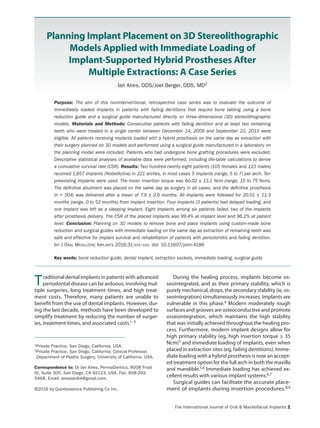
All patients received a CT scan to assess adequate
bone width and height. Digital Imaging and Commu-
nications in Medicine (DICOM) data from the CT scans
were sent electronically to a commercial laboratory
(Fig 1a). The scans were reformatted to 0.2 mm to al-
low 3D stereolithographic models (AccuDental, 3D
Systems) to be fabricated. Stereolithographic models
were based on the scans. These models showed all of
the important anatomical structures and the available
3. The International Journal of Oral Maxillofacial Implants 3
Aires/Berger
Fig 1 (a) CT image. (b) Soft tissue in-
dex. (c to e) Articulating the 3D stereo-
lithographic model. (f) Bone reduction
guide. (g) Surgical guide. (h, i) Provisional
prosthesis.
bone for implant placement. If many teeth were miss-
ing, a soft tissue index was fabricated (Fig 1b). The re-
cord base was then seated on the soft tissue index to
assist in the articulation of the 3D model (Fig 1c). The
models were articulated, and the working stone casts
were cross-articulated on the opposing models for
fabrication of the provisional prosthesis (Fig 1d). An in-
dex of the new teeth was fabricated to place on the 3D
model to help guide the placement of implants on the
3D bone model (Fig 1e).The surgeon then removed the
teeth from the model, and a precalculated amount of
bone (approximately 2 to 3 mm) was removed, which
created a flat platform and enabled a wider ridge for
implant insertion. Implant planning was accomplished
directly on the 3D bone models by the oral surgeon
without using software. During planning, the anterior-
posterior (A-P) spread was maximized by positioning
one implant in the middle of the arch and by position-
ing the most posterior implants as far as possible to the
distal sides. The surgeon then completed the surgery
a b
c d
f
h
e
g
i
4. 4 Volume 31, Number 1, 2016
Aires/Berger
on the model by drilling five to seven osteotomies for
the implant positions.
Implant analogs at the abutment level were insert-
ed into the 3D model. A bone reduction guide (Fig 1f)
and a surgical guide (Fig 1g) were then fabricated on
the stereolithographic model.
Clinical Treatment and Follow-up
All patients received full general anesthesia, and the
remaining teeth were extracted. A complete perios-
teal flap was made, which allowed seating of the bone
reduction guide over the bone, and the appropriate
amount of bone was removed. The surgical guide was
then seated on the new bone platform, anchored with
guide pins into the bone, and the implants (NobelAc-
tive, Nobel Biocare) were inserted according to the
preplanned surgical guide. Definitive abutments (mul-
tiunit abutment, Nobel Biocare) were then inserted.
These abutments were not removed and were used
as the definitive abutments for the final prostheses.
A full-arch impression with open trays was made, and
cover caps (Healing Caps, Nobel Biocare) were placed
on the abutments. The patients were then awakened
and brought to the prosthetic treatment rooms.
The caps were removed, and titanium cylinders
were attached to two implants, one on each side of
the arch. The prefabricated provisional prosthesis was
then fitted onto the cylinders, and the cylinders were
acrylized to the prosthesis‑ at the correct vertical and
centric positions. The provisional prosthesis was then
sent to the laboratory, where the remaining three cyl-
inders were attached, and the provisional prostheses
went through final processing and polishing. The pro-
visional prosthesis (Fig 1h) was delivered the same day,
and the prosthetic screws were torqued to 15 Ncm (Fig
1i). The occlusion was carefully evaluated. A modified
group function was obtained, in which the lateral ex-
cursions obtained contact on the canines and two pre-
molars but not on the molars. Patients were instructed
to eat a soft diet for 6 weeks. After 4 months, a defini-
tive prosthesis was fabricated with a milled titanium
bar and denture teeth. No study-specific follow-up
visits were requested, as treatment and recall followed
the standard procedure of the clinic.
Retrospective Analysis
All consecutive patients who met the following cri-
teria were included in the analysis: (1) patients pre-
sented with at least two existing teeth; (2) patients
had these teeth extracted, and 5 to 8 implants were
inserted immediately and loaded on the same day;
and (3) patients underwent the procedure using the
laboratory-made surgical guide described earlier. The
data, dropouts, withdrawals, and implant failures were
extracted by the treating dentists and were deidenti-
fied by the clinician.
Variables Assessed
The following variables were retrieved and analyzed:
• Patient-related data including age, sex, and smok-
ing history.
• Details of the surgical procedure, including wheth-
er the surgery used a flap or was flapless and was
one-stage or two-stage. The two-stage procedure
utilized a cover screw and primary flap closure with
a subsequent surgery for uncovering the implant.
The one-stage procedure used an abutment
Table 1 Patient Characteristics (n = 228)
Characteristics
No. failed
implants
Age (y)
Mean 63.1
Range 30-89
Sex, n (%)
Female 105 (46.1%) 2
Male 123 (53.9%) 4
History of Smoking, n (%)
Smoker 4 (1.8%) 4
Nonsmoker 224 (98.2%) 2
Table 2 Maxilla and Mandible Implant
Characteristics
Arch treated per patient n (%)
No. experiencing
failure
Maxilla only 77 (33.8%)
Mandible only 58 (25.4%)
Both arches 93 (40.8%)
No. of implants per patienta
5 118 (51.8%)
6 13 (5.7%)
7 4 (1.8%)
10 64 (28.1%)
11 19 (8.3%)
12 8 (3.5%)
13 2 (0.9%)
No. of implants per arch: maxilla/mandible
5b 140/130 2/2
6c 20/20 2/0
7d 10/1 1/0
aIncludes 10 preexisting implants and one submerged implant.
bIncludes one maxilla and one mandible in which preexisting implants
were used.
cIncludes two mandibles in which preexisting implants were used.
One implant in one mandible was left submerged.
dIncludes one maxilla in which preexisting implants were used.
5. The International Journal of Oral Maxillofacial Implants 5
Aires/Berger
(healing, provisional, or definitive) placed on the
day of surgery.
• Insertion torque as measured with a torque
wrench.
• The type of prosthesis, the type of abutment, and if
it was screw- or cement-retained.
The CSR was assessed by the actuarial life table
method according to Altman on implant basis and on
patient basis (ie, time to first failure). Statistical analysis
was performed using SPSS Statistics, version 19 (IBM).
RESULTS
Patient and Implant Characteristics
Two hundred twenty-eight patients (105 women
and 123 men) were consecutively treated with this
new planning method at the same clinic between
December 14, 2009 and September 23, 2013. Patients
ranged from 30 to 89 years of age at the time of surgery
(Table 1), and the majority of patients were nonsmok-
ers. Three hundred twenty-one surgical guides were
used to insert 1,657 implants in both arches (Table 2).
All implants were placed without bone grafting and by
raising a full flap. Implants were placed in either fresh
extraction or healed sites. In addition, 10 preexisting
implants were used (Table 3) and remained in func-
tion over follow-up; however, they were excluded from
implant-level analysis. The mean insertion torque was
60.02 ± 13.1 (range, 13 to 75) Ncm, and 49.8% of im-
plants (n = 817) were inserted with an insertion torque
of 70 Ncm.
The technique allowed for placement of all implant
widths and lengths ranging from 3.5 to 5.0 mm in diam-
eter and 8.5 to 18 mm in length (Table 4). In all surger-
ies, a hybrid prosthesis could be inserted the same day.
Table 3 Implant Failures and Deviations
Patient ID Sex
Age
(y) Smoker
Implant
site
Dimension
(mm)
Insertion
torque
(Ncm)
No.
implants
in arch
Multiunit
abutment
(deg)
Time to
failure
(mo)
Definitive
prosthesis
Implant failures
2 M 56 Y 15 4.3 × 15 50 7 17 27 Before
14 M 58 N 13 4.3 × 13 35 5 0 20 After
19 M 45 Y 15
23
5 × 8.5
4.3 × 13
50
50
5 0
17
8
8
After
After
93 F 70 N 45
35
5 × 15
5 × 15
35
35
5 0
0
5
5
Before
Before
133 M 30 Y 31 3.5 × 15 70 5 17 8 Before
158 F 39 Y 25 4.3 × 11.5 70 5 17 11 Before
Deviations
Brand Comments
19 M 45 Y 13 4.3 × 18 NR 5 NobelActive 2-stage, delayed loading,
included in prosthesis
24 F 60 N 23 4.3 × 13 NR 5 NobelActive 2-stage, delayed loading,
included in prosthesis
47 M 65 N 11
25
3.5 × 11
3.5 × 11
NR 6 NobelActive 2-stage, delayed loading,
included in prosthesis
76 M 63 N 43 4.3 × 15 30 6 NobelActive Left submerged, not
included in definitive
prosthesis (cover screw)
112 M 60 N 45
43
41
NR NR 6 NR Preexisting implant(s)
124 F 88 N 15 NR NR 5 NR Preexisting implant(s)
134 M 56 N 45 NR NR 5 NobelActive Preexisting implant(s)
186 M 80 N 15
13
NR NR 7 Straumann Preexisting implant(s)
226 F 76 N 32
34
36
NR NR 6 NobelActive Preexisting implant(s)
NR = not reported.
aFDI tooth-numbering system.
6. 6 Volume 31, Number 1, 2016
Aires/Berger
However, in four patients, a total of five implants were not
subjected to immediate loading. Four of those delayed
loaded implants (0.2%) were loaded between 4 and 7
months following surgery, and one unloaded implant was
not uncovered after the healing period but remained as
a sleeper implant (Table 3).
All of the definitive abutments on the NobelActive
implants were multiunit abutments (Nobel Biocare) in
0 degrees (n = 585, 35.5% and 4 failures), 17 degrees (n
= 972, 59.1% and 4 failures), or 30 degrees (n = 89, 5.4%
and no failures) and attached at the day of loading. Only
screw-retained hybrid prostheses were used for definitive
restoration. Several patients had not received a definitive
prosthesis yet. Among 321 arches, 304 received a defini-
tive prosthesis.The mean time from surgery to definitive
prosthesis was 7.9 ± 2.6 months (range, 1 to 22 months),
and the mean time from definitive prosthesis to the last
follow-up was 11.6 ± 11.8 months (range, 0 to 46 months).
Survival Rate
The mean follow-up time for all 1,657 implants was
20.01 ± 11.3 months (range, 0 to 52 months). The CSR
of the implant was 99.4% at the implant level (Table 5).
Because this was a retrospective study, one patient was
lost to follow-up directly after implant insertion.The mean
follow-up time for all 228 patients was 19.37 ± 11.1 months
(range, 0 to 52 months). The CSR of the implants at the
patient level was 96.2%. The CSR was determined based
on the first failure in a patient. Further failed implants or
further follow-up results of other implants in the same
patient were not taken into account.
The mean follow-up time of all definitive prostheses
was 11.58 ± 11.8 months (range, 0 to 46 months).The CSR
of the prosthesis at the level of the definitive prosthesis
was 98.8% at 3 years of follow-up (Table 5). Provisional
prostheses were not taken into account for this analysis.
Adverse Events
Eight implant failures occurred among six patients (Ta-
ble 3). Five maxillary implants among four patients and
three mandible implants among two patients failed. One
provisional prosthesis failed 6 weeks postoperatively,
but all others were successful. Twenty-two provisional
prostheses developed factures: six fractures during the
healing phase requiring repair in the laboratory and 16
fractures at the distal aspect of the cantilever sites, some
of which were trimmed and polished and others repaired
in the laboratory. Two definitive prostheses had to be
removed due to failures of implants and were classified as
failed restorations. One definitive prosthesis developed
a clear fracture of the titanium frame distal to the most
distal abutment. Multiple tooth fractures and debond-
ing occurred, and all were repaired in the laboratory in
≤ 2 hours.
DISCUSSION
Accurate stereolithographic bone models developed 5
to 6 years ago, with regard to occlusion, improved to the
point where the authors could articulate the models.
Here, the authors conducted a retrospective study of
implants placed and immediately loaded using 3D ste-
reolithographic models. Patients with failing dentition
received implants and final multiunit abutments on the
same day of surgery.
In addition to the benefits to the patients, 3D model-
ing is viewed as being particularly user-friendly among
craniofacial surgeons12 and highly reliable and accurate.13
Three-dimensional modeling in contrast to computer
planning of implant surgery does not require specific
software. Furthermore, while computer planning only
allows the surgeon to see the bone structure on a two-
dimensional screen, 3D models enable the surgeon to
physically examine the bone structure at all angles and use
this information to formulate a step-by-step approach to
the implant placement.The data presented here support
the concept of 3D stereolithographic models, demon-
strating feasibility and a high implant survival rate. In
this study, nearly all implants were immediately loaded
successfully. No serious adverse events were reported,
Table 4 Implant Dimensions
Diameter
(mm) Length (mm)
8.5 10.0 11.5 13.0 15.0 18.0
3.5 0 3 9 9 16 (1) 3
4.3 2 19 43 (1) 323 (2) 916 (1) 99
5.0 5 (1) 23 40 46 89 (2) 12
Numbers of patients experiencing failures are given in parentheses.
Table 5 Survival of Implants and Definitive
Prostheses
Time
period
No. of implants/
prostheses
No.
failed
No.
withdrawn CSR (%)
Implants
1 y 1,657 6 421 99.6
1–2 y 1,230 1 693 99.6
2–3 y 536 1 327 99.4
3–4 y 208 0 157 99.4
4 y 51 – – 99.4
Definitive prostheses
1 y 304a 1 184 99.7
1–2 y 119 1 62 98.8
2–3 y 56 0 40 98.8
3 y 16 – – 98.8
aOne implant had failed before insertion of prostheses.
7. The International Journal of Oral Maxillofacial Implants 7
Aires/Berger
and the minor adverse events that were reported were
mainly technical complications with the provisional or
definitive prostheses.
Immediate loading of dental implants in patients with
advanced periodontal disease is an accepted treatment
option.4,6,7,14 Presurgical planning directly on 3D stereo-
lithographic models can facilitate treatment of patients
in a timely manner and reduce or eliminate the need for
subsequent surgeries.13 Additionally, patients with a fail-
ing dentition can undergo extraction and accurate bone
reduction followed by immediate implant placement (in
healed and extraction sites) loaded with a prosthesis on
the same day.
Implants that enable high insertion torques allow for
immediate loading; the threshold value for immediate
loading is 35 Ncm. Only 3.2% of the implants reported
here were below that value, and by splinting, immedi-
ate loading was also feasible in these cases. Implants
restored with angulated multiunit abutments was a safe
procedure as previously reported for tilted implants.15
The present results build on earlier data that showed
favorable survival rates of 97% to 100% at 1 to 3 years
of follow-up using titanium implants with an oxidized
surface,1,9,16,17 including those of a previous study that
reported a 99.6% CSR for NobelActive implants up to 29
months after loading.5 This study has some limitations,
which include its retrospective nature, lack of compari-
son to other methods, limited follow-up, and limited
number of endpoints. However, a substantial number
of patients were followed, and a clear benefit was found.
Further research is needed to better assess the accuracy
of the method, costs, and time involved in using a 3D
stereolithographic model and compare these with those
of other digitally planned and widely accepted stereo-
lithographic guides that were not assessed in this study.
CONCLUSIONS
This retrospective investigation showed that planning
of custom-made bone reduction guides and custom-
made surgical guides fabricated on 3D stereolithographic
models facilitated implant placement in patients with
failing dentition.The treatment regimen of extraction of
failing dentition and loading on the same day using this
approach was safe and effective with respect to implant
survival rate.
ACKNOWLEDGMENTS
Medical writing services for this investigator-initiated study were
supported by Nobel Biocare Services AG, Kloten (ZH), Switzer-
land, study number 2014-1274. The authors report no conflicts
of interest related to this study.
REFERENCES
1. Degidi M, Perrotti V, Piattelli A. Immediately loaded titanium
implants with a porous anodized surface with at least 36 months of
follow-up. Clin Implant Dent Relat Res 2006;8:169–177.
2. Randow K, Ericsson I, Nilner K, Petersson A, Glantz PO. Immediate
functional loading of Brånemark dental implants. An 18-month
clinical follow-up study. Clin Oral Implants Res 1999;10:8–15.
3. Tarnow DP, Emtiaz S, Classi A. Immediate loading of threaded
implants at stage 1 surgery in edentulous arches: Ten consecutive
case reports with 1- to 5-year data. Int J Oral Maxillofac Implants
1997;12:319–324.
4. Cochran DL, Morton D, Weber HP. Consensus statements and
recommended clinical procedures regarding loading protocols
for endosseous dental implants. Int J Oral Maxillofac Implants
2004;19(suppl):109–113.
5. Babbush CA, Kutsko GT, Brokloff J. The all-on-four immediate func-
tion treatment concept with NobelActive implants: A retrospective
study. J Oral Implantol 2011;37:431–445.
6. Papaspyridakos P, Chen CJ, Chuang SK, Weber HP. Implant load-
ing protocols for edentulous patients with fixed prostheses: A
systematic review and meta-analysis. Int J Oral Maxillofac Implants
2014;29(suppl):256–270.
7. Capelli M, Zuffetti F, Del Fabbro M, Testori T. Immediate rehabilita-
tion of the completely edentulous jaw with fixed prostheses sup-
ported by either upright or tilted implants: A multicenter clinical
study. Int J Oral Maxillofac Implants 2007;22:639–644.
8. Sarment DP, Sukovic P, Clinthorne N. Accuracy of implant place-
ment with a stereolithographic surgical guide. Int J Oral Maxillofac
Implants 2003;18:571–577.
9. Malo P, de Araujo Nobre M, Lopes A. The use of computer-guided
flapless implant surgery and four implants placed in immedi-
ate function to support a fixed denture: Preliminary results after
a mean follow-up period of thirteen months. J Prosthet Dent
2007;97:S26–S34.
10. Peñarrocha-Oltra D, Peñarrocha-Diago M, Canullo L, Covani U,
Peñarrocha M. Patient-reported outcomes of immediate versus
conventional loading with fixed full-arch prostheses in the maxilla:
A nonrandomized controlled prospective study. Int J Oral Maxil-
lofac Implants 2014;29:690–698.
11. Kim KS, Kim YL, Bae JM, Cho HW. Biomechanical comparison of axial
and tilted implants for mandibular full-arch fixed prostheses. Int J
Oral Maxillofac Implants 2011;26:976–984.
12. D’Urso PS, Atkinson RL, Lanigan MW, et al. Stereolithographic
(SL) biomodelling in craniofacial surgery. Br J Plast Surg
1998;51:522–530.
13. Rosenfeld AL, Mandelaris GA, Tardieu PB. Prosthetically directed
implant placement using computer software to ensure precise
placement and predictable prosthetic outcomes. Part 3: Stereo-
lithographic drilling guides that do not require bone exposure and
the immediate delivery of teeth. Int J Periodontics Restorative Dent
2006;26:493–499.
14. Chen ST, Wilson TG Jr, Hämmerle CH. Immediate or early placement
of implants following tooth extraction: Review of biologic basis,
clinical procedures, and outcomes. Int J Oral Maxillofac Implants
2004;19(suppl):12–25.
15. Agliardi EL, Pozzi A, Stappert CF, et al. Immediate fixed rehabilita-
tion of the edentulous maxilla: A prospective clinical and radio-
logical study after 3 years of loading. Clin Implant Dent Relat Res
2014;16:292–302.
16. Kolinski ML, Cherry JE, McAllister BS, et al. Evaluation of a variable-
thread tapered implant in extraction sites with immediate
temporization: A 3-year multicenter clinical study. J Periodontol
2014;85:386–394.
17. McAllister BS, Cherry JE, Kolinski ML, et al. Two-year evaluation of a
variable-thread tapered implant in extraction sites with immedi-
ate temporization: A multicenter clinical trial. Int J Oral Maxillofac
Implants 2012;27:611–618.