
Recommandé
Contenu connexe
Tendances
Tendances (20)
Similaire à EMBRYOLOGY.pptx
Similaire à EMBRYOLOGY.pptx (20)
Plus de DentalYoutube
Plus de DentalYoutube (20)
Dernier
Dernier (20)
EMBRYOLOGY.pptx
- 1. DR. RAVI.S.PATIL PROFESSOR DEPT. ORAL SURGERY NAVODAYA DENTAL COLLEGE AND EMBRYOLOGY AND DEVELOPMENT OF FACE AND ASSOCIATED STRUCTURES
- 2. P R E S E N T A T I O N F L O W Facial Development Formation OfVarious Parts Of Face • Formation of Upper lip, Lower Lip, Cheek, Nose, Eye, External Ear, Nasal Cavities Development & Anomalies • Paranasal Air Sinus • Face and Palate • Mouth • Teeth • Salivary Gland • TMJ Embryology-A brief Introduction Fertilization Human embryogenesis Pharyngeal Arches – Development and Anomalies Skeletal elements – Derivatives
- 3. EMBRYOLOGY From Greek : embryon, "the unborn, embryo" is the branch of biology that studies the prenatal fusion of gametes (sex cells), fertilization, and development of embryos and foetuses. DEVELOPMENT Refers to naturally occurring, progressive, unidirectional, sequential changes in the life of an individual from its existence as a single cell to its elaboration as a multifunctional unit terminating in death. During the pregnancy, • First two months - Embryo. • Third month until birth - Foetus. Embryology- A brief Introduction
- 4. Some stages in maturation of ovum: a.) Ovum before ovulation b.) Ovum at the time of ovulation c.) Ovum at the time of fertilization d.) Ovum just after fertilization. Behavior of chromosomes during fertilization
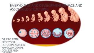
- 5. HUMAN EMBRYOGENESIS STAGES DAYS Two cell stage 2 days after fertilization Morula 3 days Blastocyst 4th day Bilaminar disc 8th day Prochordal plate and primitive streak 14th day Three layered germ disc 16th day
- 6. At a very early stage in development, the embryo proper acquires the form of three layered disc called as embryonic disc. The embryonic disc is derived from the bilaminar disc The epiblast cells when seen from the superior view have a bulge know as the primitive streak. The cells surrounding it start to degenerate and this forms the primitive groove . Fibroblast growth factor 8 acts on the groove and thus the epiblasts cells start to migrate into it , causing it to invaginate and forms a layer of cells above the hypoblast known as mesoderm. Hence epiblast-ectoderm , hypoblast-endoderm. Formation of germ layers
- 7. Appearance of Primitive streak
- 10. DEVELOPMENT OF PHARYNGEAL ARCHES They consist of mesenchymal tissue ,covered on the outside by ectoderm and inside by epithelium of endodermal origin. They are formed during fourth week. Rod like thickening present in the wall of fore gut. 6 arches arise but the 5th arch disappears.
- 11. The first four branchial arches are well developed in humans. Only the first and second arches extend to the midline, and each arch is progressively smaller from first to the last. The Mandibular Branchial Arch is the first to develop. It is located just below the stomodeum. The HYOID is the SECOND arch to develop. The IIIrd, IVth andVIth arches does not have special names. The fifth arch exists only transiently during embryogenesis , disappears soon after its formation.
- 12. Derivatives of skeletal elements 1st Arch- forms maxillary and mandibular process Supplied by the trigeminal nerve. Molds muscles related to mastication, Meckels cartilage(malleus and incus)is derived from it. 2nd Arch – innervated by the facial nerve. Facial expressions and post. Diagastric muscles are derived Styloid process of temporal bone ,hyoid bone(lesser cornu), superior part of body of hyoid,Stapes styloid process. 3rd Arch- Glossopharyngeal Molds stylopharyngeous muscle and forms the skeletal structure of greater horn and lower portion of body of hyoid bone. 4th 6th Arches-vagus nerve both arches fuse to form laryngeal cartilage.
- 14. DERIVATIVES OF PHARYNGEAL POUCHES 1st pouch- forms the eustachian tube (links the naso pharynx to the middle ear) 2nd disappers leaving the tonsillar fossa 3rd gives rise to inferior parathyroid gland and thymus. 4th ,5th – is a unique structure gives rise to parathyroid and parafollicular cells of the thyroid gland. The pharyngeal grooves mostly is obliterated and the only reminent is external auditory meatus.
- 15. DEFECTS IN BRANCHIAL ARCH DEVELOPMENT Branchial Cysts and Fistulae: Lower overhanging border of 2nd arch fuses with the tissues caudal to arches The side of the neck now becomes smooth The cavity of cervical sinus may often get obliterated Part of it may persists giving rise to swelling that lie in the neck along the ant border of SCM called as branchial cyst Such cyst opening into surface is called branchial sinus
- 16. Development of face After the formation of the head fold, the developing brain and the pericardium form the two prominent bulgings on the ventral aspect of the embryo ,separated by the stomodaeum. Floor of the stomodaeum is formed by the buccopharyngeal membrane, and it separates it from the foregut. The mesoderm covering the forebrain proliferate & a downward projection overlaps the upper part of the stomodaeum called the frontonasal process The face is derived from the following structures-1.frontonasal process 2 . 1st pharyngeal arch of each side.
- 17. The mandibular arch forms the lateral wall of stomodaeum, and it gives if a bud from the dorsal end called maxillary process. It grows ventro-medially cranial to the main part of the arch which is called mandibular process. Ectodermal overlying the frontonasal process soon shows bilateral localized thickenings situated above the stomodaeum called nasal placodes. The formation of the nasal placodes is induced by the underlying forebrain. They soon sink below the surface and form the nasal pits . The edge of each pit is raised above the surface medially and is called medial nasal process, the lateral edge is called lateral nasal process.
- 19. UPPER LIP Each maxillary process grows medially and fuses with the lateral nasal process 1st then with the medial nasal process. The medial and lateral nasal process also fuse with each in such a way that the nasal pits (external nares) are cut off from the stomodaeum. The maxillary process grows and at the same time frontonasal process narrows from side to side bringing the external nares closer together.
- 20. The mesodermal basis of the lateral part of the lip is formed from the maxillary process,the overlying skin is derived from the ectoderm. The mesodermal of the medial part of the lip (philtrum) is formed from the frontonasal process, the ectoderm of the maxillary process however grows over this mesoderm to meet the opposite maxillary process at the midline. Hence the entire upper lip is innervated by maxillary nerve.
- 21. The mandibular processes of the two sides grow towards each other and fuse in the midline, they now form the lower margins of stomodaeum, fused mandibular process will give rise to lower lip and to lower jaw. LOWER LIP Cheeks The progressive fusion of the maxillary process above and the mandibular process below with each other forms the cheeks.
- 22. NOSE • The nose receives contributions from the frontonasal process, and from the medial and lateral nasal processes of the right and left sides. • External nares are formed when the nasal pits are cut off from the stomodaeum by the fusion of the maxillary process with the medial nasal process. • External nares gradually approach each other due to the fact that the frontonasal process becomes progressively narrower and its deeper part ultimately forms the nasal septum. • Mesoderm becomes heaped up in the median plane to form the prominence of the nose.
- 23. NASOOPTIC FURROW • Maxillary process fuses with the lateral nasal process during the formation of upper lip.This fusion not only occurs in the region of the lip but also extends from the stomodaeum to the medial angle of the developing eye. • For some time this line of fusion is marked by a groove called the naso-optic furrow or nasolacrimal sulcus.
- 24. EYE • Forms At 7 weeks of IU life • The region of the eye is first seen as an ectodermal thickening, the lens placode, which appears on the ventro-lateral side of the developing forebrain, lateral and cranial to the nasal placode • The lens placode sinks below the surface and is eventually cut off from the surface ectoderm. The developing eyeball produces a bulge. The bulging of the eyes are at first directed laterally and lie in the angles between the maxillary processes and the lateral nasal processes. With the narrowing of the frontonasal process they come to face forwards. • The eyelids are derived from folds of ectoderm that are formed above and below the eyes, and by mesoderm enclosed within the folds.
- 25. Developmental anomaly of eye HYPERTELORISM. • The eyes may be widely separated • Nasal bridge may be broad. • This condition results from the presence of excessive tissue in the frontonasal process.
- 26. EXTERNAL EAR • Forms At 6 weeks of age • The External ear is formed around the dorsal part of the first ectodermal cleft . A series of mesodermal thickenings (often called tubercles or hillocks) appear on the mandibular and hyoid arches where they adjoin this cleft. • The pinna (or auricle) is formed by fusion of these thickenings.
- 27. Derivation of Parts Of Face
- 28. NASAL CAVITIES Nasal cavities are formed by the extension of the nasal pits which are in communication with stomodaeum. The primitive palate is formed from the fusion of the medial & lateral nasal process which a partition btw the pit and stomodaeum. The nasal pits deepen to form the nasal sac,the two are widely separated by the frontonasal process. But the narrowing of the process and enlargement of the nasal cavities brings it closer. The intervening tissue become thin and form the nasal septum, an dthey are separated from the mouth by the developing palate.
- 29. Developmental Anomaly of Nasal Cavity • There may be atresia of the cavity at the external nares, at the posterior nasal aperture or in the cavity proper. • This may be unilateral or bilateral. • Very rarely, there may be total absence of the nasal passages. Congenital defects in the cribriform plate of the ethmoid bone may lead to a communication between the cranial cavity and the nose.
- 30. • The nasal septum may not be in the middle line, i.e. it may be deflected to one side.The septum may be absent. • The nasal cavity may communicate with the mouth
- 31. The paranasal sinuses appear as diverticula from the nasal cavity. The diverticula gradually invade the bones after which they are named. The maxillary and sphenoidal sinuses begin to develop before birth.The other sinuses develop after birth. Enlargement of paranasal sinuses is associated with overall enlargement of the facial skeleton, including the jaws.This provides space in the jaws for growth and eruption of teeth. Growth of the facial skeleton is responsible for the gradual change in looks of a baby. PARANASAL AIR SINUSES
- 32. DEVELOPMENT OF PALATE • From each maxillary process, a plate-like shelf grows medially This is called the palatal process. • We now have three components from which the palate will be formed. • They are: 1)the two palatal processes 2)the primitive palate formed from the frontonasal process.
- 33. At a later stage, the mesoderm in the palate undergoes intramembranous ossification to form the hard palate. However, ossification does not extend into the most posterior portion, which remains as the soft palate. The part of the palate derived from the frontonasal process forms the premaxilla, which carries the incisor teeth. • Begins at the end of the 5th week • Gets completed by the end of the 12th week • The most critical period for the development of palate is from the end of 6th week to the beginning of 9th week. • Each palate fuses with the posterior margins of the primitive palate . • The two palatal process fuse with each other in the midline beginning anteriorly and proceeding backwards.
- 34. Developmental Anomalies Of Face and Palate HARELIP The upper lip of the hare normally has a cleft. Failure of fusion of maxillary process with median nasal fold. Hence the term harelip is used for defects of the lips OBLIQUE FACIAL CLEFT Non-fusion of the maxillary and lateral nasal processes gives rise to a cleft running from the medial angle of the eye to the mouth The nasolacrimal duct is not formed.
- 35. The nose may be bifid.This may be associated with median cleft lip. Both these occur due to bifurcation of the frontonasal process. Occasionally one-half of it may be absent.Very rarely the nose forms a cylindrical projection, or PROBOSCIS jetting out form just below the forehead. This anomaly may sometimes affect only one-half of the nose and is usually associated with fusion of the two eyes (CYCLOPS)
- 36. MANDIBULOFACIAL DYSOSTOSIS, TREACHER COLLINS SYNDROME OR FIRST ARCH SYNDROME. The entire first arch may remain underdeveloped on one or both sides, affecting the lower eyelid (coloboma type defect), the maxilla, the mandible and the external ear. The prominence of the cheek is absent and the ear may be displaced Abnormal face showing single median eye (cyclops). A rod-like projection is seen above the eye (proboscis). There may be presence of cleft palate and of faulty dentition.
- 37. Cleft lip MEDIAN CLEFT LIP Unilateral cleft lip and palate Cleft palate Bilateral cleft lip and palate
- 38. DEVELOPMENT OF MOUTH • The oral cavity is derived partly from the stomodaeum (ectoderm) and partly from the foregut (endoderm). • These two are separated by the buccopharyngeal membrane that disappears later.
- 39. Microstomia : small mouth opening DEVELOPMENTAL ANOMALIES of mouth Anodontia : absence of the teeth Macro stomia : large mouth opening Agnathia : absence lower jaw Micrognathia : small lower jaw
- 40. The teeth are formed in relation to the alveolar process.The epithelium overlying the convex border of this process becomes thickened and projects into the underlying mesoderm.This epithelial thickening is called the DENTAL LAMINA( 6 weeks) The dental lamina now shows a series of local thickenings, each of which is destined to form one milk tooth.These thickenings are called ENAMEL ORGANS forms at 8 weeks. DEVELOPMENT OF TEETH
- 41. As the enamel organ grows downwards into the mesenchyme (of the alveolar process) its lower end assumes a cup-shaped appearance .The cup comes to be occupied by a mass of mesenchyme called the dental papilla. The enamel organ and the dental papilla together constitute the TOOTH GERM. At this stage the developing tooth looks like a cap therefore, described as the CAP STAGE of tooth development.(At 10 weeks of age). The cells of the enamel organ that line the papilla become columnar. These are called AMELOBLASTS Mesodermal cells of the papilla that are adjacent to the ameloblasts arrange themselves as a continuous epithelium- like layer. The cells of this layer are called odontoblasts. The developing tooth now looks like a Bell Stage.
- 42. • As ossification progresses, the roots of the teeth become surrounded by bone • The root of the tooth is established by continued growth into underlying mesenchyme. • Odontoblasts in this region lay down dentine. As layers of dentine are deposited, the pulp space becomes progressively narrower and is gradually converted into a canal through which nerves and blood vessels pass into the tooth. • Ameloblasts lay down enamel on the superficial (outer) surface of the basement membrane. • The odontoblasts lay down dentine on its deeper surface. • The process of laying down of enamel and of dentine is similar to that of formation of bone by osteoblasts. • After the enamel is fully formed the ameloblasts disappear leaving a thin membrane, the DENTAL CUTICLE over the enamel.The odontoblasts, however, continue to separate the dentine from the pulp throughout the life of the tooth.
- 43. DEVELOPMENT OF TEETH • In the region of the root there are no ameloblasts. • The dentine is covered by mesenchymal cells that differentiate into CEMENTOBLASTS. • These cells lay down a layer of dense bone called the CEMENTUM(forms just before birth) , mesenchymal cells form the PERIODONTAL LIGAMENT which connects the root to the socket in the jaw bone is formed just after birth before eruption of tooth.
- 44. DEVELOPMENTAL ANOMALIES OF TEETH Anodontia, also called anodontia vera, is a rare genetic disorder characterized by the congenital absence of all primary or permanent teeth Hutchinson’s Incisors. Due to congenital syphilis.(peg shaped) Mulberry Molars .rounded rudimentary enamel cusps on permanent 1st molars.
- 45. DEVELOPMENTAL ANOMALIES OF TEETH Tooth fusion arises through union of two normally separated tooth germs, and depending upon the stage of development of the teeth at the time of union, it may be either complete or incomplete. Gemination arises when two teeth develop from one tooth bud and, as a result, the patient has an extra tooth
- 46. DENTINOGENESIS IMPERFECTA • Dentinogenesis imperfecta (hereditary Opalescent Dentin) is a genetic disorder of tooth development. •This condition causes teeth to be discolored (most often a blue-gray or yellow-brown color) and translucent. Teeth are also weaker than normal, making them prone to rapid wear, breakage, and loss.
- 47. Amelogenesis Imperfecta This causes teeth to be unusually small, discoloured, pitted, prone to wear and breakage. Inherited autosomal recessive disorder.
- 48. SUPERNUMERARY TEETH Hyperdontia is the condition of having supernumerary teeth, or teeth which appear in addition to the regular number of teeth.
- 49. DEVELOPMENT OF SALIVARY GLANDS • The salivary glands develop as outgrowths of the buccal epithelium at the age of 7 weeks • The terminal parts of the duct system develop into secretory acini • The outgrowth for the PAROTID GLAND arises in relation to the line along which the maxillary and mandibular processes fuse to form the cheek. It is generally considered to be ectodermal • The outgrowths for the SUBMANDIBULAR AND SUBLINGUAL GLANDS arise in relation to the linguogingival sulcus. They are usually considered to be of endodermal origin
- 51. Aplasia • It is congenitally absence of salivary gland. • Aplasia occurs in combination with congenital anomalies Aberrant salivary glands An aberrant or ectopic is salivary gland tissue that develops at a site where it is not normally found
- 52. Accessory ducts Most common developmental anomaly. Site- superior and anterior to normal Stenson's duct orifice. Atresia Congenital occlusion or absence of one or two major salivary gland ducts. Site- submandibular duct in floor of mouth. Causes severe xerostomia.
- 53. : The presence of previously unnoticed bilateral macroscopic salivary gland locations in the human nasopharynx. Predominantly mucous glands with multiple draining ducts, predominantly near the torus tubarius.
- 54. TEMPOROMANDIBULAR JOINT TMJ develops initially from widely separated temporal and condylar blastema that grows towards each other. TMJ has a fibrous tissue rather than hyaline cartilage on the articular facets of temporal fossa and condyle. Between 10th-12th week- mesenchyme between growing secondary cartilage of condyle and temporal bone differentiate into fibrous tissue. During 12th week 2 clefts develops in the interposed vascular fibrous connective tissue forming two joint cavities and defining intervening articular disc.
- 55. PRIMARY AND SECONDARY JOINT • Compression of central portion of articular disc occurs as it takes biconcave shape. • Tissue of disc becomes continuous with tendon of lateral pterygoid anteriorly. • Posteriorly it gets attached to portion of Meckel’s cartilage which differentiates into malleus. • A condensation of mesenchyme forms the analogue of the joint capsule thereby isolating the joint with its synovial membrane from its surrounding tissues.
- 56. Anomalies of development of TMJ Ankylosis – it is the fusion of the condyle with the glenoid fossa obliterating the normal articulation and movement of the joint . produces immobilisation and impaired mandibular formation. Due to failure in cavitation of one or both joint compartments results in uni or bi lateral ankylosis. This results in underdevelopment of mandible causing masticatory distress of varying severity.
- 57. Asymmetric development of one or more body parts Enlargement may be limited to a single limb. Increased pigmentation Hypertrichosis Telangiectasia Unilateral macroglossia. Malocclusion with open-bite. FACIAL HEMIHYPERTROPHY
- 58. Textbook of Embryology – Inderbir Singh Langman’s Medical Embryology – T.W. Sadler Textbook of Oral Medicine- Burkitt's Textbook of Anatomy- BD CHAURASIA Geoffrey H Sperber – Craniofacial Development and Growth. The Tubarial Salivary Glands : A potential new organ at risk for radiotherapy - Radiotherapy and Oncology – Sep 2020 REFERENCE S
- 59. THANK YOU