

Recommandé
Contenu connexe
Tendances
Tendances (20)
En vedette
En vedette (20)
Similaire à Tissue Freezing Presentation (2)
Similaire à Tissue Freezing Presentation (2) (20)
Tissue Freezing Presentation (2)
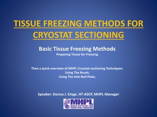
- 1. TISSUE FREEZING METHODS FOR CRYOSTAT SECTIONING Basic Tissue Freezing Methods Preparing Tissue for Freezing Then a quick overview of MHPL Cryostat sectioning Techniques: Using The Brush; Using The Anti-Roll Plate; Speaker: Donna J. Emge, HT-ASCP, MHPL Manager
- 2. Preparing Tissue For Freezing Tissue for freezing should be frozen or fixed as promptly as possible after cessation of circulation to avoid morphological distortions and damage due to: • Tissue drying artifact. • Autolysis - The destruction of tissues or cells by the action of substances, such as enzymes, that are produced within the organism. Also called self-digestion. • Putrefaction - Decomposition by microorganisms.
- 3. WHY SNAP FREEZE OR FIX AND CRYOPROTECT FOR FREEZING? Slow freezing can cause distortion of tissue due to ice crystal formation that replaces the architecture with a “Swiss Cheese” effect.
- 4. The object is to freeze so rapidly that water does not have time to form crystals and remains in a vitreous form that does not expand when solidified. Freezing artifact in sections of Brain, Muscle, and Spleen Brain Skeletal Muscle
- 5. SHORT ARTICLE ON THE SUBJECT OF WATER CRYSTAL FORMATION: “FREEZING BIOLOGICAL SAMPLES” Charles W. Scouten & Miles Cunningham http://www.myneurolab.com/global/Manuals/Tips%20an d%20Techniques%20Freezing%20Artifact.pdf
- 6. METHODS OF TISSUE FREEZING 1. Fresh tissue freezing – Tissue is in OCT and flash frozen fresh. 2. 4% PFA fixed, sucrose cryoprotected tissue freezing – Tissue is in OCT and may be frozen using dry ice or the flash frozen method. 3. Enzyme study tissue freezing – Often used for fresh muscle tissue. A fresh frozen method with no OCT matrix. Tissue protrudes from a Tragacanth or other support medium. MHPL Protocols for these methods are included in your workshop folder.
- 7. WHY NOT JUST USE LIQUID NITROGEN? • It Boils – this creates a vapor barrier that causes freezing in a slower, unpredictable pattern. • Tissue and OCT often cracks – due to this unpredictable freezing pattern. AFTER ALL: “Liquid nitrogen is one of the coldest liquids routinely available and it does not mix with tissue.” Review the article: “FREEZING BIOLOGICAL SAMPLES” Charles W. Scouten & Miles Cunningham http://www.myneurolab.com/global/Manuals/Tips%20and%20Techniques%20Freezing% 20Artifact.pdf
- 8. GETTING STARTED: Before you dissect the animal - organize and set-up:
- 9. Before you dissect the animal - organization and set-up: Choose appropriate freezing method - and depending on the method prepare liquid nitrogen, isopentane, dry ice. Label ahead of time - cryo molds, aluminum foil, specimen bags while at room temperature. Covered Foam cooler with crushed dry ice – to temporarily hold frozen samples as you work. Tools & other supplies - OCT, or Tragacanth, forceps, small labeled weigh boats or small labeled petri dishes.
- 10. FRESH TISSUE FREEZING Pros Fastest of all methods. Excellent for IHC, IF, ISH. No antigen retrieval required since no cross-linking fixative. Often easiest to section – depending upon tissue. Cons Poorest morphology. Prone to freezing artifact – must be snap frozen. ISH integrity – extreme clean techniques required or RNA will be rapidly and easily degraded.
- 11. PREPARING FRESH TISSUE FOR FREEZING (Not for enzyme study method) Acclimate tissue to OCT - cover freshly dissected tissue for a few minutes in OCT in a labeled small petri dish or small weigh boat. Transfer and orientate in fresh OCT in a labeled Cryomold – with just enough OCT to cover the tissue. Avoid bubbles in the OCT – especially near the tissue. Sectioning surface - is the bottom of the Cryomold. Begin freezing.
- 12. Fresh Tissue Freezing Procedure: A metal beaker is filled 2/3 with Isopentane and placed in a Dewar of Liquid Nitrogen enough to come up to about 1/3 of the metal beaker. Prepare at least 10 minutes before freezing sample. With 12 inch forceps freeze the cryo mold prepared sample in the clear portion of isopentane – do not fully submerge. Avoid block cracking - when there is still a small drop size of unfrozen OCT transfer sample to covered foam cooler of dry ice while continuing on to other samples.
- 13. Temporarily store frozen samples in a covered foam cooler of dry ice while continuing to freeze other samples Wrap individual samples in labeled foil, seal in a plastic bag, place in a freezer box . Store at -80° C.
- 14. 4% PFA FIXED, SUCROSE CRYOPROTECTED TISSUE FREEZING Pros Excellent morphology compared to other methods. May use a slower freeze in crushed powder dry ice alone, slush of dry ice and 100% alcohol, or in a beaker of isopentane surrounded by dry ice - without incurring freezing artifact or block cracking. Any of the freezing methods discussed can be used. Good for most IHC, IF and ISH. Cons Time consuming Most IHC will require antigen retrieval. Although the fixative cross-linking is protective for ISH techniques there is some RNA degradation.
- 15. PREPARING FIXED, SUCROSE CRYOPROTECTED TISSUE • 4% PFA transcardial perfuse animal. • Drop fix in 4% PFA for a few hours to O/N. • 15% sucrose in 1XPBS until tissue sinks. • 30% sucrose in 1XPBS until tissue sinks.
- 16. SUCROSE CRYOPROTECTED TISSUE FREEZING (Not for enzyme study method) Acclimate tissue to OCT - cover the 4% PFA fixed, sucrose cryoprotected tissue for a few minutes in OCT in a labeled small petri dish or small weigh boat. Transfer and orientate in fresh OCT in a labeled Cryomold – with just enough OCT to cover the tissue. Avoid bubbles in the OCT – especially near the tissue. Sectioning surface - is the bottom of the Cryomold. Begin freezing.
- 17. A metal beaker is ½ filled with isopentane, placed in a foam cooler or laboratory ice bucket and surround with crushed dry ice. Add a few pieces of dry ice to the isopentane and wait until boiling stops. Add more isopentane if necessary. With 12 inch forceps freeze the cryo mold prepared sample in the isopentane – do not fully submerge. Alternatively: Freeze cryomold prepared sample by surrounding it in finely powder crushed dry ice alone, or in a dry ice methanol or 100% Ethanol slurry. Transfer frozen sample to a covered foam cooler of dry ice while continuing on to other samples. Wrap all samples in labeled foil, place in a bag sealed bag, in a freezer box. Store at -80 FIXED, SUCROSE CRYOPROTECTED TISSUE FREEZING
- 18. ENZYME STUDY TISSUE FREEZING Pros • Excellent for Enzyme histochemistry and Immunohistochemistry studies. • Best method for muscle tissue. Cons • Advanced skill needed for sectioning – no supportive OCT matrix. Anti-roll plate better than brush technique. • Time and technique skill to prepare. • Extremely susceptible to any freeze thaw – leading to loss of morphologic detail in muscle or brain tissue.
- 19. ENZYME STUDY METHOD TISSUE PREP FOR FREEZING Organize and set up:
- 20. ENZYME STUDY METHOD PREP FOR TISSUE FREEZING • Prepare a small pyramid of thick Tragacanth paste on a small piece of cork. • Make a small hole in the Tragacanth paste pyramid.
- 21. ENZYME STUDY METHOD PREP FOR TISSUE FREEZING • Gently remove any surface moisture from tissue with fresh tissue wipe. • Place 1/8 to ¼ of the tissue in a hole at top of pyramid, leaving the rest stick out above the tragacanth. • Seal edges of tragacanth to the tissue.
- 22. ENZYME STUDY TISSUE FREEZING Procedure: • A metal beaker is filled 2/3 with Isopentane and placed in a Dewar of Liquid Nitrogen enough to come up to about 1/3 to ½ of the metal beaker. Prepare at least 10 minutes before freezing sample. • Hold the cork/tragacanth/sample with 12” forceps and submerge sample side down completely into the isopentane for 10 to 20 seconds.
- 23. ENZYME STUDY TISSUE FREEZING Transfer sample to covered foam cooler of crushed dry ice or immediately to a -80 freezer. Rapidly wrap all samples in pre-cooled, labeled foil, and place in a pre-cooled plastic bag, in a freezer box. Store at -80°C.
- 24. CRYOSECTIONING PREP • Remove the frozen block from the -70°C freezer and allow it to equilibrate in the cryostat chamber temperature for approximately 30 minutes. • The optimal temperature for cryostat sectioning depends on the nature of the tissue and on whether the tissues have been freshly frozen or pre-fixed with subsequent cryoprotection. • Note the reference chart for temperature setting guidelines for tissue types in your folder.
- 25. MHPL CRYOSTAT SECTIONING TECHNIQUES Using The Brush Using The Anti-Roll Plate
- 26. CRYOSTAT SECTIONING BRUSH TECHNIQUE The purpose of the brush is to grab and maneuver the section across the stage. You can buy a 1/4 inch, #2 flat, or bright brushes from an art supply store for about $3 and cut them at an angle. With this angled tip, the brush meets the tissue flat like a broom because the brush is held at an angle.
- 27. Stephen R Peters M.D. Pathology Innovations, LLC http://www.pathologyinnovations.com/frozen_section_technique.htm CRYOSTAT SECTIONING BRUSH TECHNIQUE
- 28. CRYOSTAT SECTIONING BRUSH TECHNIQUE
- 29. CRYOSTAT SECTIONING ANTI-ROLL PLATE Used to prevent frozen sections from curling upwards, after sectioning. The device is made of coated glass or plastic and is aligned parallel to the knife edge, a little above it. The following points are to be considered: a. Correct height to knife edge b. Correct angle to knife c. Top edge not damaged d. At cabinet temperature
- 30. CRYOSTAT SECTIONING ANTI-ROLL PLATE
- 31. CRYOSTAT SECTIONING ANTI-ROLL PLATE TRAGACANTH SUSPENDED TISSUE
- 32. EXCELLENT PAPER: Evaluation of the Value of Frozen Tissue Section Used as “Gold Standard” for Immunohistochemistry Shan-Rong Shi, MD, Cheng Liu, Llana Pootrakul, PhD, Laurie Tang, MS, Andrew Young, Ryan Chen,Richard J. Cote, MD, and Clive R. Taylor, MD, PhD Am J Clin Pathol 2008;129:358-366 DOI: 10.1309/7CXUYXT23E5AL8KQ http://ajcp.ascpjournals.org/content/129/3/358.full.pdf CARE AND HANDLING OF FROZEN SECTION SLIDES EXAMINES: The use of acetone- or ethanol-fixed frozen tissue sections as the “gold standard” for immunohistochemical analysis Frozen sections fixed by 6 protocols: acetone, ethanol, NBF (2 durations), and NBF + calcium chloride (2 durations). With and without AR.
- 33. Acknowledgements Warren Tourtellote, M.D., Ph.D, Director MHPL members: Lin Li, M.D., Assoc. Director Donna Emge, HT-ASCP, Manager Hong Chang, HT-ASCP Sheela Patel, HT-ASCP Ephie Bakou, MBA Administrative Volunteer Faculty Advisory Committee Anjen Chenn, MD, PhD Alexander Stegh, PhD Chyung-Ru Wang, PhD Richard M. Pope, MD Raymond Bergan, MD
Notes de l'éditeur
- Freezing artifact shown in 1. Brain 2. Muscle 3. Spleen
- MHPL Protocols for these methods are included in your workshop folder.
- MHPL uses a grease pencil aka China marker
- Clear portion of the isopentane. Sheets of cork can be purchased from VWR or Fisher and cut to appropriate size needed. After freezing the cork is mounted to the cryostat chuck with OCT.
- Quick trick to test if tissue is too cold or too warm for sectioning. Thumb for 2 seconds, then take the next section – if section is good then cryostat too cold for your tissue type. If section is not good then make colder with Cryofreeze spray then take the next section – if section is good then cryostat was too warm for your tissue type.
- Double click black area for video start.
- Double click image to start video.
- Double click image to start video.
- Double click image to start video.
- What we do at MHPL: Let air dry as we are sectioning and place in a sealed slide box at -20C.
- Special Thank you to Ephie Bakou, our MHPL administrative volunteer for coordinating much of our workshop today. Also, Leica Microsystems for their demonstration of the Tape Transfer System and for providing lunch.