2. Historical Information:
► In 1881 S. pneumoniae discovered by Leo Escolar
and was isolated by Louis Pasteur and George
Sternberg.
► In 1884 Albert Fraenkle performed gram stain where
it was found to be diplococcal.
► In 1902 Neufeld discovered swelling of the capsular
which lead to serotyping.
► The type-specific antibody development in S.
pneumoniae was shown by Lister in 1913.
► In 1944 transformation experiments were performed
by McCarty.
► Lastly in 1997 Joseph Dillard and Janet Yother cloned
common capsule DNA regions.
9. The capsule of S pneumoniae renders it
resistant to phagocytosis.
► S pneumoniae leading cause of bacterial
pneumonia beyond the neonatal period.
► Pleural effusion is the most common and
empyema (pus in the pleural space) one of
the most serious complications of S
pneumoniae.
► Most common cause of sinusitis, acute
bacterial otitis media, and conjunctivitis
beyond early childhood.
► Meningitis
► Acute septic arthritis and bone infections in
patients with sickle cell disease
► Peritonitis (especially in patients with
nephrotic syndrome) or endocarditis.
10.
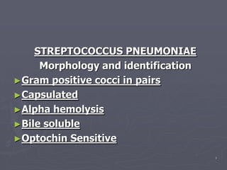
11. ►- Streptococcus pneumoniae
(previous names - Pneumococcus,
Diplococcus)
►G+, catalase-, alpha hemolytic,
URT resident.
► Possesses a large capsule to
resist phagocytosis.
►Forms short chains. Mucoid
colonies.
►>80 different serotypes based on
capsule protein as Ag.
12. ►Pathogenesis:
- Predisposition is necessary. Considered
endogenous infection. Transmission not an
issue. Examples of predisposing factors:
1. Viral infection of respiratory tract-this
causes damage to ciliated epithelial
cells as mechanical defense factor.
2. Prolonged immobilization-causes fluid
accumulation in alveoli, which
stimulates growth of St. pneumoniae.
3.
Malnutrition, alcoholism, diabetes, ext
reme prolonged fatigue.
13. ►Pathology and Symptoms:
1. Short incubation period - Difficult to
determine, since organism is already present.
2. Organism proliferates causing inflammation
and extreme edema. Packed edematous fluid
called consolidation.
3. Rapid onset of shaking, chills, fever, and chest
pains. Sputum is purulent and rust-colored
(from blood). Crisis in 5-10 days if untreated,
followed by rapid recovery or death (30%
fatality if untreated). Septicemia and meningitis
are common complications.
14. ►Diagnosis:
1. Inflammation of lung tissue visible on x-
ray.
2. Direct microscopic examination of sputum
(or CSF) shows heavily
encapsulated short chains of cocci.
3.Culture of sputum to yield
mucoid colonies of cocci also may be done.
4.Distinguished from other Streptococcus
species by optochin-sensitivity test (S.p.is
+).
15.
16. 5. Quellung Reaction – or capsular swelling
test
o Certain antibodies against capsular
material will cause a swelling of the
capsules of Streptococcus pneumoniae.
o Patient's serum is added to different
serotypes of Streptococcus pneumoniae.
If capsule swelling occurs, patient has
that particular serotype. If no response to
any of serotypes, patient does not have
pneumococcal pneumonia.
19. ► Prevention:
Correct predisposing conditions
Vaccine - contains capsular material
from 23 of most common serotypes.
Recommended for young
children, elderly, when predisposed.
20. Treatment:
1. Penicillin (97% of isolates are
susceptible).
2. Erythromycin, Klaracid, or other
Macrolides antibiotics.
3. For chronic sinusitis CECLOR oral
second generation cephalosporin
4. Quinolones etc. can be used.
21. Control
►Antibiotic Treatment
►Penicillin remains the drug of choice
for S pyogenes. It is safe, inexpensive,
and of narrow spectrum, and there is
no direct or indirect evidence of loss of
efficacy.
►Prior to the 1990's, S pneumoniae was
also uniformly sensitive to penicillin
but a recent abrupt shift in the
usefulness of penicillin has occurred.
22. ►If penicillin allergy occurs, an
alternative drug for treating
pharyngitis is
erythromycin, although sporadic
erythromycin and tetracycline
resistance has been reported, leaving
clindamycin or the newer macrolides
as possible treatments.