SlideShare une entreprise Scribd logo
1  sur  215
The Hip Complex
By DrRajal B. Sukhiyaji
MPTh in Sports Science
Introduction
• The hip joint, or coxofemoral joint, is the articulation
of the acetabulum of the pelvis and the head of the
femur.
• These two segments form a diarthrodial ball-and-socket
joint with three degrees of freedom:
1. Flexion/extension in the sagittal plane
2. Abduction/adduction in the frontal plane
3. Medial/lateral rotation in the transverse plane
The primary function of the hip joint is to support
the weight of the head, arms, and trunk (HAT)
both in static erect posture and in dynamic
postures such as ambulation, running, and stair
climbing.
Structure of the Hip Joint
Proximal Articular Surface:
• The cuplike concave socket of the hip joint is called the
acetabulum and is located on the lateral aspect of the
pelvic bone (innominate or os coxa).
• Three bones form the pelvis: the ilium, the ischium, and
the pubis.
• The pubis forms one fifth of the acetabulum, the ischium
forms two fifths, and the ilium forms the remainder.
• Until full ossification of the pelvis occurs between 20 and
25 years of age, the separate segments of the
acetabulum may remain visible on radiograph.
• The acetabulum appears to be a hemisphere, but only its
upper margin has a true circular contour, and the
roundness of the acetabulum as a whole decreases with
age.
• In actuality, only a horseshoe-shaped portion of the
periphery of the acetabulum (the lunate surface) is
covered with hyaline cartilage and articulates with the
head of the femur.
• The inferior aspect of the lunate surface (the base of the
horseshoe) is interrupted by a deep notch called the
acetabular notch.
• The acetabular notch is spanned by a fibrous band, the
transverse acetabular ligament, that connects the
two ends of the horseshoe.
• The transverse acetabular ligament also spans the
acetabular notch to create a fibro-osseous tunnel, called
the acetabular fossa, beneath the ligament, through
which blood vessels may pass into the central or
deepest portion of the acetabulum.
• The acetabulum is deepened by the fibrocartilaginous
acetabular labrum, which surrounds the periphery.
• The acetabular fossa is nonarticular; the femoral head
does not contact this surface.
• The acetabular fossa contains fibroelastic fat covered
with synovial membrane.
Center Edge Angle of the
Acetabulum
• Each acetabulum is oriented on each innominate bone
somewhat inferiorly and anteriorly.
• The magnitude of inferior orientation is assessed on
radiograph by using a line connecting the lateral rim of
the acetabulum and the center of the femoral head.
• This line forms an angle with the vertical known as the
center edge (CE) angle or the angle of Wiberg and is
the amount of inferior tilt of the acetabulum.
• CE angles in adults to average 38 in men and 35 in
women (with ranges in both sexes to be about 22 to 42).
Acetabular Anteversion
• The acetabulum faces not only somewhat inferiorly but
also anteriorly.
• The magnitude of anterior orientation of the acetabulum
may be referred to as the angle of acetabular
anteversion.
• Average value to be 18.5 for men and 21.5 for women.
• Pathologic increases in the angle of acetabular
anteversion are associated with decreased joint stability
and increased tendency for anterior dislocation of the
head of the femur.
Acetabular Labrum
• The entire periphery of the acetabulum is rimmed by a
ring of wedge-shaped fibrocartilage called the acetabular
labrum.
• The labrum is attached to the periphery of the
acetabulum by a zone of calcified cartilage.
• It not only deepens the socket but also increases the
concavity of the acetabulum through its triangular shape
and grasps the head of the femur to maintain contact
with the acetabulum.
• The labrum is not load-bearing but serves a role in
proprioception and pain sensitivity that may help protect
the rim of the acetabulum.
• Hydrostatic fluid pressure within the intra-articular space
was greater within the labrum than without, which
suggests that the labrum may also enhance joint
lubrication if the labrum adequately fits the femoral head.
• The transverse acetabular ligament is considered to be
part of the acetabular labrum.
• It protect the blood vessels which reach to the head of
femur.
Distal Articular Surface:
• The head of the femur is a fairly rounded hyaline
cartilage-covered surface that may be slightly larger than
a true hemisphere or as much as two thirds of a sphere,
depending on body type.
• The radius of curvature of the femoral head is smaller in
women than in men in comparison with the dimensions
of the pelvis.
• Just inferior to the most medial point on the femoral head
is a small roughened pit called the fovea or fovea
capitis.
• The fovea is not covered with articular cartilage and is
the point at which the ligament of the head of the femur
is attached.
• The femoral head is attached to the femoral neck; the
femoral neck is attached to the shaft of the femur
between the greater trochanter and the lesser trochanter.
• The femoral neck is, in general, only about 5 cm long.
• The femoral neck is angulated so that the femoral head
most commonly faces medially, superiorly, and anteriorly.
Angulation of the Femur
• There are two angulations made by the head and neck
of the femur in relation to the shaft.
• One angulation (angle of inclination) occurs in the frontal
plane between an axis through the femoral head and
neck and the longitudinal axis of the femoral shaft.
• The other angulation (angle of torsion) occurs in the
transverse plane between an axis through the femoral
head and neck and an axis through the distal femoral
condyles.
Angle of Inclination of the Femur:
• The angle of inclination of the femur averages 126
degrees (referencing the medial angle formed by the
axes of the head/neck and the shaft), ranging from 115
to 140 degrees in the unimpaired adult.
• In women, the angle of inclination is somewhat smaller
than it is in men, owing to the greater width of the female
pelvis.
• With a normal angle of inclination, the greater trochanter
lies at the level of the center of the femoral head.
• The angle of inclination of the femur changes across the
life span, being substantially greater in infancy and
childhood and gradually declining to about 120 degrees
in the normal elderly person.
• A pathologic increase in the medial angulation between
the neck and shaft is called coxa valga, and a
pathologic decrease is called coxa vara.
• Angle of Torsion of the Femur:
• An axis through the femoral head and neck in the
transverse plane will lie at an angle to an axis through
the femoral condyles, with the head and neck torsioned
anteriorly (laterally) with regard to an angle through the
femoral condyles.
• The angle of torsion decreases with age. In the newborn,
the angle of torsion has been estimated to be 40
degrees, decreasing substantially in the first 2 years.
• In the adult, the normal angle of torsion is considered to
be 10 to 20 degrees.
• A pathologic increase in the angle of torsion is called
anteversion, and a pathologic decrease in the angle or
reversal of torsion is known as retroversion.
Articular Congruence
• The hip joint is considered to be a congruent joint.
• However, there is substantially more articular surface on
the head of the femur than on the acetabulum.
• In the neutral or standing position, the articular surface of
the femoral head remains exposed anteriorly and
somewhat superiorly.
• The acetabulum does not fully cover the head superiorly,
and the anterior torsion of the femoral head (angle of
torsion) exposes a substantial amount of the femoral
head’s articular surface anteriorly.
• Articular contact between the femur and the acetabulum
can be increased in the normal non–weight-bearing hip
joint by a combination of flexion, abduction, and slight
lateral rotation.
• This position (also known as the frog-leg position)
corresponds to that assumed by the hip joint in a
quadruped position and is the true physiologic position of
the hip joint.
Hip Joint Capsule and
Ligaments
Hip Joint Capsule:
• The hip joint capsule is a substantial contributor to joint
stability.
• The articular capsule of the hip joint is an irregular,
dense fibrous structure with longitudinal and oblique
fibers and with three thickened regions that constitute
the capsular ligaments.
• The capsule is attached proximally to the entire
periphery of the acetabulum beyond the acetabular
labrum.
• The capsule itself is thickened anterosuperiorly, where
the predominant stresses occur; it is relatively thin and
loosely attached posteroinferiorly.
• The capsule covers the femoral head and neck like a
cylindrical sleeve and attaches to the base of the femoral
neck.
• The femoral neck is intracapsular, whereas both the
greater and lesser trochanters are extracapsular.
• The synovial membrane lines the inside of the capsule.
• Anteriorly, there are longitudinal retinacular fibers deep
in the capsule that travel along the neck toward the
femoral head.
• The retinacular fibers carry blood vessels that are the
major source of nutrition to the femoral head and neck.
• The retinacular blood vessels arise from a vascular ring
located at the base of the neck and formed by the medial
and lateral circumflex arteries (branches of the deep
femoral artery).
Hip Joint Ligaments:
 The ligamentum teres:
• It is an intra-articular but extrasynovial accessory joint
structure.
• The ligament is a triangular band attached at one end to
both sides of the peripheral edge of the acetabular
notch.
• The ligament then passes under the transverse
acetabular ligament (with which it blends) to attach at its
other end to the fovea of the femur; thus, it is also called
the ligament of the head of the femur.
• The ligamentum teres is encased in a flattened sleeve of
synovial membrane so that it does not communicate with
the synovial cavity of the joint.
• It is tensed in semiflexion and adduction.
• The ligamentum teres appears to function primarily as a
conduit for the secondary blood supply from the
obturator artery and for the nerves that travel along the
ligament to reach the head of the femur through the
fovea.
• The hip joint capsule is typically considered to have
three reinforcing capsular ligaments (two anteriorly and
one posteriorly).
• The two anterior ligaments are,
1) The iliofemoral ligament
2) The pubofemoral ligament
• The one posterior ligament is,
3) The ischiofemoral ligament
1) The iliofemoral ligament:
• It is a fan-shaped ligament that resembles an inverted
letter Y.
• It often is referred to as the Y ligament of Bigelow.
• The apex of the ligament is attached to the anterior
inferior iliac spine, and the two arms of the Y fan out to
attach along the intertrochanteric line of the femur.
• The superior band of the iliofemoral ligament is the
strongest and thickest of the hip joint ligaments.
2) The pubofemoral ligament:
• It is also anteriorly located, arising from the anterior
aspect of the pubic ramus and passing to the anterior
surface of the intertrochanteric fossa.
• The bands of the iliofemoral and the pubofemoral
ligaments form a Z on the anterior capsule.
3) The ischiofemoral ligament:
• It is the posterior capsular ligament.
• The ischiofemoral ligament attaches to the posterior
surface of the acetabular rim and the acetabulum
labrum.
• Some of its fibers spiral around the femoral neck and
blend with the fibers of the circumferential fibers of the
capsule.
• Other fibers are arranged horizontally and attach to the
inner surface of the greater trochanter.
• The hip joint capsule and the majority of its ligaments are
quite strong and that each tightens with full hip extension
(hyperextension).
• The anterior ligaments are stronger (stiffer and
withstanding greater force at failure) than the
ischiofemoral ligament.
• Under normal circumstances, the hip joint, its capsule,
and ligaments routinely support two thirds of the body
weight (the weight of head, arms, and trunk, or HAT).
• In bilateral stance, the hip joint is typically in neutral
position or slight extension.
• In this position, the capsule and ligaments are under
some tension.
• The normal line of gravity (LoG) in bilateral stance falls
behind the hip joint axis, creating a gravitational
extension moment.
Capsuloligamentous Tension:
• Hip joint extension, with slight abduction and medial rotation,
is the close-packed position for the hip joint.
• With increased extension, the ligaments twist around the
femoral head and neck, drawing the head into the
acetabulum.
• In contrast to most other joints in the body, the close-packed
and stable position for the hip joint is not the position of
optimal articular contact (congruence).
• Optimal articular contact occurs with combined flexion,
abduction, and lateral rotation.
• Under circumstances in which the joint surfaces are
neither maximally congruent nor closepacked, the hip
joint is at greatest risk for traumatic dislocation.
• A position of particular vulnerability occurs when the hip
joint is flexed and adducted (as it is when sitting with the
thighs crossed).
• In this position, a strong force up the femoral shaft
toward the hip joint (as when the knee hits the
dashboard in a car accident) may push the femoral head
out of the acetabulum.
• The capsuloligamentous tension at the hip joint is least
when the hip is in moderate flexion, slight abduction, and
midrotation.
Structural Adaptations
to Weight-Bearing
• The internal architecture of the pelvis and femur reveal
the remarkable interaction between mechanical stresses
and structural adaptation created by the transmission of
forces between the femur and the pelvis.
• The trabeculae (calcified plates of tissue within the
cancellous bone) line up along lines of stress and form
systems that normally adapt to stress requirements.
• In standing or upright weight-bearing activities, at least
half the weight of the HAT (the gravitational force)
passes down through the pelvis to the femoral head,
whereas the ground reaction force (GRF) travels up the
shaft.
• These two forces, nearly parallel and in opposite
directions, create a force couple with a moment arm
(MA) equal to the distance between the superimposed
body weight on the femoral head and the GRF up the
shaft.
• These forces create
a bending moment
(or set of shear
forces) across the
femoral neck.
• The bending stress creates a tensile force on the
superior aspect of the femoral neck and a compressive
stress on the inferior aspect.
• A complex set of forces prevents the rotation and resists
the shear forces that the force couple causes; among
these forces are the structural resistance of two major
and three minor trabecular systems.
• The medial (or principal compressive) trabecular system
arises from the medial cortex of the upper femoral shaft
and radiates through the cancellous bone to the cortical
bone of the superior aspect of the femoral head.
• The medial system of trabeculae is oriented along the
vertical compressive forces passing through the hip joint.
• The lateral (or principal tensile) trabecular system of the
femur arises from the lateral cortex of the upper femoral
shaft and, after crossing the medial system, terminates
in the cortical bone on the inferior aspect of the head of
the femur.
• The lateral trabecular system is oblique and may
develop in response to parallel (shear) forces of the
weight of HAT and the GRF.
• There are two accessory (or secondary) trabecular
systems, of which one is considered compressive and
the other is considered tensile.
• Another secondary trabecular system is confined to the
trochanteric area femur.
• The areas in which the trabecular systems cross each
other at right angles are areas that offer the greatest
resistance to stress and strain.
• There is an area in the femoral neck in which the
trabeculae are relatively thin and do not cross each
other.
• This zone of weakness has less reinforcement and thus
more potential for failure.
• The primary weight-bearing surface of the acetabulum,
or dome of the acetabulum, is located on the superior
portion of the lunate surface.
• In the normal hip, the dome lies directly over the center
of rotation of the femoral head.
• The dome shows the greatest prevalence of
degenerative changes in the acetabulum.
• The primary weight-bearing area of the femoral head is,
correspondingly, its superior portion.
• Although the primary weight-bearing area of the
acetabulum is subject to the most degenerative changes,
degenerative changes in the femoral head are most
common around or immediately below the fovea or
around the peripheral edges of the head’s articular
surface.
• The forces of HAT and GRF that act on the articular
surfaces of the hip joint and on the femoral head and
neck also act on the femoral shaft.
• The shaft of the femur is not vertical but lies at an angle
that varies considerably among individuals.
• The vertical loading on the oblique femur results in
bending stresses in the shaft.
• The medial cortical bone in the shaft (diaphysis) must
resist compressive stresses, whereas the lateral cortical
bone must resist tensile stresses.
Function of the Hip Joint
 Motion of the Femur on the Acetabulum:
• The motions of the hip joint are easiest to visualize as
movement of the convex femoral head within the
concavity of the acetabulum as the femur moves through
its three degrees of freedom: flexion/extension,
abduction/adduction, and medial/lateral rotation.
• The femoral head will glide within the acetabulum in a
direction opposite to motion of the distal end of the
femur.
• Flexion and extension of the femur occur from a neutral
position as an almost pure spin of the femoral head
around a coronal axis through the head and neck of the
femur.
• The head spins posteriorly in flexion and anteriorly in
extension.
• Flexion and extension from other positions (e.g., in
abduction or medial rotation) must include both spinning
and gliding of the articular surfaces, depending on the
combination of motions.
• The motions of abduction/adduction and medial/lateral
rotation must include both spinning and gliding of the
femoral head within the acetabulum.
 Range of motion:
• The joint’s range of motion (ROM) is influenced by
structural elements, as well as by whether the motion is
performed actively or passively and whether passive
tension in two-joint muscles is encountered or avoided.
• Flexion of the hip is generally about 90 degrees with the
knee extended and 120 degrees when the knee is flexed
and when passive tension in the two-joint hamstrings
muscle group is released.
• Hip extension is considered to have a range of 10 to 30
degrees.
• Hip extension ROM appears to diminish somewhat with
age, whereas flexion remains relatively unchanged.
• When hip extension is combined with knee flexion,
passive tension in the two-joint rectus femoris muscle
may limit the movement.
• The femur can be abducted 45 to 50 degrees and
adducted 20 to 30 degrees.
• Abduction can be limited by the two-joint gracilis
muscle and adduction limited by the tensor fascia lata
(TFL) muscle and its associated iliotibial (IT) band.
• Medial and lateral rotation of the hip are usually
measured with the hip joint in 90 degrees of flexion; the
typical range is 42 to 50 degrees.
• Femoral anteversion is correlated with decreased range
of lateral rotation and less strongly with increased range
of medial rotation.
• When the femoral head is torsioned anteriorly more than
normal, lateral rotation of the femur turns the head out
even more, both risking subluxation and encountering
capsuloligamentous and muscular restrictions on the
anterior aspect of the joint.
• Normal gait on level ground requires at least the
following hip joint ranges:
o 30 degrees flexion,
o 10 degrees hyperextension,
o 5 degrees of both abduction and adduction, and
o 5 degrees of both medial and lateral rotation.
• Walking on uneven terrain or stairs will increase the
need for joint range beyond that required for level
ground, as will activities such as sitting in a chair or
sitting cross-legged.
Motion of the Pelvis on the Femur
• Whenever the hip joint is weight-bearing, the femur is
relatively fixed, and, motion of the hip joint is produced
by movement of the pelvis on the femur.
 Anterior and Posterior Pelvic Tilt
 Lateral Pelvic Tilt
 Anterior and Posterior Pelvic Rotation
Anterior and Posterior Pelvic Tilt
• Anterior and posterior pelvic tilt are motions of the entire
pelvic ring in the sagittal plane around a coronal axis.
• In the normally aligned pelvis, the anterosuperior iliac
spines (ASISs) of the pelvis lie on a horizontal line with
the posterior superior iliac spines and on a vertical line
with the symphysis pubis.
• Anterior and posterior tilting of the pelvis on the fixed
femur produce hip flexion and extension, respectively.
• Hip joint extension through posterior tilting of the pelvis
brings the symphysis pubis up and the sacrum of the
pelvis closer to the femur, rather than moving the femur
posteriorly on the pelvis.
• Hip flexion through anterior tilting of the pelvis moves the
ASISs anteriorly and inferiorly; the inferior sacrum moves
farther from the femur, rather than moving the femur
away from the sacrum.
• Anterior and posterior tilting will result in flexion and
extension of both hip joints simultaneously in bilateral
stance or can occur at the stance hip joint alone if the
opposite limb is non–weight-bearing.
Lateral Pelvic Tilt
• Lateral pelvic tilt is a frontal plane motion of the entire
pelvis around an anteroposterior axis.
• In the normally aligned pelvis, a line through the ASISs is
horizontal.
• In lateral tilt of the pelvis in unilateral stance, one hip
joint is the pivot point or axis for motion of the opposite
side of the pelvis as it elevates (pelvic hiking) or drops
(pelvic drop).
• If a person stands on the left limb and hikes the pelvis,
the left hip joint is being abducted because the medial
angle between the femur and a line through the ASISs
increases.
• If a person stands on the left leg and drops the pelvis,
the left hip joint will adduct because the medial angle
formed by the femur and a line through the ASISs will
decrease.
• In descriptions of the hip joint motions that occur in
unilateral stance, the hip joint of the non–weightbearing
limb is in an open chain and has no motions on it.
• However, the non–weight-bearing leg typically hangs
straight down as the pelvis moves.
Lateral Shift of the Pelvis:
• Lateral pelvic tilt can also occur in bilateral stance.
• If both feet are on the ground and the hip and knee of
one limb are flexed, the opposite limb is largely the
weight-bearing limb and the terminology is the same as
for unilateral stance.
• If both limbs are weightbearing, lateral tilt of the pelvis
will cause the pelvis to shift to one side or the other.
• With pelvic shift, the pelvis cannot hike but can only
drop.
• Because there is a closed chain between the two weight-
bearing feet and the pelvis, both hip joints will move in
the frontal plane in a predictable way as the pelvic tilt (or
pelvic shift) occurs.
• If the pelvis is shifted to the right in bilateral stance, the
left side of the pelvis will drop, the right hip joint will be
adducted, and the left hip joint will be abducted.
Anterior and Posterior Pelvic
Rotation
• Pelvic rotation is motion of the entire pelvic ring in the
transverse plane around a vertical axis.
• Although rotation can occur around a vertical axis
through the middle of the pelvis in bilateral stance, it
most commonly and more importantly occurs in single-
limb support around the axis of the supporting hip joint.
• Forward rotation of the pelvis occurs in unilateral stance
when the side of the pelvis opposite to the supporting hip
joint moves anteriorly.
• Forward rotation of the pelvis produces medial rotation of
the supporting hip joint.
• Backward rotation of the pelvis occurs when the side of
the pelvis opposite the supporting hip moves posteriorly.
• Posterior rotation of the pelvis produces lateral rotation
of the supporting hip joint.
• Pelvic rotation can occur in bilateral stance as well as
unilateral stance, as is true for lateral pelvic tilt.
• If both feet are bearing weight and the axis of motion
occurs around a vertical axis through the center of the
pelvis, the terms forward rotation and backward rotation
must be used by referencing a side (e.g., forward
rotation on the right and backward rotation on the left).
Coordinated Motions of the Femur,
Pelvis, and Lumbar Spine
• When the pelvis moves on a relatively fixed femur, there
are two possible outcomes to consider.
• Either the head and trunk will follow the motion of the
pelvis (moving the head through space) or the head will
continue to remain relatively upright and vertical despite
the pelvic motions.
• These are open- and closed-chain responses,
respectively.
Pelvifemoral Motion:
• When the femur, pelvis, and spine move in a coordinated
manner to produce a larger ROM than is available to one
segment alone, the hip joint is participating in what will
predominantly (but not exclusively) be an open-chain
motion termed pelvifemoral motion.
• In the case of pelvifemoral motion, the joints may serve
either end of the chain: the foot or head.
 Moving the Head and Arms through Space:
(Example)
• If the goal is to bend forward to bring the hands (and
head) toward the floor, isolated flexion at the hip joints
(anteriorly tilting the pelvis on the femurs) is generally
insufficient to reach the ground.
• If the knees remain extended, the hips will typically flex
no more than 90 (and often less, depending on
extensibility of the hamstrings).
• The addition of flexion of the lumbar spine (and,
perhaps, flexion of the thoracic spine) will add to the total
ROM.
• The combination of hip and trunk flexion is generally
sufficient for the hands to reach the ground—as long as
the hamstrings and lumbar extensors allow sufficient
lengthening.
• The combination of hip motion and lumbar motion to
achieve a greater ROM for the hands and head is an
example of a largely open-chain response in the hips
and trunk.
 Moving the Foot through Space: (Example)
• When a person is lying on the right side, the left foot may
be moved through an arc of motion approaching 90
degrees.
• This is clearly not all from the left hip joint, which can
typically abduct only to 45 degrees; motion of the foot
through space also includes lateral tilting of the pelvis
(hiking around the right hip joint) and lateral flexion of the
lumbar spine to the left.
• The abducting limb is in an open chain; the lumbar spine
(and thoracic spine) are constrained by the body weight
and contact with the ground.
• Pelvifemoral motion has also been referred to as
pelvifemoral “rhythm”.
• The link between hip, pelvis, and lumbar motion is the
basis of using pain with active straight-leg raising as a
test for severity of dysfunction in persons with low back
pain.
Closed-Chain Hip Joint Function:
• The joints of the right and left lower limbs are part of a
true closed chain when both lower limbs are
weightbearing and the chain is defined as all the
segments between the right foot, up through the pelvis,
and down through the left foot.
• A true closed chain is formed because both ends of the
chain (both feet in this example) are “fixed” and
movement at any one joint in the chain invariably
involves movement at one or more other links in the
chain.
• For the hips (and other lower limb joints) to be in a
closed chain in standing, both ends of the chain (the
head and the feet) must be fixed.
• The feet are fixed by weight-bearing.
• The head is often (but not necessarily) functionally
“fixed.”
• When the head (one end of the chain) is held upright and
over the feet (the other end of the chain), all the
segments in the axial skeleton and lower limbs function
as part of a closed chain; movement at one joint will
create movement in at least one other linkage in the
chain.
• Consequently, in our functional closed chain, hip flexion
does not occur independently (which would move the
head forward in space) but is accompanied by motion in
one or more interposed segments to ensure that the
head remains upright over the base of support and that
the body does not become unstable.
 Closed-Chain Hip Joint Function: (Example)
• A common example of closed-chain versus open-chain
function is seen when the hip flexor musculature is tight
and the hip joint is maintained in flexion.
• A person standing with fixed hip flexion is shown in
Figure 10-27A (an open-chain response) and B (a
closed-chain response).
• A true open-chain response to isolated hip flexion would
displace the head and trunk forward, with the LoG falling
in front of the supporting feet.
• More commonly, hip flexion in stance is not isolated to
the hips but is accompanied by compensatory
movements of the vertebral column (including extension
or lordosis of the lumbar spine) that maintain the head in
the upright position and keep the LoG well within the
base of support).
• In a functional closed chain, motion at the hip (one link in
the chain) is accompanied by an essentially mandatory
lumbar extension to maintain the head over the sacrum
(see Fig. 10-27B).
• In contrast, hip flexion in open-chain pelvifemoral motion
is accompanied by lumbar flexion because the goal is to
achieve more range for the head in space (see Fig. 10-
25).
Hip Joint Musculature
• The muscles of the hip joint make their most important
contributions to function during weight-bearing.
• In weight-bearing, the muscles are called on to move or
support the HAT (approximately two thirds of body
weight).
• The hip joint muscles adapt their structure to the
required function, as can be seen in their large areas of
attachment, their length, and their large cross-section.
Flexors
• The flexors of the hip joint function primarily as mobility
muscles in open-chain function; that is, they function
primarily to bring the swinging limb forward during
ambulation or in various sports.
• The flexors may function secondarily to resist strong hip
extension forces that occur as the body passes over the
weight bearing foot.
• Nine muscles have action lines crossing the anterior
aspect of the hip joint.
• The primary muscles of hip flexion are the iliopsoas,
rectus femoris, TFL, and sartorius.
• The iliopsoas muscle is considered to be the most
important of the primary hip flexors.
• It consists of two separate muscles, the iliacus muscle
and the psoas major muscle, both of which attach to
the femur by a common tendon.
• The two components of the iliopsoas muscle have many
points of origin, including the iliac fossa and the disks,
bodies, and transverse processes of the lumbar
vertebrae.
• Given the attachments of the psoas major muscle to the
anterior vertebrae and the iliacus muscle to the iliac
fossa, activity of or passive tension in these muscles
would anteriorly tilt the pelvis (iliacus muscle) and,
apparently, pull the lumbar vertebrae anteriorly into
flexion (psoas major muscle).
• In closed-chain function (head vertical), however, these
muscles seem to create a paradoxical lumbar lordosis
(lumbar extension) that results from the body’s attempt
to keep the head over the sacrum with anterior pelvic tilt
and lower lumbar flexion.
• The role of the iliopsoas muscle in hip flexion may be
particularly critical when hip flexion from a sitting position
is required.
• The rectus femoris muscle is the only portion of the
quadriceps muscle that crosses both the hip joint and
knee joint.
• It originates on the anterior inferior iliac spine and inserts
by way of a common tendon into the tibial tuberosity.
• The rectus femoris muscle flexes the hip joint and
extends the knee joint.
• Because it is a two-joint hip flexor, the position of the
knee during hip flexion will affect its ability to generate
force at the hip.
• Simultaneous hip flexion and knee extension
considerably shorten this muscle and increase the
likelihood of active insufficiency.
• Consequently, the rectus femoris muscle makes its best
contribution to hip flexion when the knee is maintained in
flexion.
• The sartorius muscle is a straplike muscle originating on
the ASIS.
• It crosses the anterior aspect of the femur to insert into
the upper portion of the medial aspect of the tibia.
• The sartorius muscle is considered to be a flexor,
abductor, and lateral rotator of the hip, as well as a flexor
and medial rotator of the knee.
• It is a two-joint muscle, should be relatively unaffected by
the position of the knee.
• Its function is probably most important when the knee
and hip need to be flexed simultaneously (as in climbing
stairs).
• The TFL muscle originates more laterally than the
sartorius muscle.
• Its origin is on the anterolateral lip of the iliac crest.
• The muscle fibers extend only about one fourth of the
way down the lateral aspect of the thigh before inserting
into the IT band.
• The IT band or IT tract is the thickened lateral portion of
the fascia lata of the hip and thigh.
• The IT band attaches proximally to the iliac crest lateral
to the TFL muscle.
• After the tensor attaches to the IT band, the IT band
continues distally on the lateral thigh to insert into the
lateral condyle of the tibia.
• The TFL muscle is considered to flex, abduct, and
medially rotate the femur at the hip.
• The most important contribution of the TFL muscle may
be in maintaining tension in the IT band.
• The IT band assists in relieving the femur of some of the
tensile stresses imposed on the shaft by weight-bearing
forces.
• The secondary hip flexors are the pectineus, adductor
longus, adductor magnus, and the gracilis muscles.
• Each, however, is capable of contributing to hip joint
flexion, but that contribution is dependent on hip joint
position.
• The gracilis, a two-joint muscle, is active as a hip flexor
when the knee is extended but not when the knee is
flexed.
Adductors
• The hip adductor muscle group is generally considered
to include the pectineus, adductor brevis, adductor
longus, adductor magnus, and the gracilis muscles.
• The adductors are located anteromedially.
• The adductors longus, brevis, and magnus muscles
arise in a group from the body and inferior ramus of the
pubis to insert along the linea aspera.
• The gracilis muscle is the only two-joint adductor.
• It originates on the symphysis pubis and pubic arch and
inserts on the medial surface of the shaft of the tibia.
• In bilateral stance, the adductors may be synergists to
the abductor muscles when both feet are on the ground,
enhancing side-to-side stabilization of the pelvis.
• The adductors are also capable of generating a
maximum isometric torque greater than that of the
abductors.
Extensors
• The one-joint gluteus maximus muscle and the two-joint
hamstrings muscle group are the primary hip joint
extensors.
• These muscles may receive assistance from the
posterior fibers of the gluteus medius, from the posterior
adductor magnus muscle, and from the piriformis
muscle.
• The gluteus maximus is a large, quadrangular muscle
that originates from the posterior sacrum, dorsal
sacroiliac ligaments, sacrotuberous ligament, and a
small portion of the ilium.
• The gluteus maximus crosses the sacroiliac joint before
its most superior fibers insert into the IT band (as do the
fibers of the TFL muscle) and its inferior fibers insert into
the gluteal tuberosity.
• The gluteus maximus is the largest of the lower extremity
muscles; this muscle alone constituting 12.8% of the
total muscle mass of the lower extremity.
• The maximus is a strong hip extensor that appears to be
active primarily against a resistance greater than the
weight of the limb.
• The three two-joint extensors are the long head of the
biceps femoris, the semitendinosus, and the
semimembranosus muscles, known collectively as the
hamstrings.
• Each of these three muscles originates on the ischial
tuberosity.
• The biceps femoris crosses the posterior femur to insert
into the head of the fibula and lateral aspect of the lateral
tibial condyle.
• The other two hamstrings insert on the medial aspect of
the tibia.
• All three muscles extend the hip with or without
resistance, as well as serving as important knee flexors.
• If the hip is extended and the knee is flexed to 90 or
more, the hamstrings may not be able to contribute
much to hip extension force because of active
insufficiency or approaching active insufficiency.
• Extension forces in the hip increase by 30% if the knee
is extended during hip extension.
• The biceps femoris appears to contribute to lateral
rotation of the hip.
Abductors
• Active abduction of the hip is brought about
predominantly by the gluteus medius and the gluteus
minimus muscles.
• The gluteus medius originates on the lateral surface of
the wing of the ilium and inserts into the greater
trochanter, beneath the gluteus maximus.
• The gluteus medius has anterior, middle, and posterior
parts that function asynchronously during movement at
the hip.
• The gluteus minimus muscle lies deep to the gluteus
medius, arising from the outer surface of the ilium with its
fibers converging on an aponeurosis that ends in a
tendon on the greater trochanter.
• The minimus is consistently an abductor and flexor of the
hip, with its rotator function dependent on hip position.
• The minimus is a medial rotator in hip flexion.
• The gluteus minimus and medius muscles function
together to either abduct the femur (distal level free) or,
more important, to stabilize the pelvis (and
superimposed HAT) in unilateral stance against the
effects of gravity.
Lateral Rotators
• Six short muscles have lateral rotation as a primary
function.
• These muscles are the obturator internus and externus,
the gemellus superior and inferior, the quadratus
femoris, and the piriformis muscles.
• Other muscles that have fibers posterior to the axis of
motion at the hip (the posterior fibers of the gluteus
medius and minimus and the gluteus maximus) may
produce lateral rotation combined with the primary action
of the muscle.
• The obturator internus muscle originates from the inside
(posterior aspect) of the obturator foramen and emerges
through the lesser sciatic foramen to insert on the medial
aspect (inside) of the greater trochanter.
• The gemellus superior and gemellus inferior muscles
arise from the ischium of the pelvis, just above and just
below the point at which the obturator internus passes
through the lesser sciatic notch.
• Both gemelli follow and blend with the obturator internus
tendon to insert with the internus tendon into the greater
trochanter.
• The obturator externus muscle is sometimes considered
to be an anteromedial muscle of the thigh because it
originates on the external (anterior) surface of the
obturator foramen.
• However, it crosses the posterior aspect of the hip joint
and inserts on the medial aspect of the greater
trochanter in the trochanteric fossa.
• The quadratus femoris muscle is a small quadrangular
muscle that originates on the ischial tuberosity and
inserts on the posterior femur between the greater and
lesser trochanters.
• The piriformis muscle originates largely on the anterior
surface of the sacrum, passes through the greater sciatic
notch, and follows the inferior border of the posterior
gluteus medius to insert above the other lateral rotators
into the medial aspect of the greater trochanter.
• The piriformis and gluteus maximus are the only two
muscles that cross the sacroiliac joint.
• The sciatic nerve, the largest nerve in the body, enters
the gluteal region just inferior to the piriformis muscle.
• The lateral rotator muscles are positioned to perform
their rotatory function effectively, given the nearly
perpendicular orientation to the shaft of the femur.
• These muscles would certainly appear to be effective
joint compressors because their combined action line
parallels the head and neck of the femur.
• Of the primary lateral rotators, each inserts either on or
in the vicinity of the greater trochanter.
Medial Rotators
• There are no muscles with the primary function of
producing medial rotation of the hip joint.
• The more consistent medial rotators are the anterior
portion of the gluteus medius, gluteus minimus, and the
TFL muscles.
Hip Joint Forces and Muscle
Function in Stance
 Bilateral Stance:
• In erect bilateral stance, both hips are in neutral or slight
hyperextension, and weight is evenly distributed
between both legs.
• The LoG falls just posterior to axis for flexion/extension
of the hip joint.
• The posterior location of the LoG creates an extension
moment of force around the hip that tends to posteriorly
tilt the pelvis on the femoral heads.
• The gravitational extension moment is largely checked
by passive tension in the hip joint capsuloligamentous
structures, although slight or intermittent activity in the
iliopsoas muscles in relaxed standing may assist the
passive structures.
• In the frontal plane during bilateral stance, the body
weight is transmitted through the sacroiliac joints and
pelvis to the right and left femoral heads.
• Hypothetically, the weight of the HAT (two thirds of body
weight) should be distributed so that each femoral head
receives approximately half of the weight.
• The joint axis of each hip lies at an equal distance from
the LoG of HAT; that is, the gravitational MAs for the
right hip (DR) and the left hip (DL) are equal.
• Because the body weight (W) on each femoral head is
the same (WR=WL), the magnitude of the gravitational
torques around each hip must be identical (WR
*DR=WL* DL).
• The gravitational torques on the right and left hips,
however, occur in opposite directions.
• The weight of the body acting around the right hip tends
to drop the pelvis down on the left (right adduction
moment), whereas the weight acting around the left hip
tends to drop the pelvis down on the right (left adduction
moment).
• These two opposing gravitational moments of equal
magnitude balance each other, and the pelvis is
maintained in equilibrium in the frontal plane without the
assistance of active muscles.
• When bilateral stance is not symmetrical, frontal plane
muscle activity will be necessary to either control the
side-to-side motion or to return the hips to symmetrical
stance.
• The pelvis is shifted to the right, resulting in relative
adduction of the right hip and abduction of the left hip.
• To return to neutral position, an active contraction of the
right hip abductors would be expected.
• However, a contraction of the left hip adductors would
accomplish the same goal.
• Under the condition that both extremities bear at least
some of the superimposed body weight, the adductors
may assist the abductors in control of the pelvis against
the force of gravity or the GRF.
• In unilateral stance, activity of the adductors either in the
weight-bearing or non–weight-bearing hip cannot
contribute to stability of the stance limb.
• In the absence of adequate hip abductor function, the
adductors can contribute to stability—but only in bilateral
stance.
 Unilateral Stance:
• The left leg has been lifted from the ground and the full
superimposed body weight is being supported by the
right hip joint.
• Rather than sharing the compressive force of the
superimposed body weight with the left limb, the right hip
joint must now carry the full burden.
• In addition, the weight of the non–weight bearing left limb
that is hanging on the left side of the pelvis must be
supported along with the weight of HAT.
• Of the one-third portion of the body weight found in the
lower extremities, the non-supporting limb must account
for half of that, or one sixth of the full body weight.
• The magnitude of body weight (W) compressing the right
hip joint in right unilateral stance, therefore, is:
Right hip joint compression body weight = [2/3*W]+[1/6*W]
Right hip joint compression body weight =5/6*W
• The force of gravity acting on HAT and the non–weight-
bearing left lower limb (HATLL) will create an adduction
torque around the supporting hip joint; that is, gravity will
attempt to drop the pelvis around the right weight-
bearing hip joint axis.
• The abduction counter torque will have to be supplied by
the hip abductor musculature.
• The result will be joint compression or a joint reaction
force that is a combination of both body weight and
abductor muscular compression.
• The actual MA is likely to be slightly greater because the
weight of the hanging left leg will pull the center of
gravity of the superimposed weight slightly to the left,
although the LoG will simultaneously be shifted slightly
right to get the LoG within the single foot base of
support.
Reduction of Muscle Forces
in Unilateral Stance
• If the hip joint undergoes osteoarthritic changes that lead
to pain on weight-bearing, the joint reaction force must
be reduced to avoid pain.
• For most painful hip joints, however, the reductions in
compression generally required are greater than can be
achieved through weight loss.
• The solution must be in a reduction of abductor muscle
force requirements.
• If less muscular countertorque is needed to offset the
effects of gravity, there will be a decrease in the amount
of muscular compression across the joint, although the
body weight compression will remain unchanged.
• The need to diminish abductor force requirements also
occurs when the abductor muscles are weakened
through paralysis, through structural changes in the
femur that reduce biomechanical efficiency of the
muscles, or through degenerative changes producing
tears at the greater trochanter.
• Hip abductor muscle weakness will affect gait, whereas
paralysis of other hip joint muscles in the presence of
intact abductors will permit someone to walk or even run
with relatively little disability.
• Several options are available when there is a need to
decrease abductor muscle force requirements.
• Some compression reduction strategies occur
automatically, but at a cost of extra energy expenditure
and structural stress.
• Other strategies require intervention such as assistive
devices but minimize the energy cost.
Compensatory Lateral Lean of
the Trunk
• Gravitational torque at the pelvis is the product of body
weight and the distance that the LoG lies from the hip
joint axis (MA).
• If there is a need to reduce the torque of gravity in
unilateral stance and if body weight cannot be reduced,
the MA of the gravitational force can be reduced by
laterally leaning the trunk over the pelvis toward the side
of pain or weakness when in unilateral stance on the
painful limb.
• The compensatory lateral lean of the trunk toward the
painful stance limb will swing the LoG closer to the hip
joint, thereby reducing the gravitational MA.
• Because the weight of HATLL must pass through the
weight bearing hip joint regardless of trunk position,
leaning toward the painful or weak supporting hip does
not increase the joint compression caused by body
weight.
• However, it does reduce the gravitational torque.
• Whether a lateral trunk lean is due to muscular
weakness or pain, a lateral lean of the trunk during
walking still uses more energy than ordinarily required
for single-limb support and may result in stress changes
within the lumbar spine if used over an extended time
period.
• Use of a cane or some other assistive device offers a
realistic alternative to the person with hip pain or
weakness.
Use of a Cane Ipsilaterally
• Pushing downward on a cane held in the hand on the
side of pain or weakness should reduce the
superimposed body weight by the amount of downward
thrust; that is, some of the weight of HATLL would follow
the arm to the cane, rather than arriving on the sacrum
and the weight-bearing hip joint.
• It is realistic to expect that someone can push down on a
cane with approximately 15% of his body weight.
• The proportion of body weight that passes through the
cane will not pass through the hip joint and will not
create an adduction torque around the supporting hip
joint.
• Although a cane used ipsilaterally provides some
benefits in energy expenditure and structural stress
reduction, it is not as effective in reducing hip joint
compression as the undesirable lateral lean of the trunk.
• Moving the cane to the opposite hand produces
substantially different and better results.
Use of a Cane Contralaterally
• When the cane is moved to the side opposite the painful
or weak hip joint, the reduction in HATLL is the same as
it is when the cane is used on the same side as the
painful hip joint; that is, the superimposed body weight
passing through the weight-bearing hip joint is reduced
by approximately 15% of body weight.
• The cane is now farther from the painful supporting hip
joint; that is, in addition to relieving some of the
superimposed body weight, the cane is now in a position
to assist the abductor muscles in providing a
countertorque to the torque of gravity.
Adjustment of a Carried Load
• When someone with hip joint pain or weakness carries a
load in the hand or on the trunk (as with a backpack or
purse), there is a potential for increasing the demands
on the hip abductors and increasing the hip joint
compression.
• The added external load will increase the superimposed
weight acting through the affected supporting hip in
unilateral stance.
• The gravitational torque may increase, resulting in an
increased demand on the supporting hip abductors to
prevent drop of the pelvis.
• Although the increase in superimposed weight when a
load is carried cannot be avoided, it is possible to
minimize the demand on the abductor muscles on the
side of a painful or weak hip.
• If the external load is carried in the arm or on the side of
the trunk ipsilateral to the painful or weak hip, the
asymmetrical external load will cause a shift in the
combined force of HAT/external load center of mass
(CoM) toward the painful hip.
• Any shift of the combined CoM (or resulting LoG) toward
the painful hip will reduce the MA of the HAT/external
load.
• If the external load is not too great, the reduction in MA
of the HAT/external load can result in a reduction in
adduction torque.
• With a reduction in adduction torque, the demand on the
hip abductors is reduced.
• The reverse effect will occur if the load is carried on the
side opposite to the weak or painful hip.
• In that scenario, the external load both increases
superimposed body weight and increases the
gravitational MA around the weak or painful hip when in
unilateral stance on that hip.
Hip Joint Pathology
• The very large active and passive forces crossing the hip
joint make the joint’s structures susceptible to wear and
tear of normal components and to failure of weakened
components.
• Small changes in the biomechanics of the femur or the
acetabulum can result in increases in passive forces
above normal levels or in weakness of the dynamic joint
stabilizers.
Arthrosis
• The most common painful condition of the hip is due to
deterioration of the articular cartilage and to subsequent
related changes in articular tissues.
• It is known as osteoarthritis, degenerative arthritis, or
hip joint arthrosis.
• Its prevalence rates are about 10% to 15% in those
older than 55 years, with approximately equal distribution
among men and women.
• Changes may be due to subtle deviations present from
birth, to tissue changes inherent in aging, to the
repetitive mechanical stress of loading the body weight
on the hip joint over a prolonged period, to impingement
between the femur and labrum or adjacent acetabulum,
or to interactions of each of these factors.
• The factors most closely associated with hip joint
arthrosis are increased age and increased weight/height
ratio.
• Forces in excess of half the body weight are needed to
fully compress the femoral head into congruent contact
with the dome of the acetabulum.
• The more common degenerative changes in the femur
are at the periphery of the head and the perifoveal area.
• The periphery of the head receives only about one third
the compressive force of the superior portion of the
head, whereas the superior portion of the head is
compressed not only in standing but is also in contact
with the posterior acetabulum during sitting activities.
• The area of the femoral head around the fovea is most
commonly in the non–weight-bearing acetabular notch
and would undergo compression relatively infrequently.
Fracture
• The bony components must also be of sufficient strength
to withstand the forces that are acting around and
through the hip joint.
• The vertical weight-bearing forces that pass down
through the superior margin of the acetabulum in both
unilateral and bilateral stance act at some distance from
GRF up the shaft of the femur.
• The result is a bending force across the femoral neck.
• Normally the trabecular systems are capable of resisting
the bending forces, but abnormal increases in the
magnitude of the force or weakening of the bone can
lead to bony failure.
• The site of failure is likely to be in areas of thinner
trabecular distribution such as the zone of weakness.
• Bony failure in the femoral neck is uncommon in the
child or young adult.
• Of middle-aged people, women actually suffer fewer hip
fractures than do men, although the fractures in this age
group are usually attributable to trauma.
• The precipitating factor appears to be moderate trauma
such as that caused by a fall from standing, from a chair,
or from a bed.
• Hip fracture is associated with diminished bone density.
• Bone density decreases about 2% per year after age 50
and trabeculae clearly thin and disappear with aging.
• Not only is the condition painful, but malunion of the
fracture can lead to joint instability or cartilaginous
deterioration (or both) as a result of poorly aligned bony
segments.
• Although the femoral head may receive some blood
supply via the ligament of the femoral head, an absent or
diminished supply through the ligament of the head (as
occurs with aging) means reliance on anastomoses from
the circumflex arteries.
• This circumflex arterial supply may be disrupted by
femoral neck fracture, which leaves the femoral head
susceptible to avascular necrosis and necessitates
replacement of the head of the femur with an artificial
implant.
Bony Abnormalities of the Femur
 Coxa Valga/Coxa Vara:
• In coxa valga, the angle of inclination in the femur is
greater than the normal adult angle of 125 degrees.
• The increased angle brings the vertical weight bearing
line closer to the shaft of the femur, diminishing the
shear, or bending, force across the femoral neck.
• The reduction in force is actually reflected in a reduction
in density of the lateral trabecular system.
• The decreased distance between the femoral head and
the greater trochanter also decreases the length of the
MA of the hip abductor muscles.
• The decreased muscular MA results in an increased
demand for muscular force generation to maintain
sufficient abduction torque to counterbalance the
gravitational adduction moment acting around the
supporting hip joint during single-limb support.
• Coxa valga also decreases the amount of femoral
articular surface in contact with the dome of the
acetabulum.
• As the femoral head points more superiorly, there is a
decreasing amount of coverage from the acetabulum
superiorly.
• Consequently, coxa valga decreases the stability of the
hip and predisposes the hip to dislocation.
• Coxa vara is considered to give the advantage of
improved hip joint stability (if angle reduction is not too
extreme).
• The apparent improvement in congruence occurs
because the decreased angle between the neck and
shaft of the femur will turn the femoral head deeper into
the acetabulum, decreasing the amount of articular
surface exposed superiorly and increasing coverage
from the acetabulum.
• A varus femur, if not caused by trauma, may also
increase the length of the MA of the hip abductor
muscles by increasing the distance between the femoral
head and the greater trochanter.
• The increased MA decreases the amount of force that
must be generated by the abductor muscles in single-
limb support and reduces the joint reaction force.
• Coxa vara has the disadvantage of increasing the
bending moment along the femoral head and neck.
• Coxa vara may increase the likelihood in the adolescent
child that the femoral head will slide on the cartilaginous
epiphysis of the head of the femur.
 Anteversion/Retroversion:
• Anteversion of the femoral head reduces hip joint
stability because the femoral articular surface is more
exposed anteriorly.
• The line of the hip abductors may fall more posterior to
the joint, reducing the MA for abduction.
• The effect of femoral anteversion may also be seen at
the knee joint.
• When the femoral head is anteverted, pressure from the
anterior capsuloligamentous structures and the anterior
musculature may push the femoral head back into the
acetabulum, causing the entire femur to rotate medially.
• Although the medial rotation of the femur improves the
congruence in the acetabulum, the knee joint axis
through the femoral condyles is now turned medially,
altering the plane of knee flexion/extension and resulting,
at least initially, in a toe-in gait.
• The abnormal position of the knee joint axis is commonly
labeled medial femoral torsion.
• Medial femoral torsion and femoral anteversion are the
same abnormal condition of the femur.
• Femoral retroversion is the opposite of anteversion and
creates opposite problems from femoral anteversion.
Thank you

Contenu connexe

Tendances

BIOMECHANICS OF HIP JOINT BY Dr. VIKRAM
BIOMECHANICS OF HIP JOINT BY Dr. VIKRAMBIOMECHANICS OF HIP JOINT BY Dr. VIKRAM
BIOMECHANICS OF HIP JOINT BY Dr. VIKRAMVicky Vikram
 
Biomechanics of lumbar spine
Biomechanics of lumbar spineBiomechanics of lumbar spine
Biomechanics of lumbar spineVenus Pagare
 
Biomechanics of posture
Biomechanics of postureBiomechanics of posture
Biomechanics of posturekumarkirekha
 
Cervical spine: anatomy, biomechanics and pathomechanics
Cervical spine: anatomy, biomechanics and pathomechanicsCervical spine: anatomy, biomechanics and pathomechanics
Cervical spine: anatomy, biomechanics and pathomechanicsRadhika Chintamani
 
Kinesiology of the Shoulder
Kinesiology of the ShoulderKinesiology of the Shoulder
Kinesiology of the ShoulderSado Anatomist
 
Active movements
Active movementsActive movements
Active movementsSunil kumar
 
Biomechanics of wrist complex
Biomechanics of wrist complexBiomechanics of wrist complex
Biomechanics of wrist complexNeeti Christian
 
Kinetics and kimematics of the hip
Kinetics and kimematics of the hipKinetics and kimematics of the hip
Kinetics and kimematics of the hipKumarpal Singh
 
Joint Mobilization Review
Joint Mobilization ReviewJoint Mobilization Review
Joint Mobilization Reviewcaseychristyatc
 

Tendances (20)

Scapulohumeral rhythm ppt
Scapulohumeral rhythm pptScapulohumeral rhythm ppt
Scapulohumeral rhythm ppt
 
BIOMECHANICS OF HIP JOINT BY Dr. VIKRAM
BIOMECHANICS OF HIP JOINT BY Dr. VIKRAMBIOMECHANICS OF HIP JOINT BY Dr. VIKRAM
BIOMECHANICS OF HIP JOINT BY Dr. VIKRAM
 
Biomechanics of lumbar spine
Biomechanics of lumbar spineBiomechanics of lumbar spine
Biomechanics of lumbar spine
 
Biomechanics of thorax
Biomechanics of thoraxBiomechanics of thorax
Biomechanics of thorax
 
Biomechanics of hip complex 1
Biomechanics of hip complex 1Biomechanics of hip complex 1
Biomechanics of hip complex 1
 
Bio-mechanics of the Elbow Joint
Bio-mechanics of the Elbow Joint Bio-mechanics of the Elbow Joint
Bio-mechanics of the Elbow Joint
 
Biomechanics of posture
Biomechanics of postureBiomechanics of posture
Biomechanics of posture
 
Bio-mechanics of the hip joint
Bio-mechanics of the hip jointBio-mechanics of the hip joint
Bio-mechanics of the hip joint
 
Biomechanics spine
Biomechanics spineBiomechanics spine
Biomechanics spine
 
Bio-mechanics of the wrist joint
Bio-mechanics of the wrist jointBio-mechanics of the wrist joint
Bio-mechanics of the wrist joint
 
Cervical spine: anatomy, biomechanics and pathomechanics
Cervical spine: anatomy, biomechanics and pathomechanicsCervical spine: anatomy, biomechanics and pathomechanics
Cervical spine: anatomy, biomechanics and pathomechanics
 
Biomechanics of Posture
Biomechanics of PostureBiomechanics of Posture
Biomechanics of Posture
 
Kinesiology of the Shoulder
Kinesiology of the ShoulderKinesiology of the Shoulder
Kinesiology of the Shoulder
 
Active movements
Active movementsActive movements
Active movements
 
Biomechanics of wrist complex
Biomechanics of wrist complexBiomechanics of wrist complex
Biomechanics of wrist complex
 
Kinetics and kimematics of the hip
Kinetics and kimematics of the hipKinetics and kimematics of the hip
Kinetics and kimematics of the hip
 
Muscle biomechanics
Muscle biomechanicsMuscle biomechanics
Muscle biomechanics
 
Knee biomechanic
Knee biomechanicKnee biomechanic
Knee biomechanic
 
S d curve
S d curveS d curve
S d curve
 
Joint Mobilization Review
Joint Mobilization ReviewJoint Mobilization Review
Joint Mobilization Review
 

Similaire à The hip complex

HIP JOINTS-1.pptx.........................
HIP JOINTS-1.pptx.........................HIP JOINTS-1.pptx.........................
HIP JOINTS-1.pptx.........................IshaKanojiya1
 
OS COXAE.pptx
OS COXAE.pptxOS COXAE.pptx
OS COXAE.pptxSwaroopR16
 
RIBS.pptx
RIBS.pptxRIBS.pptx
RIBS.pptxsraffi
 
Fetal skull and maternal pelvis
Fetal skull and maternal pelvisFetal skull and maternal pelvis
Fetal skull and maternal pelvisjagan _jaggi
 
fetal skull and maternal pelvis diameters.pdf
fetal skull and maternal pelvis diameters.pdffetal skull and maternal pelvis diameters.pdf
fetal skull and maternal pelvis diameters.pdfShubhaSiraRavi
 
Cervical spine anatomy.pptx
Cervical spine anatomy.pptxCervical spine anatomy.pptx
Cervical spine anatomy.pptxHMU
 
Vertebral Colmn and Thorex.pptx
Vertebral Colmn and Thorex.pptxVertebral Colmn and Thorex.pptx
Vertebral Colmn and Thorex.pptxssusere3aa49
 
Vertebral Colmn and Thorex.pptx
Vertebral Colmn and Thorex.pptxVertebral Colmn and Thorex.pptx
Vertebral Colmn and Thorex.pptxRCGaur1
 
Lower limb complete
Lower limb completeLower limb complete
Lower limb completeLucidante1
 
clavicle Anatomy.pptx
clavicle Anatomy.pptxclavicle Anatomy.pptx
clavicle Anatomy.pptxmhmad farooq
 
applied aspects of shoulder joint
applied aspects of shoulder jointapplied aspects of shoulder joint
applied aspects of shoulder jointmrinal joshi
 
Surgical approaches to hip joint
Surgical approaches to hip jointSurgical approaches to hip joint
Surgical approaches to hip jointadityachakri
 
fetalskullandmaternalpelvis-by teju.pptx
fetalskullandmaternalpelvis-by teju.pptxfetalskullandmaternalpelvis-by teju.pptx
fetalskullandmaternalpelvis-by teju.pptxDrTejaswini7
 

Similaire à The hip complex (20)

HIP JOINTS-1.pptx.........................
HIP JOINTS-1.pptx.........................HIP JOINTS-1.pptx.........................
HIP JOINTS-1.pptx.........................
 
Joints of lower limb
Joints of lower limbJoints of lower limb
Joints of lower limb
 
Hip joint
Hip jointHip joint
Hip joint
 
OS COXAE.pptx
OS COXAE.pptxOS COXAE.pptx
OS COXAE.pptx
 
Hip Joint.pptx
Hip Joint.pptxHip Joint.pptx
Hip Joint.pptx
 
Biomechanics of hip
Biomechanics of hipBiomechanics of hip
Biomechanics of hip
 
Hip joint biomecanics
Hip joint biomecanicsHip joint biomecanics
Hip joint biomecanics
 
anatomy of spine
anatomy of spineanatomy of spine
anatomy of spine
 
RIBS.pptx
RIBS.pptxRIBS.pptx
RIBS.pptx
 
Fetal skull and maternal pelvis
Fetal skull and maternal pelvisFetal skull and maternal pelvis
Fetal skull and maternal pelvis
 
fetal skull and maternal pelvis diameters.pdf
fetal skull and maternal pelvis diameters.pdffetal skull and maternal pelvis diameters.pdf
fetal skull and maternal pelvis diameters.pdf
 
Cervical spine anatomy.pptx
Cervical spine anatomy.pptxCervical spine anatomy.pptx
Cervical spine anatomy.pptx
 
Vertebral Colmn and Thorex.pptx
Vertebral Colmn and Thorex.pptxVertebral Colmn and Thorex.pptx
Vertebral Colmn and Thorex.pptx
 
Vertebral Colmn and Thorex.pptx
Vertebral Colmn and Thorex.pptxVertebral Colmn and Thorex.pptx
Vertebral Colmn and Thorex.pptx
 
Lower limb complete
Lower limb completeLower limb complete
Lower limb complete
 
clavicle Anatomy.pptx
clavicle Anatomy.pptxclavicle Anatomy.pptx
clavicle Anatomy.pptx
 
applied aspects of shoulder joint
applied aspects of shoulder jointapplied aspects of shoulder joint
applied aspects of shoulder joint
 
Surgical approaches to hip joint
Surgical approaches to hip jointSurgical approaches to hip joint
Surgical approaches to hip joint
 
Hip Joint.pptx
Hip Joint.pptxHip Joint.pptx
Hip Joint.pptx
 
fetalskullandmaternalpelvis-by teju.pptx
fetalskullandmaternalpelvis-by teju.pptxfetalskullandmaternalpelvis-by teju.pptx
fetalskullandmaternalpelvis-by teju.pptx
 

Plus de Dr.Rajal Sukhiyaji

Plus de Dr.Rajal Sukhiyaji (14)

Ankle and foot complex
Ankle and foot complexAnkle and foot complex
Ankle and foot complex
 
Fitness and strength testing in sports
Fitness and strength testing in sportsFitness and strength testing in sports
Fitness and strength testing in sports
 
Fitness training
Fitness trainingFitness training
Fitness training
 
Sports and young athletes
Sports and young athletesSports and young athletes
Sports and young athletes
 
Principles of pt in post operative cases (general
Principles of pt in post operative cases (generalPrinciples of pt in post operative cases (general
Principles of pt in post operative cases (general
 
Pre and post operative management in tendon transfer
Pre and post operative management in tendon transferPre and post operative management in tendon transfer
Pre and post operative management in tendon transfer
 
Labyrinthitis
LabyrinthitisLabyrinthitis
Labyrinthitis
 
Ergonomics principles in rehabilitation
Ergonomics principles in rehabilitationErgonomics principles in rehabilitation
Ergonomics principles in rehabilitation
 
Ergonomics in sitting posture
Ergonomics in sitting postureErgonomics in sitting posture
Ergonomics in sitting posture
 
Isokenetic testing in sports
Isokenetic testing in sportsIsokenetic testing in sports
Isokenetic testing in sports
 
Imaging in sports injury
Imaging in sports injuryImaging in sports injury
Imaging in sports injury
 
Osteoporosis
OsteoporosisOsteoporosis
Osteoporosis
 
Otitis media
Otitis mediaOtitis media
Otitis media
 
Hydrotherapy in knee osteoarthrities
Hydrotherapy in knee osteoarthritiesHydrotherapy in knee osteoarthrities
Hydrotherapy in knee osteoarthrities
 

Dernier

Jual Obat Aborsi Di Dubai UAE Wa 0838-4800-7379 Obat Penggugur Kandungan Cytotec
Jual Obat Aborsi Di Dubai UAE Wa 0838-4800-7379 Obat Penggugur Kandungan CytotecJual Obat Aborsi Di Dubai UAE Wa 0838-4800-7379 Obat Penggugur Kandungan Cytotec
Jual Obat Aborsi Di Dubai UAE Wa 0838-4800-7379 Obat Penggugur Kandungan Cytotecjualobat34
 
Control of Local Blood Flow: acute and chronic
Control of Local Blood Flow: acute and chronicControl of Local Blood Flow: acute and chronic
Control of Local Blood Flow: acute and chronicMedicoseAcademics
 
❤️Call Girl Service In Chandigarh☎️9814379184☎️ Call Girl in Chandigarh☎️ Cha...
❤️Call Girl Service In Chandigarh☎️9814379184☎️ Call Girl in Chandigarh☎️ Cha...❤️Call Girl Service In Chandigarh☎️9814379184☎️ Call Girl in Chandigarh☎️ Cha...
❤️Call Girl Service In Chandigarh☎️9814379184☎️ Call Girl in Chandigarh☎️ Cha...Sheetaleventcompany
 
Race Course Road } Book Call Girls in Bangalore | Whatsapp No 6378878445 VIP ...
Race Course Road } Book Call Girls in Bangalore | Whatsapp No 6378878445 VIP ...Race Course Road } Book Call Girls in Bangalore | Whatsapp No 6378878445 VIP ...
Race Course Road } Book Call Girls in Bangalore | Whatsapp No 6378878445 VIP ...dishamehta3332
 
Gastric Cancer: Сlinical Implementation of Artificial Intelligence, Synergeti...
Gastric Cancer: Сlinical Implementation of Artificial Intelligence, Synergeti...Gastric Cancer: Сlinical Implementation of Artificial Intelligence, Synergeti...
Gastric Cancer: Сlinical Implementation of Artificial Intelligence, Synergeti...Oleg Kshivets
 
Call 8250092165 Patna Call Girls ₹4.5k Cash Payment With Room Delivery
Call 8250092165 Patna Call Girls ₹4.5k Cash Payment With Room DeliveryCall 8250092165 Patna Call Girls ₹4.5k Cash Payment With Room Delivery
Call 8250092165 Patna Call Girls ₹4.5k Cash Payment With Room DeliveryJyoti singh
 
Kolkata Call Girls Shobhabazar 💯Call Us 🔝 8005736733 🔝 💃 Top Class Call Gir...
Kolkata Call Girls Shobhabazar  💯Call Us 🔝 8005736733 🔝 💃  Top Class Call Gir...Kolkata Call Girls Shobhabazar  💯Call Us 🔝 8005736733 🔝 💃  Top Class Call Gir...
Kolkata Call Girls Shobhabazar 💯Call Us 🔝 8005736733 🔝 💃 Top Class Call Gir...Namrata Singh
 
Cardiac Output, Venous Return, and Their Regulation
Cardiac Output, Venous Return, and Their RegulationCardiac Output, Venous Return, and Their Regulation
Cardiac Output, Venous Return, and Their RegulationMedicoseAcademics
 
7 steps How to prevent Thalassemia : Dr Sharda Jain & Vandana Gupta
7 steps How to prevent Thalassemia : Dr Sharda Jain & Vandana Gupta7 steps How to prevent Thalassemia : Dr Sharda Jain & Vandana Gupta
7 steps How to prevent Thalassemia : Dr Sharda Jain & Vandana GuptaLifecare Centre
 
Whitefield { Call Girl in Bangalore ₹7.5k Pick Up & Drop With Cash Payment 63...
Whitefield { Call Girl in Bangalore ₹7.5k Pick Up & Drop With Cash Payment 63...Whitefield { Call Girl in Bangalore ₹7.5k Pick Up & Drop With Cash Payment 63...
Whitefield { Call Girl in Bangalore ₹7.5k Pick Up & Drop With Cash Payment 63...dishamehta3332
 
Call Girl In Indore 📞9235973566📞 Just📲 Call Inaaya Indore Call Girls Service ...
Call Girl In Indore 📞9235973566📞 Just📲 Call Inaaya Indore Call Girls Service ...Call Girl In Indore 📞9235973566📞 Just📲 Call Inaaya Indore Call Girls Service ...
Call Girl In Indore 📞9235973566📞 Just📲 Call Inaaya Indore Call Girls Service ...Sheetaleventcompany
 
Circulatory Shock, types and stages, compensatory mechanisms
Circulatory Shock, types and stages, compensatory mechanismsCirculatory Shock, types and stages, compensatory mechanisms
Circulatory Shock, types and stages, compensatory mechanismsMedicoseAcademics
 
❤️Chandigarh Escorts Service☎️9814379184☎️ Call Girl service in Chandigarh☎️ ...
❤️Chandigarh Escorts Service☎️9814379184☎️ Call Girl service in Chandigarh☎️ ...❤️Chandigarh Escorts Service☎️9814379184☎️ Call Girl service in Chandigarh☎️ ...
❤️Chandigarh Escorts Service☎️9814379184☎️ Call Girl service in Chandigarh☎️ ...Sheetaleventcompany
 
Goa Call Girl Service 📞9xx000xx09📞Just Call Divya📲 Call Girl In Goa No💰Advanc...
Goa Call Girl Service 📞9xx000xx09📞Just Call Divya📲 Call Girl In Goa No💰Advanc...Goa Call Girl Service 📞9xx000xx09📞Just Call Divya📲 Call Girl In Goa No💰Advanc...
Goa Call Girl Service 📞9xx000xx09📞Just Call Divya📲 Call Girl In Goa No💰Advanc...Sheetaleventcompany
 
Bandra East [ best call girls in Mumbai Get 50% Off On VIP Escorts Service 90...
Bandra East [ best call girls in Mumbai Get 50% Off On VIP Escorts Service 90...Bandra East [ best call girls in Mumbai Get 50% Off On VIP Escorts Service 90...
Bandra East [ best call girls in Mumbai Get 50% Off On VIP Escorts Service 90...Angel
 
Nagpur Call Girl Service 📞9xx000xx09📞Just Call Divya📲 Call Girl In Nagpur No💰...
Nagpur Call Girl Service 📞9xx000xx09📞Just Call Divya📲 Call Girl In Nagpur No💰...Nagpur Call Girl Service 📞9xx000xx09📞Just Call Divya📲 Call Girl In Nagpur No💰...
Nagpur Call Girl Service 📞9xx000xx09📞Just Call Divya📲 Call Girl In Nagpur No💰...Sheetaleventcompany
 
Bhawanipatna Call Girls 📞9332606886 Call Girls in Bhawanipatna Escorts servic...
Bhawanipatna Call Girls 📞9332606886 Call Girls in Bhawanipatna Escorts servic...Bhawanipatna Call Girls 📞9332606886 Call Girls in Bhawanipatna Escorts servic...
Bhawanipatna Call Girls 📞9332606886 Call Girls in Bhawanipatna Escorts servic...Dipal Arora
 
Call Girls Shahdol Just Call 8250077686 Top Class Call Girl Service Available
Call Girls Shahdol Just Call 8250077686 Top Class Call Girl Service AvailableCall Girls Shahdol Just Call 8250077686 Top Class Call Girl Service Available
Call Girls Shahdol Just Call 8250077686 Top Class Call Girl Service AvailableDipal Arora
 
Dehradun Call Girl Service ❤️🍑 8854095900 👄🫦Independent Escort Service Dehradun
Dehradun Call Girl Service ❤️🍑 8854095900 👄🫦Independent Escort Service DehradunDehradun Call Girl Service ❤️🍑 8854095900 👄🫦Independent Escort Service Dehradun
Dehradun Call Girl Service ❤️🍑 8854095900 👄🫦Independent Escort Service DehradunSheetaleventcompany
 
💚Chandigarh Call Girls 💯Riya 📲🔝8868886958🔝Call Girls In Chandigarh No💰Advance...
💚Chandigarh Call Girls 💯Riya 📲🔝8868886958🔝Call Girls In Chandigarh No💰Advance...💚Chandigarh Call Girls 💯Riya 📲🔝8868886958🔝Call Girls In Chandigarh No💰Advance...
💚Chandigarh Call Girls 💯Riya 📲🔝8868886958🔝Call Girls In Chandigarh No💰Advance...Sheetaleventcompany
 

Dernier (20)

Jual Obat Aborsi Di Dubai UAE Wa 0838-4800-7379 Obat Penggugur Kandungan Cytotec
Jual Obat Aborsi Di Dubai UAE Wa 0838-4800-7379 Obat Penggugur Kandungan CytotecJual Obat Aborsi Di Dubai UAE Wa 0838-4800-7379 Obat Penggugur Kandungan Cytotec
Jual Obat Aborsi Di Dubai UAE Wa 0838-4800-7379 Obat Penggugur Kandungan Cytotec
 
Control of Local Blood Flow: acute and chronic
Control of Local Blood Flow: acute and chronicControl of Local Blood Flow: acute and chronic
Control of Local Blood Flow: acute and chronic
 
❤️Call Girl Service In Chandigarh☎️9814379184☎️ Call Girl in Chandigarh☎️ Cha...
❤️Call Girl Service In Chandigarh☎️9814379184☎️ Call Girl in Chandigarh☎️ Cha...❤️Call Girl Service In Chandigarh☎️9814379184☎️ Call Girl in Chandigarh☎️ Cha...
❤️Call Girl Service In Chandigarh☎️9814379184☎️ Call Girl in Chandigarh☎️ Cha...
 
Race Course Road } Book Call Girls in Bangalore | Whatsapp No 6378878445 VIP ...
Race Course Road } Book Call Girls in Bangalore | Whatsapp No 6378878445 VIP ...Race Course Road } Book Call Girls in Bangalore | Whatsapp No 6378878445 VIP ...
Race Course Road } Book Call Girls in Bangalore | Whatsapp No 6378878445 VIP ...
 
Gastric Cancer: Сlinical Implementation of Artificial Intelligence, Synergeti...
Gastric Cancer: Сlinical Implementation of Artificial Intelligence, Synergeti...Gastric Cancer: Сlinical Implementation of Artificial Intelligence, Synergeti...
Gastric Cancer: Сlinical Implementation of Artificial Intelligence, Synergeti...
 
Call 8250092165 Patna Call Girls ₹4.5k Cash Payment With Room Delivery
Call 8250092165 Patna Call Girls ₹4.5k Cash Payment With Room DeliveryCall 8250092165 Patna Call Girls ₹4.5k Cash Payment With Room Delivery
Call 8250092165 Patna Call Girls ₹4.5k Cash Payment With Room Delivery
 
Kolkata Call Girls Shobhabazar 💯Call Us 🔝 8005736733 🔝 💃 Top Class Call Gir...
Kolkata Call Girls Shobhabazar  💯Call Us 🔝 8005736733 🔝 💃  Top Class Call Gir...Kolkata Call Girls Shobhabazar  💯Call Us 🔝 8005736733 🔝 💃  Top Class Call Gir...
Kolkata Call Girls Shobhabazar 💯Call Us 🔝 8005736733 🔝 💃 Top Class Call Gir...
 
Cardiac Output, Venous Return, and Their Regulation
Cardiac Output, Venous Return, and Their RegulationCardiac Output, Venous Return, and Their Regulation
Cardiac Output, Venous Return, and Their Regulation
 
7 steps How to prevent Thalassemia : Dr Sharda Jain & Vandana Gupta
7 steps How to prevent Thalassemia : Dr Sharda Jain & Vandana Gupta7 steps How to prevent Thalassemia : Dr Sharda Jain & Vandana Gupta
7 steps How to prevent Thalassemia : Dr Sharda Jain & Vandana Gupta
 
Whitefield { Call Girl in Bangalore ₹7.5k Pick Up & Drop With Cash Payment 63...
Whitefield { Call Girl in Bangalore ₹7.5k Pick Up & Drop With Cash Payment 63...Whitefield { Call Girl in Bangalore ₹7.5k Pick Up & Drop With Cash Payment 63...
Whitefield { Call Girl in Bangalore ₹7.5k Pick Up & Drop With Cash Payment 63...
 
Call Girl In Indore 📞9235973566📞 Just📲 Call Inaaya Indore Call Girls Service ...
Call Girl In Indore 📞9235973566📞 Just📲 Call Inaaya Indore Call Girls Service ...Call Girl In Indore 📞9235973566📞 Just📲 Call Inaaya Indore Call Girls Service ...
Call Girl In Indore 📞9235973566📞 Just📲 Call Inaaya Indore Call Girls Service ...
 
Circulatory Shock, types and stages, compensatory mechanisms
Circulatory Shock, types and stages, compensatory mechanismsCirculatory Shock, types and stages, compensatory mechanisms
Circulatory Shock, types and stages, compensatory mechanisms
 
❤️Chandigarh Escorts Service☎️9814379184☎️ Call Girl service in Chandigarh☎️ ...
❤️Chandigarh Escorts Service☎️9814379184☎️ Call Girl service in Chandigarh☎️ ...❤️Chandigarh Escorts Service☎️9814379184☎️ Call Girl service in Chandigarh☎️ ...
❤️Chandigarh Escorts Service☎️9814379184☎️ Call Girl service in Chandigarh☎️ ...
 
Goa Call Girl Service 📞9xx000xx09📞Just Call Divya📲 Call Girl In Goa No💰Advanc...
Goa Call Girl Service 📞9xx000xx09📞Just Call Divya📲 Call Girl In Goa No💰Advanc...Goa Call Girl Service 📞9xx000xx09📞Just Call Divya📲 Call Girl In Goa No💰Advanc...
Goa Call Girl Service 📞9xx000xx09📞Just Call Divya📲 Call Girl In Goa No💰Advanc...
 
Bandra East [ best call girls in Mumbai Get 50% Off On VIP Escorts Service 90...
Bandra East [ best call girls in Mumbai Get 50% Off On VIP Escorts Service 90...Bandra East [ best call girls in Mumbai Get 50% Off On VIP Escorts Service 90...
Bandra East [ best call girls in Mumbai Get 50% Off On VIP Escorts Service 90...
 
Nagpur Call Girl Service 📞9xx000xx09📞Just Call Divya📲 Call Girl In Nagpur No💰...
Nagpur Call Girl Service 📞9xx000xx09📞Just Call Divya📲 Call Girl In Nagpur No💰...Nagpur Call Girl Service 📞9xx000xx09📞Just Call Divya📲 Call Girl In Nagpur No💰...
Nagpur Call Girl Service 📞9xx000xx09📞Just Call Divya📲 Call Girl In Nagpur No💰...
 
Bhawanipatna Call Girls 📞9332606886 Call Girls in Bhawanipatna Escorts servic...
Bhawanipatna Call Girls 📞9332606886 Call Girls in Bhawanipatna Escorts servic...Bhawanipatna Call Girls 📞9332606886 Call Girls in Bhawanipatna Escorts servic...
Bhawanipatna Call Girls 📞9332606886 Call Girls in Bhawanipatna Escorts servic...
 
Call Girls Shahdol Just Call 8250077686 Top Class Call Girl Service Available
Call Girls Shahdol Just Call 8250077686 Top Class Call Girl Service AvailableCall Girls Shahdol Just Call 8250077686 Top Class Call Girl Service Available
Call Girls Shahdol Just Call 8250077686 Top Class Call Girl Service Available
 
Dehradun Call Girl Service ❤️🍑 8854095900 👄🫦Independent Escort Service Dehradun
Dehradun Call Girl Service ❤️🍑 8854095900 👄🫦Independent Escort Service DehradunDehradun Call Girl Service ❤️🍑 8854095900 👄🫦Independent Escort Service Dehradun
Dehradun Call Girl Service ❤️🍑 8854095900 👄🫦Independent Escort Service Dehradun
 
💚Chandigarh Call Girls 💯Riya 📲🔝8868886958🔝Call Girls In Chandigarh No💰Advance...
💚Chandigarh Call Girls 💯Riya 📲🔝8868886958🔝Call Girls In Chandigarh No💰Advance...💚Chandigarh Call Girls 💯Riya 📲🔝8868886958🔝Call Girls In Chandigarh No💰Advance...
💚Chandigarh Call Girls 💯Riya 📲🔝8868886958🔝Call Girls In Chandigarh No💰Advance...
 

The hip complex

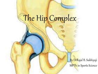
  • 1. The Hip Complex By DrRajal B. Sukhiyaji MPTh in Sports Science
  • 2. Introduction • The hip joint, or coxofemoral joint, is the articulation of the acetabulum of the pelvis and the head of the femur. • These two segments form a diarthrodial ball-and-socket joint with three degrees of freedom: 1. Flexion/extension in the sagittal plane 2. Abduction/adduction in the frontal plane 3. Medial/lateral rotation in the transverse plane
  • 3.
  • 4. The primary function of the hip joint is to support the weight of the head, arms, and trunk (HAT) both in static erect posture and in dynamic postures such as ambulation, running, and stair climbing.
  • 5. Structure of the Hip Joint Proximal Articular Surface: • The cuplike concave socket of the hip joint is called the acetabulum and is located on the lateral aspect of the pelvic bone (innominate or os coxa). • Three bones form the pelvis: the ilium, the ischium, and the pubis.
  • 6. • The pubis forms one fifth of the acetabulum, the ischium forms two fifths, and the ilium forms the remainder. • Until full ossification of the pelvis occurs between 20 and 25 years of age, the separate segments of the acetabulum may remain visible on radiograph.
  • 7.
  • 8. • The acetabulum appears to be a hemisphere, but only its upper margin has a true circular contour, and the roundness of the acetabulum as a whole decreases with age. • In actuality, only a horseshoe-shaped portion of the periphery of the acetabulum (the lunate surface) is covered with hyaline cartilage and articulates with the head of the femur.
  • 9.
  • 10. • The inferior aspect of the lunate surface (the base of the horseshoe) is interrupted by a deep notch called the acetabular notch. • The acetabular notch is spanned by a fibrous band, the transverse acetabular ligament, that connects the two ends of the horseshoe. • The transverse acetabular ligament also spans the acetabular notch to create a fibro-osseous tunnel, called the acetabular fossa, beneath the ligament, through which blood vessels may pass into the central or deepest portion of the acetabulum.
  • 11.
  • 12.
  • 13. • The acetabulum is deepened by the fibrocartilaginous acetabular labrum, which surrounds the periphery. • The acetabular fossa is nonarticular; the femoral head does not contact this surface. • The acetabular fossa contains fibroelastic fat covered with synovial membrane.
  • 14. Center Edge Angle of the Acetabulum • Each acetabulum is oriented on each innominate bone somewhat inferiorly and anteriorly. • The magnitude of inferior orientation is assessed on radiograph by using a line connecting the lateral rim of the acetabulum and the center of the femoral head.
  • 15. • This line forms an angle with the vertical known as the center edge (CE) angle or the angle of Wiberg and is the amount of inferior tilt of the acetabulum. • CE angles in adults to average 38 in men and 35 in women (with ranges in both sexes to be about 22 to 42).
  • 16.
  • 17. Acetabular Anteversion • The acetabulum faces not only somewhat inferiorly but also anteriorly. • The magnitude of anterior orientation of the acetabulum may be referred to as the angle of acetabular anteversion.
  • 18. • Average value to be 18.5 for men and 21.5 for women. • Pathologic increases in the angle of acetabular anteversion are associated with decreased joint stability and increased tendency for anterior dislocation of the head of the femur.
  • 19. Acetabular Labrum • The entire periphery of the acetabulum is rimmed by a ring of wedge-shaped fibrocartilage called the acetabular labrum. • The labrum is attached to the periphery of the acetabulum by a zone of calcified cartilage. • It not only deepens the socket but also increases the concavity of the acetabulum through its triangular shape and grasps the head of the femur to maintain contact with the acetabulum.
  • 20.
  • 21. • The labrum is not load-bearing but serves a role in proprioception and pain sensitivity that may help protect the rim of the acetabulum. • Hydrostatic fluid pressure within the intra-articular space was greater within the labrum than without, which suggests that the labrum may also enhance joint lubrication if the labrum adequately fits the femoral head.
  • 22. • The transverse acetabular ligament is considered to be part of the acetabular labrum. • It protect the blood vessels which reach to the head of femur.
  • 23. Distal Articular Surface: • The head of the femur is a fairly rounded hyaline cartilage-covered surface that may be slightly larger than a true hemisphere or as much as two thirds of a sphere, depending on body type. • The radius of curvature of the femoral head is smaller in women than in men in comparison with the dimensions of the pelvis.
  • 24. • Just inferior to the most medial point on the femoral head is a small roughened pit called the fovea or fovea capitis. • The fovea is not covered with articular cartilage and is the point at which the ligament of the head of the femur is attached.
  • 25.
  • 26. • The femoral head is attached to the femoral neck; the femoral neck is attached to the shaft of the femur between the greater trochanter and the lesser trochanter. • The femoral neck is, in general, only about 5 cm long. • The femoral neck is angulated so that the femoral head most commonly faces medially, superiorly, and anteriorly.
  • 27. Angulation of the Femur • There are two angulations made by the head and neck of the femur in relation to the shaft. • One angulation (angle of inclination) occurs in the frontal plane between an axis through the femoral head and neck and the longitudinal axis of the femoral shaft. • The other angulation (angle of torsion) occurs in the transverse plane between an axis through the femoral head and neck and an axis through the distal femoral condyles.
  • 28. Angle of Inclination of the Femur: • The angle of inclination of the femur averages 126 degrees (referencing the medial angle formed by the axes of the head/neck and the shaft), ranging from 115 to 140 degrees in the unimpaired adult.
  • 29.
  • 30. • In women, the angle of inclination is somewhat smaller than it is in men, owing to the greater width of the female pelvis. • With a normal angle of inclination, the greater trochanter lies at the level of the center of the femoral head. • The angle of inclination of the femur changes across the life span, being substantially greater in infancy and childhood and gradually declining to about 120 degrees in the normal elderly person.
  • 31. • A pathologic increase in the medial angulation between the neck and shaft is called coxa valga, and a pathologic decrease is called coxa vara.
  • 32.
  • 33. • Angle of Torsion of the Femur: • An axis through the femoral head and neck in the transverse plane will lie at an angle to an axis through the femoral condyles, with the head and neck torsioned anteriorly (laterally) with regard to an angle through the femoral condyles.
  • 34.
  • 35. • The angle of torsion decreases with age. In the newborn, the angle of torsion has been estimated to be 40 degrees, decreasing substantially in the first 2 years. • In the adult, the normal angle of torsion is considered to be 10 to 20 degrees. • A pathologic increase in the angle of torsion is called anteversion, and a pathologic decrease in the angle or reversal of torsion is known as retroversion.
  • 36.
  • 37.
  • 38. Articular Congruence • The hip joint is considered to be a congruent joint. • However, there is substantially more articular surface on the head of the femur than on the acetabulum. • In the neutral or standing position, the articular surface of the femoral head remains exposed anteriorly and somewhat superiorly.
  • 39.
  • 40. • The acetabulum does not fully cover the head superiorly, and the anterior torsion of the femoral head (angle of torsion) exposes a substantial amount of the femoral head’s articular surface anteriorly. • Articular contact between the femur and the acetabulum can be increased in the normal non–weight-bearing hip joint by a combination of flexion, abduction, and slight lateral rotation. • This position (also known as the frog-leg position) corresponds to that assumed by the hip joint in a quadruped position and is the true physiologic position of the hip joint.
  • 41.
  • 42. Hip Joint Capsule and Ligaments Hip Joint Capsule: • The hip joint capsule is a substantial contributor to joint stability. • The articular capsule of the hip joint is an irregular, dense fibrous structure with longitudinal and oblique fibers and with three thickened regions that constitute the capsular ligaments. • The capsule is attached proximally to the entire periphery of the acetabulum beyond the acetabular labrum.
  • 43.
  • 44. • The capsule itself is thickened anterosuperiorly, where the predominant stresses occur; it is relatively thin and loosely attached posteroinferiorly. • The capsule covers the femoral head and neck like a cylindrical sleeve and attaches to the base of the femoral neck. • The femoral neck is intracapsular, whereas both the greater and lesser trochanters are extracapsular. • The synovial membrane lines the inside of the capsule.
  • 45. • Anteriorly, there are longitudinal retinacular fibers deep in the capsule that travel along the neck toward the femoral head. • The retinacular fibers carry blood vessels that are the major source of nutrition to the femoral head and neck. • The retinacular blood vessels arise from a vascular ring located at the base of the neck and formed by the medial and lateral circumflex arteries (branches of the deep femoral artery).
  • 46. Hip Joint Ligaments:  The ligamentum teres: • It is an intra-articular but extrasynovial accessory joint structure. • The ligament is a triangular band attached at one end to both sides of the peripheral edge of the acetabular notch.
  • 47.
  • 48. • The ligament then passes under the transverse acetabular ligament (with which it blends) to attach at its other end to the fovea of the femur; thus, it is also called the ligament of the head of the femur. • The ligamentum teres is encased in a flattened sleeve of synovial membrane so that it does not communicate with the synovial cavity of the joint. • It is tensed in semiflexion and adduction.
  • 49.
  • 50. • The ligamentum teres appears to function primarily as a conduit for the secondary blood supply from the obturator artery and for the nerves that travel along the ligament to reach the head of the femur through the fovea.
  • 51. • The hip joint capsule is typically considered to have three reinforcing capsular ligaments (two anteriorly and one posteriorly). • The two anterior ligaments are, 1) The iliofemoral ligament 2) The pubofemoral ligament • The one posterior ligament is, 3) The ischiofemoral ligament
  • 52. 1) The iliofemoral ligament: • It is a fan-shaped ligament that resembles an inverted letter Y. • It often is referred to as the Y ligament of Bigelow. • The apex of the ligament is attached to the anterior inferior iliac spine, and the two arms of the Y fan out to attach along the intertrochanteric line of the femur. • The superior band of the iliofemoral ligament is the strongest and thickest of the hip joint ligaments.
  • 53.
  • 54. 2) The pubofemoral ligament: • It is also anteriorly located, arising from the anterior aspect of the pubic ramus and passing to the anterior surface of the intertrochanteric fossa. • The bands of the iliofemoral and the pubofemoral ligaments form a Z on the anterior capsule.
  • 55. 3) The ischiofemoral ligament: • It is the posterior capsular ligament. • The ischiofemoral ligament attaches to the posterior surface of the acetabular rim and the acetabulum labrum. • Some of its fibers spiral around the femoral neck and blend with the fibers of the circumferential fibers of the capsule. • Other fibers are arranged horizontally and attach to the inner surface of the greater trochanter.
  • 56.
  • 57. • The hip joint capsule and the majority of its ligaments are quite strong and that each tightens with full hip extension (hyperextension). • The anterior ligaments are stronger (stiffer and withstanding greater force at failure) than the ischiofemoral ligament.
  • 58. • Under normal circumstances, the hip joint, its capsule, and ligaments routinely support two thirds of the body weight (the weight of head, arms, and trunk, or HAT). • In bilateral stance, the hip joint is typically in neutral position or slight extension. • In this position, the capsule and ligaments are under some tension. • The normal line of gravity (LoG) in bilateral stance falls behind the hip joint axis, creating a gravitational extension moment.
  • 59.
  • 60. Capsuloligamentous Tension: • Hip joint extension, with slight abduction and medial rotation, is the close-packed position for the hip joint. • With increased extension, the ligaments twist around the femoral head and neck, drawing the head into the acetabulum. • In contrast to most other joints in the body, the close-packed and stable position for the hip joint is not the position of optimal articular contact (congruence). • Optimal articular contact occurs with combined flexion, abduction, and lateral rotation.
  • 61. • Under circumstances in which the joint surfaces are neither maximally congruent nor closepacked, the hip joint is at greatest risk for traumatic dislocation. • A position of particular vulnerability occurs when the hip joint is flexed and adducted (as it is when sitting with the thighs crossed).
  • 62. • In this position, a strong force up the femoral shaft toward the hip joint (as when the knee hits the dashboard in a car accident) may push the femoral head out of the acetabulum. • The capsuloligamentous tension at the hip joint is least when the hip is in moderate flexion, slight abduction, and midrotation.
  • 63. Structural Adaptations to Weight-Bearing • The internal architecture of the pelvis and femur reveal the remarkable interaction between mechanical stresses and structural adaptation created by the transmission of forces between the femur and the pelvis. • The trabeculae (calcified plates of tissue within the cancellous bone) line up along lines of stress and form systems that normally adapt to stress requirements.
  • 64.
  • 65.
  • 66. • In standing or upright weight-bearing activities, at least half the weight of the HAT (the gravitational force) passes down through the pelvis to the femoral head, whereas the ground reaction force (GRF) travels up the shaft. • These two forces, nearly parallel and in opposite directions, create a force couple with a moment arm (MA) equal to the distance between the superimposed body weight on the femoral head and the GRF up the shaft.
  • 67. • These forces create a bending moment (or set of shear forces) across the femoral neck.
  • 68. • The bending stress creates a tensile force on the superior aspect of the femoral neck and a compressive stress on the inferior aspect. • A complex set of forces prevents the rotation and resists the shear forces that the force couple causes; among these forces are the structural resistance of two major and three minor trabecular systems.
  • 69.
  • 70. • The medial (or principal compressive) trabecular system arises from the medial cortex of the upper femoral shaft and radiates through the cancellous bone to the cortical bone of the superior aspect of the femoral head. • The medial system of trabeculae is oriented along the vertical compressive forces passing through the hip joint.
  • 71. • The lateral (or principal tensile) trabecular system of the femur arises from the lateral cortex of the upper femoral shaft and, after crossing the medial system, terminates in the cortical bone on the inferior aspect of the head of the femur. • The lateral trabecular system is oblique and may develop in response to parallel (shear) forces of the weight of HAT and the GRF.
  • 72. • There are two accessory (or secondary) trabecular systems, of which one is considered compressive and the other is considered tensile. • Another secondary trabecular system is confined to the trochanteric area femur.
  • 73. • The areas in which the trabecular systems cross each other at right angles are areas that offer the greatest resistance to stress and strain. • There is an area in the femoral neck in which the trabeculae are relatively thin and do not cross each other. • This zone of weakness has less reinforcement and thus more potential for failure.
  • 74. • The primary weight-bearing surface of the acetabulum, or dome of the acetabulum, is located on the superior portion of the lunate surface. • In the normal hip, the dome lies directly over the center of rotation of the femoral head. • The dome shows the greatest prevalence of degenerative changes in the acetabulum.
  • 75. • The primary weight-bearing area of the femoral head is, correspondingly, its superior portion. • Although the primary weight-bearing area of the acetabulum is subject to the most degenerative changes, degenerative changes in the femoral head are most common around or immediately below the fovea or around the peripheral edges of the head’s articular surface.
  • 76. • The forces of HAT and GRF that act on the articular surfaces of the hip joint and on the femoral head and neck also act on the femoral shaft. • The shaft of the femur is not vertical but lies at an angle that varies considerably among individuals. • The vertical loading on the oblique femur results in bending stresses in the shaft. • The medial cortical bone in the shaft (diaphysis) must resist compressive stresses, whereas the lateral cortical bone must resist tensile stresses.
  • 77.
  • 78. Function of the Hip Joint  Motion of the Femur on the Acetabulum: • The motions of the hip joint are easiest to visualize as movement of the convex femoral head within the concavity of the acetabulum as the femur moves through its three degrees of freedom: flexion/extension, abduction/adduction, and medial/lateral rotation. • The femoral head will glide within the acetabulum in a direction opposite to motion of the distal end of the femur.
  • 79. • Flexion and extension of the femur occur from a neutral position as an almost pure spin of the femoral head around a coronal axis through the head and neck of the femur. • The head spins posteriorly in flexion and anteriorly in extension.
  • 80.
  • 81.
  • 82. • Flexion and extension from other positions (e.g., in abduction or medial rotation) must include both spinning and gliding of the articular surfaces, depending on the combination of motions. • The motions of abduction/adduction and medial/lateral rotation must include both spinning and gliding of the femoral head within the acetabulum.
  • 83.  Range of motion: • The joint’s range of motion (ROM) is influenced by structural elements, as well as by whether the motion is performed actively or passively and whether passive tension in two-joint muscles is encountered or avoided.
  • 84. • Flexion of the hip is generally about 90 degrees with the knee extended and 120 degrees when the knee is flexed and when passive tension in the two-joint hamstrings muscle group is released. • Hip extension is considered to have a range of 10 to 30 degrees.
  • 85. • Hip extension ROM appears to diminish somewhat with age, whereas flexion remains relatively unchanged. • When hip extension is combined with knee flexion, passive tension in the two-joint rectus femoris muscle may limit the movement.
  • 86. • The femur can be abducted 45 to 50 degrees and adducted 20 to 30 degrees. • Abduction can be limited by the two-joint gracilis muscle and adduction limited by the tensor fascia lata (TFL) muscle and its associated iliotibial (IT) band.
  • 87. • Medial and lateral rotation of the hip are usually measured with the hip joint in 90 degrees of flexion; the typical range is 42 to 50 degrees. • Femoral anteversion is correlated with decreased range of lateral rotation and less strongly with increased range of medial rotation.
  • 88. • When the femoral head is torsioned anteriorly more than normal, lateral rotation of the femur turns the head out even more, both risking subluxation and encountering capsuloligamentous and muscular restrictions on the anterior aspect of the joint.
  • 89.
  • 90. • Normal gait on level ground requires at least the following hip joint ranges: o 30 degrees flexion, o 10 degrees hyperextension, o 5 degrees of both abduction and adduction, and o 5 degrees of both medial and lateral rotation. • Walking on uneven terrain or stairs will increase the need for joint range beyond that required for level ground, as will activities such as sitting in a chair or sitting cross-legged.
  • 91. Motion of the Pelvis on the Femur • Whenever the hip joint is weight-bearing, the femur is relatively fixed, and, motion of the hip joint is produced by movement of the pelvis on the femur.  Anterior and Posterior Pelvic Tilt  Lateral Pelvic Tilt  Anterior and Posterior Pelvic Rotation
  • 92. Anterior and Posterior Pelvic Tilt • Anterior and posterior pelvic tilt are motions of the entire pelvic ring in the sagittal plane around a coronal axis. • In the normally aligned pelvis, the anterosuperior iliac spines (ASISs) of the pelvis lie on a horizontal line with the posterior superior iliac spines and on a vertical line with the symphysis pubis.
  • 93.
  • 94. • Anterior and posterior tilting of the pelvis on the fixed femur produce hip flexion and extension, respectively. • Hip joint extension through posterior tilting of the pelvis brings the symphysis pubis up and the sacrum of the pelvis closer to the femur, rather than moving the femur posteriorly on the pelvis.
  • 95. • Hip flexion through anterior tilting of the pelvis moves the ASISs anteriorly and inferiorly; the inferior sacrum moves farther from the femur, rather than moving the femur away from the sacrum. • Anterior and posterior tilting will result in flexion and extension of both hip joints simultaneously in bilateral stance or can occur at the stance hip joint alone if the opposite limb is non–weight-bearing.
  • 96. Lateral Pelvic Tilt • Lateral pelvic tilt is a frontal plane motion of the entire pelvis around an anteroposterior axis. • In the normally aligned pelvis, a line through the ASISs is horizontal. • In lateral tilt of the pelvis in unilateral stance, one hip joint is the pivot point or axis for motion of the opposite side of the pelvis as it elevates (pelvic hiking) or drops (pelvic drop).
  • 97. • If a person stands on the left limb and hikes the pelvis, the left hip joint is being abducted because the medial angle between the femur and a line through the ASISs increases. • If a person stands on the left leg and drops the pelvis, the left hip joint will adduct because the medial angle formed by the femur and a line through the ASISs will decrease.
  • 98.
  • 99. • In descriptions of the hip joint motions that occur in unilateral stance, the hip joint of the non–weightbearing limb is in an open chain and has no motions on it. • However, the non–weight-bearing leg typically hangs straight down as the pelvis moves.
  • 100. Lateral Shift of the Pelvis: • Lateral pelvic tilt can also occur in bilateral stance. • If both feet are on the ground and the hip and knee of one limb are flexed, the opposite limb is largely the weight-bearing limb and the terminology is the same as for unilateral stance.
  • 101. • If both limbs are weightbearing, lateral tilt of the pelvis will cause the pelvis to shift to one side or the other. • With pelvic shift, the pelvis cannot hike but can only drop. • Because there is a closed chain between the two weight- bearing feet and the pelvis, both hip joints will move in the frontal plane in a predictable way as the pelvic tilt (or pelvic shift) occurs. • If the pelvis is shifted to the right in bilateral stance, the left side of the pelvis will drop, the right hip joint will be adducted, and the left hip joint will be abducted.
  • 102.
  • 103. Anterior and Posterior Pelvic Rotation • Pelvic rotation is motion of the entire pelvic ring in the transverse plane around a vertical axis. • Although rotation can occur around a vertical axis through the middle of the pelvis in bilateral stance, it most commonly and more importantly occurs in single- limb support around the axis of the supporting hip joint.
  • 104. • Forward rotation of the pelvis occurs in unilateral stance when the side of the pelvis opposite to the supporting hip joint moves anteriorly. • Forward rotation of the pelvis produces medial rotation of the supporting hip joint. • Backward rotation of the pelvis occurs when the side of the pelvis opposite the supporting hip moves posteriorly. • Posterior rotation of the pelvis produces lateral rotation of the supporting hip joint.
  • 105.
  • 106. • Pelvic rotation can occur in bilateral stance as well as unilateral stance, as is true for lateral pelvic tilt. • If both feet are bearing weight and the axis of motion occurs around a vertical axis through the center of the pelvis, the terms forward rotation and backward rotation must be used by referencing a side (e.g., forward rotation on the right and backward rotation on the left).
  • 107. Coordinated Motions of the Femur, Pelvis, and Lumbar Spine • When the pelvis moves on a relatively fixed femur, there are two possible outcomes to consider. • Either the head and trunk will follow the motion of the pelvis (moving the head through space) or the head will continue to remain relatively upright and vertical despite the pelvic motions. • These are open- and closed-chain responses, respectively.
  • 108. Pelvifemoral Motion: • When the femur, pelvis, and spine move in a coordinated manner to produce a larger ROM than is available to one segment alone, the hip joint is participating in what will predominantly (but not exclusively) be an open-chain motion termed pelvifemoral motion. • In the case of pelvifemoral motion, the joints may serve either end of the chain: the foot or head.
  • 109.  Moving the Head and Arms through Space: (Example) • If the goal is to bend forward to bring the hands (and head) toward the floor, isolated flexion at the hip joints (anteriorly tilting the pelvis on the femurs) is generally insufficient to reach the ground. • If the knees remain extended, the hips will typically flex no more than 90 (and often less, depending on extensibility of the hamstrings). • The addition of flexion of the lumbar spine (and, perhaps, flexion of the thoracic spine) will add to the total ROM.
  • 110.
  • 111. • The combination of hip and trunk flexion is generally sufficient for the hands to reach the ground—as long as the hamstrings and lumbar extensors allow sufficient lengthening. • The combination of hip motion and lumbar motion to achieve a greater ROM for the hands and head is an example of a largely open-chain response in the hips and trunk.
  • 112.  Moving the Foot through Space: (Example) • When a person is lying on the right side, the left foot may be moved through an arc of motion approaching 90 degrees. • This is clearly not all from the left hip joint, which can typically abduct only to 45 degrees; motion of the foot through space also includes lateral tilting of the pelvis (hiking around the right hip joint) and lateral flexion of the lumbar spine to the left. • The abducting limb is in an open chain; the lumbar spine (and thoracic spine) are constrained by the body weight and contact with the ground.
  • 113.
  • 114. • Pelvifemoral motion has also been referred to as pelvifemoral “rhythm”. • The link between hip, pelvis, and lumbar motion is the basis of using pain with active straight-leg raising as a test for severity of dysfunction in persons with low back pain.
  • 115. Closed-Chain Hip Joint Function: • The joints of the right and left lower limbs are part of a true closed chain when both lower limbs are weightbearing and the chain is defined as all the segments between the right foot, up through the pelvis, and down through the left foot. • A true closed chain is formed because both ends of the chain (both feet in this example) are “fixed” and movement at any one joint in the chain invariably involves movement at one or more other links in the chain.
  • 116. • For the hips (and other lower limb joints) to be in a closed chain in standing, both ends of the chain (the head and the feet) must be fixed. • The feet are fixed by weight-bearing. • The head is often (but not necessarily) functionally “fixed.”
  • 117. • When the head (one end of the chain) is held upright and over the feet (the other end of the chain), all the segments in the axial skeleton and lower limbs function as part of a closed chain; movement at one joint will create movement in at least one other linkage in the chain. • Consequently, in our functional closed chain, hip flexion does not occur independently (which would move the head forward in space) but is accompanied by motion in one or more interposed segments to ensure that the head remains upright over the base of support and that the body does not become unstable.
  • 118.  Closed-Chain Hip Joint Function: (Example) • A common example of closed-chain versus open-chain function is seen when the hip flexor musculature is tight and the hip joint is maintained in flexion. • A person standing with fixed hip flexion is shown in Figure 10-27A (an open-chain response) and B (a closed-chain response).
  • 119.
  • 120. • A true open-chain response to isolated hip flexion would displace the head and trunk forward, with the LoG falling in front of the supporting feet. • More commonly, hip flexion in stance is not isolated to the hips but is accompanied by compensatory movements of the vertebral column (including extension or lordosis of the lumbar spine) that maintain the head in the upright position and keep the LoG well within the base of support).
  • 121. • In a functional closed chain, motion at the hip (one link in the chain) is accompanied by an essentially mandatory lumbar extension to maintain the head over the sacrum (see Fig. 10-27B). • In contrast, hip flexion in open-chain pelvifemoral motion is accompanied by lumbar flexion because the goal is to achieve more range for the head in space (see Fig. 10- 25).
  • 122. Hip Joint Musculature • The muscles of the hip joint make their most important contributions to function during weight-bearing. • In weight-bearing, the muscles are called on to move or support the HAT (approximately two thirds of body weight). • The hip joint muscles adapt their structure to the required function, as can be seen in their large areas of attachment, their length, and their large cross-section.
  • 123. Flexors • The flexors of the hip joint function primarily as mobility muscles in open-chain function; that is, they function primarily to bring the swinging limb forward during ambulation or in various sports. • The flexors may function secondarily to resist strong hip extension forces that occur as the body passes over the weight bearing foot. • Nine muscles have action lines crossing the anterior aspect of the hip joint.
  • 124. • The primary muscles of hip flexion are the iliopsoas, rectus femoris, TFL, and sartorius. • The iliopsoas muscle is considered to be the most important of the primary hip flexors. • It consists of two separate muscles, the iliacus muscle and the psoas major muscle, both of which attach to the femur by a common tendon.
  • 125.
  • 126. • The two components of the iliopsoas muscle have many points of origin, including the iliac fossa and the disks, bodies, and transverse processes of the lumbar vertebrae. • Given the attachments of the psoas major muscle to the anterior vertebrae and the iliacus muscle to the iliac fossa, activity of or passive tension in these muscles would anteriorly tilt the pelvis (iliacus muscle) and, apparently, pull the lumbar vertebrae anteriorly into flexion (psoas major muscle).
  • 127. • In closed-chain function (head vertical), however, these muscles seem to create a paradoxical lumbar lordosis (lumbar extension) that results from the body’s attempt to keep the head over the sacrum with anterior pelvic tilt and lower lumbar flexion. • The role of the iliopsoas muscle in hip flexion may be particularly critical when hip flexion from a sitting position is required.
  • 128. • The rectus femoris muscle is the only portion of the quadriceps muscle that crosses both the hip joint and knee joint. • It originates on the anterior inferior iliac spine and inserts by way of a common tendon into the tibial tuberosity. • The rectus femoris muscle flexes the hip joint and extends the knee joint.
  • 129. • Because it is a two-joint hip flexor, the position of the knee during hip flexion will affect its ability to generate force at the hip. • Simultaneous hip flexion and knee extension considerably shorten this muscle and increase the likelihood of active insufficiency. • Consequently, the rectus femoris muscle makes its best contribution to hip flexion when the knee is maintained in flexion.
  • 130. • The sartorius muscle is a straplike muscle originating on the ASIS. • It crosses the anterior aspect of the femur to insert into the upper portion of the medial aspect of the tibia. • The sartorius muscle is considered to be a flexor, abductor, and lateral rotator of the hip, as well as a flexor and medial rotator of the knee.
  • 131. • It is a two-joint muscle, should be relatively unaffected by the position of the knee. • Its function is probably most important when the knee and hip need to be flexed simultaneously (as in climbing stairs).
  • 132. • The TFL muscle originates more laterally than the sartorius muscle. • Its origin is on the anterolateral lip of the iliac crest. • The muscle fibers extend only about one fourth of the way down the lateral aspect of the thigh before inserting into the IT band.
  • 133. • The IT band or IT tract is the thickened lateral portion of the fascia lata of the hip and thigh. • The IT band attaches proximally to the iliac crest lateral to the TFL muscle. • After the tensor attaches to the IT band, the IT band continues distally on the lateral thigh to insert into the lateral condyle of the tibia.
  • 134. • The TFL muscle is considered to flex, abduct, and medially rotate the femur at the hip. • The most important contribution of the TFL muscle may be in maintaining tension in the IT band. • The IT band assists in relieving the femur of some of the tensile stresses imposed on the shaft by weight-bearing forces.
  • 135. • The secondary hip flexors are the pectineus, adductor longus, adductor magnus, and the gracilis muscles. • Each, however, is capable of contributing to hip joint flexion, but that contribution is dependent on hip joint position. • The gracilis, a two-joint muscle, is active as a hip flexor when the knee is extended but not when the knee is flexed.
  • 136. Adductors • The hip adductor muscle group is generally considered to include the pectineus, adductor brevis, adductor longus, adductor magnus, and the gracilis muscles. • The adductors are located anteromedially.
  • 137.
  • 138. • The adductors longus, brevis, and magnus muscles arise in a group from the body and inferior ramus of the pubis to insert along the linea aspera. • The gracilis muscle is the only two-joint adductor. • It originates on the symphysis pubis and pubic arch and inserts on the medial surface of the shaft of the tibia.
  • 139. • In bilateral stance, the adductors may be synergists to the abductor muscles when both feet are on the ground, enhancing side-to-side stabilization of the pelvis. • The adductors are also capable of generating a maximum isometric torque greater than that of the abductors.
  • 140. Extensors • The one-joint gluteus maximus muscle and the two-joint hamstrings muscle group are the primary hip joint extensors. • These muscles may receive assistance from the posterior fibers of the gluteus medius, from the posterior adductor magnus muscle, and from the piriformis muscle.
  • 141.
  • 142. • The gluteus maximus is a large, quadrangular muscle that originates from the posterior sacrum, dorsal sacroiliac ligaments, sacrotuberous ligament, and a small portion of the ilium. • The gluteus maximus crosses the sacroiliac joint before its most superior fibers insert into the IT band (as do the fibers of the TFL muscle) and its inferior fibers insert into the gluteal tuberosity.
  • 143. • The gluteus maximus is the largest of the lower extremity muscles; this muscle alone constituting 12.8% of the total muscle mass of the lower extremity. • The maximus is a strong hip extensor that appears to be active primarily against a resistance greater than the weight of the limb.
  • 144. • The three two-joint extensors are the long head of the biceps femoris, the semitendinosus, and the semimembranosus muscles, known collectively as the hamstrings. • Each of these three muscles originates on the ischial tuberosity.
  • 145. • The biceps femoris crosses the posterior femur to insert into the head of the fibula and lateral aspect of the lateral tibial condyle. • The other two hamstrings insert on the medial aspect of the tibia. • All three muscles extend the hip with or without resistance, as well as serving as important knee flexors.
  • 146. • If the hip is extended and the knee is flexed to 90 or more, the hamstrings may not be able to contribute much to hip extension force because of active insufficiency or approaching active insufficiency. • Extension forces in the hip increase by 30% if the knee is extended during hip extension. • The biceps femoris appears to contribute to lateral rotation of the hip.
  • 147. Abductors • Active abduction of the hip is brought about predominantly by the gluteus medius and the gluteus minimus muscles. • The gluteus medius originates on the lateral surface of the wing of the ilium and inserts into the greater trochanter, beneath the gluteus maximus. • The gluteus medius has anterior, middle, and posterior parts that function asynchronously during movement at the hip.
  • 148.
  • 149. • The gluteus minimus muscle lies deep to the gluteus medius, arising from the outer surface of the ilium with its fibers converging on an aponeurosis that ends in a tendon on the greater trochanter. • The minimus is consistently an abductor and flexor of the hip, with its rotator function dependent on hip position. • The minimus is a medial rotator in hip flexion.
  • 150. • The gluteus minimus and medius muscles function together to either abduct the femur (distal level free) or, more important, to stabilize the pelvis (and superimposed HAT) in unilateral stance against the effects of gravity.
  • 151. Lateral Rotators • Six short muscles have lateral rotation as a primary function. • These muscles are the obturator internus and externus, the gemellus superior and inferior, the quadratus femoris, and the piriformis muscles. • Other muscles that have fibers posterior to the axis of motion at the hip (the posterior fibers of the gluteus medius and minimus and the gluteus maximus) may produce lateral rotation combined with the primary action of the muscle.
  • 152.
  • 153. • The obturator internus muscle originates from the inside (posterior aspect) of the obturator foramen and emerges through the lesser sciatic foramen to insert on the medial aspect (inside) of the greater trochanter. • The gemellus superior and gemellus inferior muscles arise from the ischium of the pelvis, just above and just below the point at which the obturator internus passes through the lesser sciatic notch. • Both gemelli follow and blend with the obturator internus tendon to insert with the internus tendon into the greater trochanter.
  • 154. • The obturator externus muscle is sometimes considered to be an anteromedial muscle of the thigh because it originates on the external (anterior) surface of the obturator foramen. • However, it crosses the posterior aspect of the hip joint and inserts on the medial aspect of the greater trochanter in the trochanteric fossa.
  • 155. • The quadratus femoris muscle is a small quadrangular muscle that originates on the ischial tuberosity and inserts on the posterior femur between the greater and lesser trochanters. • The piriformis muscle originates largely on the anterior surface of the sacrum, passes through the greater sciatic notch, and follows the inferior border of the posterior gluteus medius to insert above the other lateral rotators into the medial aspect of the greater trochanter.
  • 156. • The piriformis and gluteus maximus are the only two muscles that cross the sacroiliac joint. • The sciatic nerve, the largest nerve in the body, enters the gluteal region just inferior to the piriformis muscle.
  • 157. • The lateral rotator muscles are positioned to perform their rotatory function effectively, given the nearly perpendicular orientation to the shaft of the femur. • These muscles would certainly appear to be effective joint compressors because their combined action line parallels the head and neck of the femur.
  • 158. • Of the primary lateral rotators, each inserts either on or in the vicinity of the greater trochanter.
  • 159. Medial Rotators • There are no muscles with the primary function of producing medial rotation of the hip joint. • The more consistent medial rotators are the anterior portion of the gluteus medius, gluteus minimus, and the TFL muscles.
  • 160. Hip Joint Forces and Muscle Function in Stance  Bilateral Stance: • In erect bilateral stance, both hips are in neutral or slight hyperextension, and weight is evenly distributed between both legs. • The LoG falls just posterior to axis for flexion/extension of the hip joint.
  • 161. • The posterior location of the LoG creates an extension moment of force around the hip that tends to posteriorly tilt the pelvis on the femoral heads. • The gravitational extension moment is largely checked by passive tension in the hip joint capsuloligamentous structures, although slight or intermittent activity in the iliopsoas muscles in relaxed standing may assist the passive structures.
  • 162. • In the frontal plane during bilateral stance, the body weight is transmitted through the sacroiliac joints and pelvis to the right and left femoral heads. • Hypothetically, the weight of the HAT (two thirds of body weight) should be distributed so that each femoral head receives approximately half of the weight.
  • 163. • The joint axis of each hip lies at an equal distance from the LoG of HAT; that is, the gravitational MAs for the right hip (DR) and the left hip (DL) are equal. • Because the body weight (W) on each femoral head is the same (WR=WL), the magnitude of the gravitational torques around each hip must be identical (WR *DR=WL* DL). • The gravitational torques on the right and left hips, however, occur in opposite directions.
  • 164.
  • 165. • The weight of the body acting around the right hip tends to drop the pelvis down on the left (right adduction moment), whereas the weight acting around the left hip tends to drop the pelvis down on the right (left adduction moment). • These two opposing gravitational moments of equal magnitude balance each other, and the pelvis is maintained in equilibrium in the frontal plane without the assistance of active muscles.
  • 166. • When bilateral stance is not symmetrical, frontal plane muscle activity will be necessary to either control the side-to-side motion or to return the hips to symmetrical stance. • The pelvis is shifted to the right, resulting in relative adduction of the right hip and abduction of the left hip. • To return to neutral position, an active contraction of the right hip abductors would be expected. • However, a contraction of the left hip adductors would accomplish the same goal.
  • 167.
  • 168. • Under the condition that both extremities bear at least some of the superimposed body weight, the adductors may assist the abductors in control of the pelvis against the force of gravity or the GRF. • In unilateral stance, activity of the adductors either in the weight-bearing or non–weight-bearing hip cannot contribute to stability of the stance limb. • In the absence of adequate hip abductor function, the adductors can contribute to stability—but only in bilateral stance.
  • 169.  Unilateral Stance: • The left leg has been lifted from the ground and the full superimposed body weight is being supported by the right hip joint. • Rather than sharing the compressive force of the superimposed body weight with the left limb, the right hip joint must now carry the full burden.
  • 170. • In addition, the weight of the non–weight bearing left limb that is hanging on the left side of the pelvis must be supported along with the weight of HAT. • Of the one-third portion of the body weight found in the lower extremities, the non-supporting limb must account for half of that, or one sixth of the full body weight.
  • 171.
  • 172. • The magnitude of body weight (W) compressing the right hip joint in right unilateral stance, therefore, is: Right hip joint compression body weight = [2/3*W]+[1/6*W] Right hip joint compression body weight =5/6*W
  • 173. • The force of gravity acting on HAT and the non–weight- bearing left lower limb (HATLL) will create an adduction torque around the supporting hip joint; that is, gravity will attempt to drop the pelvis around the right weight- bearing hip joint axis. • The abduction counter torque will have to be supplied by the hip abductor musculature. • The result will be joint compression or a joint reaction force that is a combination of both body weight and abductor muscular compression.
  • 174. • The actual MA is likely to be slightly greater because the weight of the hanging left leg will pull the center of gravity of the superimposed weight slightly to the left, although the LoG will simultaneously be shifted slightly right to get the LoG within the single foot base of support.
  • 175. Reduction of Muscle Forces in Unilateral Stance • If the hip joint undergoes osteoarthritic changes that lead to pain on weight-bearing, the joint reaction force must be reduced to avoid pain. • For most painful hip joints, however, the reductions in compression generally required are greater than can be achieved through weight loss.
  • 176. • The solution must be in a reduction of abductor muscle force requirements. • If less muscular countertorque is needed to offset the effects of gravity, there will be a decrease in the amount of muscular compression across the joint, although the body weight compression will remain unchanged.
  • 177. • The need to diminish abductor force requirements also occurs when the abductor muscles are weakened through paralysis, through structural changes in the femur that reduce biomechanical efficiency of the muscles, or through degenerative changes producing tears at the greater trochanter. • Hip abductor muscle weakness will affect gait, whereas paralysis of other hip joint muscles in the presence of intact abductors will permit someone to walk or even run with relatively little disability.
  • 178. • Several options are available when there is a need to decrease abductor muscle force requirements. • Some compression reduction strategies occur automatically, but at a cost of extra energy expenditure and structural stress. • Other strategies require intervention such as assistive devices but minimize the energy cost.
  • 179. Compensatory Lateral Lean of the Trunk • Gravitational torque at the pelvis is the product of body weight and the distance that the LoG lies from the hip joint axis (MA). • If there is a need to reduce the torque of gravity in unilateral stance and if body weight cannot be reduced, the MA of the gravitational force can be reduced by laterally leaning the trunk over the pelvis toward the side of pain or weakness when in unilateral stance on the painful limb.
  • 180. • The compensatory lateral lean of the trunk toward the painful stance limb will swing the LoG closer to the hip joint, thereby reducing the gravitational MA. • Because the weight of HATLL must pass through the weight bearing hip joint regardless of trunk position, leaning toward the painful or weak supporting hip does not increase the joint compression caused by body weight. • However, it does reduce the gravitational torque.
  • 181. • Whether a lateral trunk lean is due to muscular weakness or pain, a lateral lean of the trunk during walking still uses more energy than ordinarily required for single-limb support and may result in stress changes within the lumbar spine if used over an extended time period. • Use of a cane or some other assistive device offers a realistic alternative to the person with hip pain or weakness.
  • 182. Use of a Cane Ipsilaterally • Pushing downward on a cane held in the hand on the side of pain or weakness should reduce the superimposed body weight by the amount of downward thrust; that is, some of the weight of HATLL would follow the arm to the cane, rather than arriving on the sacrum and the weight-bearing hip joint. • It is realistic to expect that someone can push down on a cane with approximately 15% of his body weight.
  • 183. • The proportion of body weight that passes through the cane will not pass through the hip joint and will not create an adduction torque around the supporting hip joint. • Although a cane used ipsilaterally provides some benefits in energy expenditure and structural stress reduction, it is not as effective in reducing hip joint compression as the undesirable lateral lean of the trunk. • Moving the cane to the opposite hand produces substantially different and better results.
  • 184. Use of a Cane Contralaterally • When the cane is moved to the side opposite the painful or weak hip joint, the reduction in HATLL is the same as it is when the cane is used on the same side as the painful hip joint; that is, the superimposed body weight passing through the weight-bearing hip joint is reduced by approximately 15% of body weight.
  • 185. • The cane is now farther from the painful supporting hip joint; that is, in addition to relieving some of the superimposed body weight, the cane is now in a position to assist the abductor muscles in providing a countertorque to the torque of gravity.
  • 186.
  • 187. Adjustment of a Carried Load • When someone with hip joint pain or weakness carries a load in the hand or on the trunk (as with a backpack or purse), there is a potential for increasing the demands on the hip abductors and increasing the hip joint compression. • The added external load will increase the superimposed weight acting through the affected supporting hip in unilateral stance.
  • 188. • The gravitational torque may increase, resulting in an increased demand on the supporting hip abductors to prevent drop of the pelvis. • Although the increase in superimposed weight when a load is carried cannot be avoided, it is possible to minimize the demand on the abductor muscles on the side of a painful or weak hip.
  • 189. • If the external load is carried in the arm or on the side of the trunk ipsilateral to the painful or weak hip, the asymmetrical external load will cause a shift in the combined force of HAT/external load center of mass (CoM) toward the painful hip. • Any shift of the combined CoM (or resulting LoG) toward the painful hip will reduce the MA of the HAT/external load.
  • 190. • If the external load is not too great, the reduction in MA of the HAT/external load can result in a reduction in adduction torque. • With a reduction in adduction torque, the demand on the hip abductors is reduced.
  • 191. • The reverse effect will occur if the load is carried on the side opposite to the weak or painful hip. • In that scenario, the external load both increases superimposed body weight and increases the gravitational MA around the weak or painful hip when in unilateral stance on that hip.
  • 192. Hip Joint Pathology • The very large active and passive forces crossing the hip joint make the joint’s structures susceptible to wear and tear of normal components and to failure of weakened components. • Small changes in the biomechanics of the femur or the acetabulum can result in increases in passive forces above normal levels or in weakness of the dynamic joint stabilizers.
  • 193. Arthrosis • The most common painful condition of the hip is due to deterioration of the articular cartilage and to subsequent related changes in articular tissues. • It is known as osteoarthritis, degenerative arthritis, or hip joint arthrosis. • Its prevalence rates are about 10% to 15% in those older than 55 years, with approximately equal distribution among men and women.
  • 194. • Changes may be due to subtle deviations present from birth, to tissue changes inherent in aging, to the repetitive mechanical stress of loading the body weight on the hip joint over a prolonged period, to impingement between the femur and labrum or adjacent acetabulum, or to interactions of each of these factors. • The factors most closely associated with hip joint arthrosis are increased age and increased weight/height ratio.
  • 195. • Forces in excess of half the body weight are needed to fully compress the femoral head into congruent contact with the dome of the acetabulum. • The more common degenerative changes in the femur are at the periphery of the head and the perifoveal area.
  • 196. • The periphery of the head receives only about one third the compressive force of the superior portion of the head, whereas the superior portion of the head is compressed not only in standing but is also in contact with the posterior acetabulum during sitting activities. • The area of the femoral head around the fovea is most commonly in the non–weight-bearing acetabular notch and would undergo compression relatively infrequently.
  • 197. Fracture • The bony components must also be of sufficient strength to withstand the forces that are acting around and through the hip joint. • The vertical weight-bearing forces that pass down through the superior margin of the acetabulum in both unilateral and bilateral stance act at some distance from GRF up the shaft of the femur. • The result is a bending force across the femoral neck.
  • 198.
  • 199. • Normally the trabecular systems are capable of resisting the bending forces, but abnormal increases in the magnitude of the force or weakening of the bone can lead to bony failure. • The site of failure is likely to be in areas of thinner trabecular distribution such as the zone of weakness.
  • 200. • Bony failure in the femoral neck is uncommon in the child or young adult. • Of middle-aged people, women actually suffer fewer hip fractures than do men, although the fractures in this age group are usually attributable to trauma.
  • 201. • The precipitating factor appears to be moderate trauma such as that caused by a fall from standing, from a chair, or from a bed. • Hip fracture is associated with diminished bone density. • Bone density decreases about 2% per year after age 50 and trabeculae clearly thin and disappear with aging.
  • 202. • Not only is the condition painful, but malunion of the fracture can lead to joint instability or cartilaginous deterioration (or both) as a result of poorly aligned bony segments. • Although the femoral head may receive some blood supply via the ligament of the femoral head, an absent or diminished supply through the ligament of the head (as occurs with aging) means reliance on anastomoses from the circumflex arteries.
  • 203. • This circumflex arterial supply may be disrupted by femoral neck fracture, which leaves the femoral head susceptible to avascular necrosis and necessitates replacement of the head of the femur with an artificial implant.
  • 204. Bony Abnormalities of the Femur  Coxa Valga/Coxa Vara: • In coxa valga, the angle of inclination in the femur is greater than the normal adult angle of 125 degrees. • The increased angle brings the vertical weight bearing line closer to the shaft of the femur, diminishing the shear, or bending, force across the femoral neck.
  • 205. • The reduction in force is actually reflected in a reduction in density of the lateral trabecular system. • The decreased distance between the femoral head and the greater trochanter also decreases the length of the MA of the hip abductor muscles. • The decreased muscular MA results in an increased demand for muscular force generation to maintain sufficient abduction torque to counterbalance the gravitational adduction moment acting around the supporting hip joint during single-limb support.
  • 206. • Coxa valga also decreases the amount of femoral articular surface in contact with the dome of the acetabulum. • As the femoral head points more superiorly, there is a decreasing amount of coverage from the acetabulum superiorly. • Consequently, coxa valga decreases the stability of the hip and predisposes the hip to dislocation.
  • 207.
  • 208. • Coxa vara is considered to give the advantage of improved hip joint stability (if angle reduction is not too extreme). • The apparent improvement in congruence occurs because the decreased angle between the neck and shaft of the femur will turn the femoral head deeper into the acetabulum, decreasing the amount of articular surface exposed superiorly and increasing coverage from the acetabulum.
  • 209. • A varus femur, if not caused by trauma, may also increase the length of the MA of the hip abductor muscles by increasing the distance between the femoral head and the greater trochanter. • The increased MA decreases the amount of force that must be generated by the abductor muscles in single- limb support and reduces the joint reaction force.
  • 210. • Coxa vara has the disadvantage of increasing the bending moment along the femoral head and neck. • Coxa vara may increase the likelihood in the adolescent child that the femoral head will slide on the cartilaginous epiphysis of the head of the femur.
  • 211.  Anteversion/Retroversion: • Anteversion of the femoral head reduces hip joint stability because the femoral articular surface is more exposed anteriorly. • The line of the hip abductors may fall more posterior to the joint, reducing the MA for abduction.
  • 212. • The effect of femoral anteversion may also be seen at the knee joint. • When the femoral head is anteverted, pressure from the anterior capsuloligamentous structures and the anterior musculature may push the femoral head back into the acetabulum, causing the entire femur to rotate medially.
  • 213. • Although the medial rotation of the femur improves the congruence in the acetabulum, the knee joint axis through the femoral condyles is now turned medially, altering the plane of knee flexion/extension and resulting, at least initially, in a toe-in gait. • The abnormal position of the knee joint axis is commonly labeled medial femoral torsion. • Medial femoral torsion and femoral anteversion are the same abnormal condition of the femur.
  • 214. • Femoral retroversion is the opposite of anteversion and creates opposite problems from femoral anteversion.