SlideShare une entreprise Scribd logo
1  sur  103
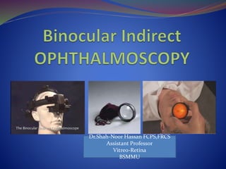
Dr.Shah-Noor Hassan FCPS,FRCS
Assistant Professor
Vitreo-Retina
BSMMU
History of ophthalmoscope
 Mery in 1704 made first
ophthalmoscopic
observation of a normal
fundus in a drowning cat
 Cumming and Brucke in
1846 explained the
principles of
ophthalmoscopy
2
 THREE basic principles
described by Hermann von
Helmholtz
 Patient and observer should
be made emmetropic
 Retina of the patient should
be sufficiently illuminated
 Optical alignment of light
source and observer’s pupil
3
• Ruete in 1852 designed first
monocular indirect
ophthalmoscope
4
 Marc-Antoine Giraud-
Teulon of France
(1861)
 Weak source of
illumination.
5
• 1911-Thorner and Allvor
Gullstrand – Reflex free
ophthalmoscopy
• 1946 – Charles Schepens-
modern binocular indirect
ophthalmoscope
6
 The illuminating
and viewing beams
must be totally
separated through
the cornea, pupillary
aperture, and lens
(to avoid reflections)
but must coincide
on the retina to
permit viewing
7
Direct Indirect
Monocular view Binocular view
Limited field of view (10-15 degrees) Wide field of view (35 degrees)
Poor view in hazy media Better view in hazy media
One has to go very close to the patient Working distance is about 35-40 cms
Drawing of retinal lesions is difficult &
incomplete
Drawing of retinal lesions are easier
Difficult to use during surgery Can be used for fundus examination during
surgery
Illumination: 0.5 – 2 Watts Illumination: 15 – 18 Watts
15 times magnification 2-5 times magnification
Virtual and erect image Real and inverted image 8
Instrument:
 Magnifying eyepiece
 Relay system re-inverts
image to a real one
 Image is focused using eye
piece
Indication of use:
 Small pupils
 Uncooperative children
 Patients intolerant to
bright illumination
9
 Headpiece
illumination
condensing
oculars
 Convex lenses in the
eyepieces of +2.00 D
to relax the
accommodation
and view aerial
image
 Condensing hand
held lens ( +30D;
+20D; +14D)
 Scleral depressors
10
 HEAD MOUNTED
 SPECTACLE MOUNTED
11
• Three types
• Biconvex
• Plano convex
• Aspheric
• Two different curved surfaces -
to avoid spherical aberration
• Steeper curvature faces the
examiner
• + 20 ,+30 , +14 D
12
Dioptric power 30 D 20 D 14 D
Magnification
Field
Stereopsis
Focal Length
2
60o
½ normal
3.3 cm
3
37o
¾ normal
5 cm
4
30o
1 normal
7 cm
13
 For viewing the fundus
periphery and oral region
 Suggested by Trantas in
1900- used nail
 Thimble depressor –
Schepens
 Articulated scleral
depressor
 Hand held scleral
depressor
14
15
 To make the eye highly myopic by placing a strong
convex lens in front of patients eye
 The emergent rays forms a real inverted image
between the lens and observer’s eye
16
17
 Binocularity is
achieved by
artificially reducing
the observer’s IPD to
approximately 15mm
by the help of
prisms/mirrors
18
 More in myopia and less in hypermetropia as
compared to emmetropia
19
 EMMETROPIA
 MYOPIA
 HYPERMETROPIA
20
 Emmetropic eye, rays from fundus are parallel,
brought to a focus by the condensing lens
 Image formed at the principal focus of the lens
 Hence, size of image remains the same, no matter
the position of lens.
21
 Rays are convergent
 Image formed in front of the eye
 Final image by condensing lens within its own focal
length
 Image is smaller when lens is nearer to anterior
focus of the eye and larger when away
22
 Rays divergent and appear to come from behind the
retina
 Image by condensing lens in front of its principle focus
 Image is larger when lens is nearer to the anterior focus
of the eye and smaller when away.
23
 In Emmetropia: - at the principal focus
 In Myopia: - Nearer to the lens than its principal focus
 In Hypermetropia: - Farther away from the principal
focus
24
 Patient's pupil size
 Power of the condensing lens
 Over all size of the condensing lens
 Refractive error (very small amount )
 Distance the condensing lens is held from the patient's
eye
25
 Real, inverted and magnified
 Magnification depends on: -
 Dioptric power of the convex lens
 Position of lens in relation to the eyeball
 Refractive state of the eyeball
26
• Explain the procedure
• At least one attendant in examination room
• Make the patient feel comfortable
• Dilate pupils
• Darken the room
• Keep both eyes open
27
 Adjust head band
 Eye pieces are as close to the
pupil as possible (+2.0D in eye
piece to compensate for the
accommodation)
 Eye pieces should be
perpendicular to pupillary axis
28
Adjust IPD
Face a wall approximately 40 cms away,
and adjust the illumination mirror such that
the illumination field is vertically centralized
to the observation ports
 Sitting position
a. First
b. Opacities may move out of
the way in one position
c. Change in retinal folds and
expose retinal breaks which
may not be otherwise visible
 Lying down position
a. Easier for the patient
b. Examination of periphery
30
 Hold the condensing lens with non-dominant hand
 Dominant hand for multiple functions which requires
dexterity like: -
 Keeping patients eyelids apart when necessary
 Using scleral depressor
 Adjusting the knobs of the ophthalmoscope
 AND MOST IMPORTANT SKETCHING FUNDUS
DETAILS
31
• Condensing lens grasped
between bulb of thumb & tip of
flexed index finger
• Middle finger holds one lid &
thumb of other hand, the other
lid
• Flex the wrist
• Most lenses are coded either with
a white or silver ring, this side is
placed toward the patient's eye
32
 Start with minimum intensity
 Brief examination in sitting position from disc to
equator
 Then patient lies down for detailed fundus
examination and fundus charting
33
 Both eyes of the patient should be open
 Throw light into the patient’s eye from an arm’s distance
and observe for red reflex
 Interpose the condensing lens, with more convex side
towards the examiner in the path of the beam of light,
keeping a watch on the reflex close to the patient’s eye
 Slowly move the lens away from the eye till the image of the
retina is clearly seen
 This is usually at the focal length of the lens
34
 Move around the head of the patient
to examine different quadrant
 Stand opposite the clock hour to be
examined
 Ask the patient to look in extreme
gaze to see the more periphery of the
fundus
 Correct position of the eye: -
 Provide a target like patient’s
thumb
 Non seeing eye: - proprioceptive
impulses
35
 Maintain a common line of sight by imagining that the
fundus under examination, the centre of the patient’s
pupil, the centre of the condensing lens and the
examiners visual axis are all connected by an
imaginary line.
36
 Shape of pupil and retro-illumination changes with
change in gaze
 With this changes the amount and extent of peripheral
retina seen
37
 Stereopsis is good when the images of the observer’s both
pupil are far apart in the patient’s pupil
 During examination of fundus periphery, the patient’s
pupil appears elliptic to the observer
 The observer’s view becomes monocular
39
 While viewing fundus periphery much of the light is
imaged outside the patient’s pupil
 The light source should be adjusted to bring the image
of the light source inside the elliptic pupil
40
 Using variable pupil
function and altering the
covergence angle of right
and left image steropsis
can be achived.
41
 Eye is rotated in the direction of the quadrant to be
examined
 Stand 180° away from the quadrant to be examined
 Observer should align his head with the long axis of
the pupil. This will allow wider exit pupil for
stereoscopic view
 Use scleral indenter
42
Change the patients gaze in 20 - 30° increments
Observe all the parts of Retina
(‘Sweeping of the fundus’)
43
 Examination of both eyes at the same time
 For quick comparison of both peripheral fundi
pigmentation and appearance
44
• Tilt the BIO lens to remove undesirable
reflections
• Adjust the illumination slightly higher or
lower than center
• Moving closer towards the image will
magnify the view but decrease the field
• Moving away from the image will increase
the field of view but decrease the
magnification 45
46
 Adjunct to see the peripheral/anterior
parts of the fundus
 Dynamic examination (Rolling of
lesion)
 Usually worn in middle finger of
dominant hand
 Better control by holding between
thumb and index finger
47
Differentiate between a
retinal tear and
hemorrhage
Hemorrhage will
become elevated with
indentation, holes will
either gape open, look
larger and/or appear
darker with a
surrounding
edematous (white)
cuff.
48
 Place the tip of indenter on the skin on eyelid
tarsal plate over the area of sclera to be indented
 While examining upper fundus
 Close the eyelids
 Apply depressor tip to the upper lid at the upper edge of the
tarsus
 Ask the patient to open the eyelids and look up
 Depressor slides easily under the orbital margins
49
 For 3 or 9 o’clock: -
 Sometimes necessary to apply pressure over
the bulbar conjunctiva directly
 Topical anaesthesia
 Depressor should be introduced and
removed from the conjunctival sac very
slowly
 Perform this examination last as
proparacaine may cause corneal
epithelial oedema
 Use a 70% isopropyl alcohol swab to clean
the depressor
50
 Use indenter tangentially
to the globe, with gentle
pressure
 If used perpendicularly,
causes pain and squeezing
of eyelids
51
 Axis of the indenter along
the meridian of the globe-
This ensures tip more
likely to be in proper
meridian
 If introduced obliquely- tip
may not be in the observed
meridian
52
 Shine your BIO in the pupil and
observe the red-orange reflex
 Have the patient look in the direction
where you have placed the depressor.
Apply a light amount of pressure with
the depressor. If the depressor is
properly aligned along the correct
axis, a darkening or change in the
quality of the red- orange reflex is
seen
 Insert the condensing lens and adjust
the illumination such that the light
shines into the eye in the direction of
the depressor
53
• Again apply a light amount of pressure
with the depressor. Pay attention to the
lower part of the condensing lens
• The examiner should see an elevated
possibly "grayish mound" of the indented
retina. So called “Mouse under the
Blanket” phenomena
• Indicates that the indenter is in correct
position
54
 Indentation beyond the Tarsal Plate
 Ora Serrata is 7mm from Limbus.
Indenting too anteriorly is useless
counter productive
If mound of fundus not seen on indentation, its in
another location
55
 Don’t apply too much pressure
 Be careful in patients who have IOL specifically AC
IOL or Iris Supported IOL
 Procedure may be painful in patients with high IOP
56
Scleral indentation
Retinal breaks in detached
retina without indentation Enhanced visualization of
breaks with indentation
Recent or suspected penetrating injuries
Orbital injuries
Intraocular surgery within 8 weeks
Correct indentation is not believed to enlarge retinal
holes or cause RD
58
59
 Best Chart Papers are the ones
 Avoids Glare
 Photographic reproducibility better
 Clipped on rigid board which rests on
patient’s chest
 Oriented upside down so that 12 o’ clock on
the chart is towards patients feet
60
Fundus drawing
• Place chart upside down
• Draw what you see
Technique
3 Concentric Circle
Innermost – Equator
Middle – Ora Serrata
Outermost – Pars plana
Radial lines to describe
the location of fundus
finding in clock hours
Posterior pole – in the 1st
circle
62
Ora serrata on chart has
a larger circumference
than the equator, while
actually the equator has
a greater circumference
Centre of the chart:
Optic nerve [O]
Fovea [+]
63
64
65
• Ora – Dentate processes
• Ampulla of vortex veins (red
Octopus) – approximately at
equator 1,5,7,11 o’clock
• Long post. ciliary vessels &
nerves – 3 & 9 o’clock
• Dividing line between
anterior and posterior
portions of the fundus :
Equator
66
67
• Calculations in mm : 1 DD = 1.5 mm
• Elevation: +3DD = 4.5 mm
• Distance between each clock hour in the eye
• Ora serrata : 3 DD
• Equator : 6 DD
• Total distance from the
• Equator to Ora serrata : 4 DD (6 mm)
• Equator to Macula : 6 DD (9 mm)
68
• Enter patient details
• Chart placed with the 12-00 meridian facing patient’s
feet at 6-00 meridian facing patient’s chin
• Stand on the same side as the eye being examined
• Stand 1800 from the site to be observed
• First observe: Disc, Macula and Post. pole
• Trace the major blood vessels as far anteriorly as
possible
69
 Whatever meridian we see, its as if we are standing at the
ora at that meridian and looking at the post. Pole
 Examine a meridian standing 180 degrees away
 Constantly check orientation by removing the condensing
lens to verify the position of the eye
 Draw exactly what is seen
 Repeat the examination using scleral indentation
 Look for fundus landmarks
 Start drawing from disc towards periphery
70
 Direct Ophthalmoscopy easy to learn than indirect
 Inversion of image with indirect method of
ophthalmoscopy- requires some practice to overcome
 INSTRUMENT DIPLOPIA in learners who accommodate
on inverted image and necessarily converge as well causing
homonymous diplopia
 Less magnification
 Patient is more uncomfortable with intense bright light
71
 Feared if Indirect Ophthalmoscope used at full intensity
for prolonged time
 In experimental animals it is seen that damage to outer
segment of the photoreceptors and RPE cells does take
place
 Heat is an important element in this damage
 Damage to macula occurs when light thrown more than
7 min
72
 In clinical conditions: -
 Light is seldom focused - same area for more than 30-60 seconds
 Patient’s slight but constant eye movements
 These factors protect against accumulation of heat
 Avoid examining macula for prolonged period with full
intensity of indirect light
 Caution is to be exercised while examining patient’s with
high fever since difference of 2° or 3 ° C may sensitize the
RPE and retina to photo-damage
73
Filters
Green light – Nerve fibre layer, Blood
vessels, microaneurysms
Red light – Subtle pigmentary
abnormalities
Blue light – Angioscopy
Yellow filter – Reduces photophobia
(1) Clean the lens using contact lens cleaner and warm
tepid water, NOT HOT WATER. Then dry with a soft lint
free cloth or paper towel.
(2) Never autoclave or boil a condensing lens.
(3) Place the lens completely in
(1) 3% hydrogen peroxide solution
(2) 2% Glutaraldehyde aqueous solution 20-25 mins
(3) Sodium Hypochlorite 1:10 parts 10 mins
(4) Pure 70% Isopropyl Alcohol for 5-10 minutes.
75
INDIRECT OPHTHALMOSCOPY
 Binocular view
 Use of condensing lens
captures peripheral rays
 Wide field of view 25° or
more depending on lens
INDIRECT OPHTHALMOSCOPY
 Check correct interpupillary
distance
 Beam in centre of viewing
frame
 Lens flat surface facing the
patient
 Patient asked to move eyes and
head into optimal positions for
examination
NORMAL FUNDUS
 Pink optic disc with cup
in centre
 Arteries lighter in colour
and narrower than veins
 Red background due to
choroidal vessels and
retinal pigment
epithelium
 Central macula
The Indirect Ophthalmoscope
Gullstrand Indirect
Ophthalmoscope
ca. 1910
George T. Timberlake, Ph.D.
Department of Ophthalmology
University of Kansas Medical Center
If the retina could light up….
Emmetropic
eye
Image of retina
on distant surface
GTT 04
Fundamental Principle of the
Indirect Ophthalmoscope
Ophthalmoscopic lens
Aerial image of retina
Fundamental Principle of
Indirect Ophthalmoscope
GTT 04
Viewing the aerial image with a magnifier
GTT 04
Allvar Gullstrand
Swedish Ophthalmologist
1862 - 1930
Professor of Physical &
Physiological Optics,
University of Uppsala
Nobel Prize 1911 for
work on optics of eye
First “reflex free”
ophthamoscope
GTT 04
FIRST ATTEMPT AT BINOCULAR VIEW
Obs. L eye
Obs. R eye
S’s eye
Combine L and R eye views
Observer’s eyes have to be too close
GTT 05
IMAGE ORIENTATION
MAGNIFICATION
FIELD OF VIEW
Subject’s eye
Observer
R Eye
L exit
pupil
R exit
pupil
Observer
L Eye
aerial image left-to-right
reversed
subject’s retina appears
reversed L to R
SUBJECT’S RETINAAPPEARS REVERSED
LEFT TO RIGHT
TOP VIEW
Subject’s eye
SIDE VIEW
sup
inf
RIP
Observer’s eye
aerial image top-to-bottom
reversed
subject’s retina appears
reversed top-to-bottom
SUBJECT’S RETINA APPEARS REVERSED
TOP-TO-BOTTOM
42
40 mm
50 mm
20 D
1 mm dia exit pupil
2.0 mm
MONOCULAR FIELD OF VIEW
GTT 04
20 D
40
Area of binocular view
BINOCULAR FIELD OF VIEW
GTT 04
SUMMARY
Draw a simplified diagram of the optics of the
binocular indirect ophthalmoscope.
Illumination planes
Pupil planes
Retinal image planes
Be able to explain:
Image orientation
Field of view
Magnification
BIO MAGNIFICATION
Maerial image =
Peye
Plens
GTT/98
Indirect ophthalmology
Condensing lenses
Slitlamp biomicroscopy
Goldmann triple-mirror lens
• Image is upside down
View of peripheral fundus
GTT/98
Confocal Scanning Laser Ophthalmoscope
History of ophthalmoscope
 Mery in 1704 made first
ophthalmoscopic
observation of a normal
fundus in a drowning cat
 Cumming and Brucke in
1846 explained the
principles of
ophthalmoscopy
102
Binocular Indirect Ophthalmoscopy

Contenu connexe

Tendances (20)

Gonioscopy presentation
Gonioscopy presentationGonioscopy presentation
Gonioscopy presentation
 
Cover tests
Cover testsCover tests
Cover tests
 
Indirect ophthalmoscopy
Indirect ophthalmoscopy Indirect ophthalmoscopy
Indirect ophthalmoscopy
 
Aphakia
AphakiaAphakia
Aphakia
 
Direct ophthalmoscope
Direct ophthalmoscopeDirect ophthalmoscope
Direct ophthalmoscope
 
A scan biometry
A scan biometryA scan biometry
A scan biometry
 
Hess chart, diplopia chart, cover tests
Hess chart, diplopia chart, cover testsHess chart, diplopia chart, cover tests
Hess chart, diplopia chart, cover tests
 
Visual field testing and interpretation
Visual field testing and interpretationVisual field testing and interpretation
Visual field testing and interpretation
 
Specular microscopy
Specular microscopySpecular microscopy
Specular microscopy
 
Trial set
Trial setTrial set
Trial set
 
Acaratio
AcaratioAcaratio
Acaratio
 
Ocular motility test
Ocular motility testOcular motility test
Ocular motility test
 
Subjective refraction
Subjective refractionSubjective refraction
Subjective refraction
 
Maddox rod and double maddox rod
Maddox rod and double maddox rodMaddox rod and double maddox rod
Maddox rod and double maddox rod
 
Astigmatism 2
Astigmatism 2Astigmatism 2
Astigmatism 2
 
Hirschberg test
Hirschberg testHirschberg test
Hirschberg test
 
Low Vision Aids
Low Vision AidsLow Vision Aids
Low Vision Aids
 
Amsler grid
Amsler gridAmsler grid
Amsler grid
 
Slit lamp examination lecture
Slit lamp examination lectureSlit lamp examination lecture
Slit lamp examination lecture
 
Maddox Rod test
Maddox Rod testMaddox Rod test
Maddox Rod test
 

Similaire à Binocular Indirect Ophthalmoscopy

Presentation Retina.pptx
Presentation Retina.pptxPresentation Retina.pptx
Presentation Retina.pptxmahendra singh
 
Presentation Retina.pptx
Presentation Retina.pptxPresentation Retina.pptx
Presentation Retina.pptxmahendra singh
 
Indirect Ophthalmoscopy and slit lamp biomicroscopy
Indirect Ophthalmoscopy and slit lamp biomicroscopyIndirect Ophthalmoscopy and slit lamp biomicroscopy
Indirect Ophthalmoscopy and slit lamp biomicroscopyLakshmi Murthy
 
Direct & indirect ophthalmoscopy
Direct & indirect ophthalmoscopyDirect & indirect ophthalmoscopy
Direct & indirect ophthalmoscopyDR. ROHIT AGRAWAL
 
Slit Lamp Biomicroscopy.
Slit Lamp Biomicroscopy.Slit Lamp Biomicroscopy.
Slit Lamp Biomicroscopy.Mystic Dider
 
ppt on direct opthalmoscopy
 ppt on direct opthalmoscopy ppt on direct opthalmoscopy
ppt on direct opthalmoscopymanisaikoduri
 
directophthalmoscope-180730142501 (2).pptx
directophthalmoscope-180730142501 (2).pptxdirectophthalmoscope-180730142501 (2).pptx
directophthalmoscope-180730142501 (2).pptxanju468752
 
directophthalmoscope-180730142501 (2).pdf
directophthalmoscope-180730142501 (2).pdfdirectophthalmoscope-180730142501 (2).pdf
directophthalmoscope-180730142501 (2).pdfanju468752
 
ilovepdf_merged.pdf
ilovepdf_merged.pdfilovepdf_merged.pdf
ilovepdf_merged.pdfanju468752
 
Binocular Indirect Ophthalmoscopy
Binocular Indirect OphthalmoscopyBinocular Indirect Ophthalmoscopy
Binocular Indirect Ophthalmoscopyabhishek ghelani
 
Ophthalmoscopy
OphthalmoscopyOphthalmoscopy
OphthalmoscopySalalKhan5
 
ophthalmoscopy.pptx
ophthalmoscopy.pptxophthalmoscopy.pptx
ophthalmoscopy.pptxDivya785180
 
SLIT LAMP AND ITS DIFFERENT ILLUMINATION TECHNIQUES.pptx
SLIT LAMP AND ITS DIFFERENT ILLUMINATION TECHNIQUES.pptxSLIT LAMP AND ITS DIFFERENT ILLUMINATION TECHNIQUES.pptx
SLIT LAMP AND ITS DIFFERENT ILLUMINATION TECHNIQUES.pptxAbhishek Kashyap
 
Retinoscopy @adi
Retinoscopy @adiRetinoscopy @adi
Retinoscopy @adiFarhana Adi
 

Similaire à Binocular Indirect Ophthalmoscopy (20)

Ophthalmoscopy
OphthalmoscopyOphthalmoscopy
Ophthalmoscopy
 
Presentation Retina.pptx
Presentation Retina.pptxPresentation Retina.pptx
Presentation Retina.pptx
 
Presentation Retina.pptx
Presentation Retina.pptxPresentation Retina.pptx
Presentation Retina.pptx
 
Indirect Ophthalmoscopy and slit lamp biomicroscopy
Indirect Ophthalmoscopy and slit lamp biomicroscopyIndirect Ophthalmoscopy and slit lamp biomicroscopy
Indirect Ophthalmoscopy and slit lamp biomicroscopy
 
Ophthalmoscopy
OphthalmoscopyOphthalmoscopy
Ophthalmoscopy
 
Ophthalmoscopy
OphthalmoscopyOphthalmoscopy
Ophthalmoscopy
 
OPTHALMOSCOPY.pdf
OPTHALMOSCOPY.pdfOPTHALMOSCOPY.pdf
OPTHALMOSCOPY.pdf
 
Direct & indirect ophthalmoscopy
Direct & indirect ophthalmoscopyDirect & indirect ophthalmoscopy
Direct & indirect ophthalmoscopy
 
Slit Lamp Biomicroscopy.
Slit Lamp Biomicroscopy.Slit Lamp Biomicroscopy.
Slit Lamp Biomicroscopy.
 
ppt on direct opthalmoscopy
 ppt on direct opthalmoscopy ppt on direct opthalmoscopy
ppt on direct opthalmoscopy
 
directophthalmoscope-180730142501 (2).pptx
directophthalmoscope-180730142501 (2).pptxdirectophthalmoscope-180730142501 (2).pptx
directophthalmoscope-180730142501 (2).pptx
 
directophthalmoscope-180730142501 (2).pdf
directophthalmoscope-180730142501 (2).pdfdirectophthalmoscope-180730142501 (2).pdf
directophthalmoscope-180730142501 (2).pdf
 
ilovepdf_merged.pdf
ilovepdf_merged.pdfilovepdf_merged.pdf
ilovepdf_merged.pdf
 
Binocular Indirect Ophthalmoscopy
Binocular Indirect OphthalmoscopyBinocular Indirect Ophthalmoscopy
Binocular Indirect Ophthalmoscopy
 
Ophthalmoscopy
OphthalmoscopyOphthalmoscopy
Ophthalmoscopy
 
ophthalmoscopy.pptx
ophthalmoscopy.pptxophthalmoscopy.pptx
ophthalmoscopy.pptx
 
Indirect Ophthalmoscopy .pptx
Indirect Ophthalmoscopy .pptxIndirect Ophthalmoscopy .pptx
Indirect Ophthalmoscopy .pptx
 
INDIRECT OPHTHALMOSCOPY ppt
INDIRECT OPHTHALMOSCOPY pptINDIRECT OPHTHALMOSCOPY ppt
INDIRECT OPHTHALMOSCOPY ppt
 
SLIT LAMP AND ITS DIFFERENT ILLUMINATION TECHNIQUES.pptx
SLIT LAMP AND ITS DIFFERENT ILLUMINATION TECHNIQUES.pptxSLIT LAMP AND ITS DIFFERENT ILLUMINATION TECHNIQUES.pptx
SLIT LAMP AND ITS DIFFERENT ILLUMINATION TECHNIQUES.pptx
 
Retinoscopy @adi
Retinoscopy @adiRetinoscopy @adi
Retinoscopy @adi
 

Plus de Dr. Shah Noor Hassan

Refractory Diabetic Macular Edema - A Case Report
Refractory Diabetic Macular Edema - A Case ReportRefractory Diabetic Macular Edema - A Case Report
Refractory Diabetic Macular Edema - A Case ReportDr. Shah Noor Hassan
 
Surgical management of Uveal Effusion Syndrome-A Case Report
Surgical management of Uveal Effusion Syndrome-A Case Report Surgical management of Uveal Effusion Syndrome-A Case Report
Surgical management of Uveal Effusion Syndrome-A Case Report Dr. Shah Noor Hassan
 
How To Make a Cataract Surgery Perfect - Posterior Segment Perspective
How To Make a Cataract Surgery Perfect - Posterior Segment PerspectiveHow To Make a Cataract Surgery Perfect - Posterior Segment Perspective
How To Make a Cataract Surgery Perfect - Posterior Segment PerspectiveDr. Shah Noor Hassan
 
Approach to Disc Pallor and Automated Fields in Neuro-ophthalmology
Approach to Disc Pallor and Automated Fields in Neuro-ophthalmology  Approach to Disc Pallor and Automated Fields in Neuro-ophthalmology
Approach to Disc Pallor and Automated Fields in Neuro-ophthalmology Dr. Shah Noor Hassan
 
Optical Coherence Tomography (OCT)
Optical Coherence Tomography (OCT)Optical Coherence Tomography (OCT)
Optical Coherence Tomography (OCT)Dr. Shah Noor Hassan
 

Plus de Dr. Shah Noor Hassan (20)

Vitrectomy Principles
Vitrectomy PrinciplesVitrectomy Principles
Vitrectomy Principles
 
Vitreous hemorrhage
Vitreous hemorrhageVitreous hemorrhage
Vitreous hemorrhage
 
Anatomy of Retina
Anatomy of RetinaAnatomy of Retina
Anatomy of Retina
 
Prophylaxis of RD
Prophylaxis of RDProphylaxis of RD
Prophylaxis of RD
 
Refractory Diabetic Macular Edema - A Case Report
Refractory Diabetic Macular Edema - A Case ReportRefractory Diabetic Macular Edema - A Case Report
Refractory Diabetic Macular Edema - A Case Report
 
Retinoblastoma (Rb)
Retinoblastoma (Rb)Retinoblastoma (Rb)
Retinoblastoma (Rb)
 
Surgical management of Uveal Effusion Syndrome-A Case Report
Surgical management of Uveal Effusion Syndrome-A Case Report Surgical management of Uveal Effusion Syndrome-A Case Report
Surgical management of Uveal Effusion Syndrome-A Case Report
 
How To Make a Cataract Surgery Perfect - Posterior Segment Perspective
How To Make a Cataract Surgery Perfect - Posterior Segment PerspectiveHow To Make a Cataract Surgery Perfect - Posterior Segment Perspective
How To Make a Cataract Surgery Perfect - Posterior Segment Perspective
 
Hypertensive Retinopathy
Hypertensive RetinopathyHypertensive Retinopathy
Hypertensive Retinopathy
 
Pars Planitis
Pars PlanitisPars Planitis
Pars Planitis
 
Fluorescein Angiography
Fluorescein AngiographyFluorescein Angiography
Fluorescein Angiography
 
Endophthalmitis
EndophthalmitisEndophthalmitis
Endophthalmitis
 
Retinal Arterial Obstructions
Retinal Arterial ObstructionsRetinal Arterial Obstructions
Retinal Arterial Obstructions
 
Coats' Disease
Coats' DiseaseCoats' Disease
Coats' Disease
 
Approach to Disc Pallor and Automated Fields in Neuro-ophthalmology
Approach to Disc Pallor and Automated Fields in Neuro-ophthalmology  Approach to Disc Pallor and Automated Fields in Neuro-ophthalmology
Approach to Disc Pallor and Automated Fields in Neuro-ophthalmology
 
Retinoblastoma
Retinoblastoma Retinoblastoma
Retinoblastoma
 
Endophthalmitis
EndophthalmitisEndophthalmitis
Endophthalmitis
 
Diabetic Maculopathy
Diabetic MaculopathyDiabetic Maculopathy
Diabetic Maculopathy
 
Optical Coherence Tomography (OCT)
Optical Coherence Tomography (OCT)Optical Coherence Tomography (OCT)
Optical Coherence Tomography (OCT)
 
Cryotherapy
CryotherapyCryotherapy
Cryotherapy
 

Dernier

VIP Call Girls Indore Kirti 💚😋 9256729539 🚀 Indore Escorts
VIP Call Girls Indore Kirti 💚😋  9256729539 🚀 Indore EscortsVIP Call Girls Indore Kirti 💚😋  9256729539 🚀 Indore Escorts
VIP Call Girls Indore Kirti 💚😋 9256729539 🚀 Indore Escortsaditipandeya
 
♛VVIP Hyderabad Call Girls Chintalkunta🖕7001035870🖕Riya Kappor Top Call Girl ...
♛VVIP Hyderabad Call Girls Chintalkunta🖕7001035870🖕Riya Kappor Top Call Girl ...♛VVIP Hyderabad Call Girls Chintalkunta🖕7001035870🖕Riya Kappor Top Call Girl ...
♛VVIP Hyderabad Call Girls Chintalkunta🖕7001035870🖕Riya Kappor Top Call Girl ...astropune
 
Call Girls Aurangabad Just Call 9907093804 Top Class Call Girl Service Available
Call Girls Aurangabad Just Call 9907093804 Top Class Call Girl Service AvailableCall Girls Aurangabad Just Call 9907093804 Top Class Call Girl Service Available
Call Girls Aurangabad Just Call 9907093804 Top Class Call Girl Service AvailableDipal Arora
 
(Rocky) Jaipur Call Girl - 09521753030 Escorts Service 50% Off with Cash ON D...
(Rocky) Jaipur Call Girl - 09521753030 Escorts Service 50% Off with Cash ON D...(Rocky) Jaipur Call Girl - 09521753030 Escorts Service 50% Off with Cash ON D...
(Rocky) Jaipur Call Girl - 09521753030 Escorts Service 50% Off with Cash ON D...indiancallgirl4rent
 
Best Rate (Hyderabad) Call Girls Jahanuma ⟟ 8250192130 ⟟ High Class Call Girl...
Best Rate (Hyderabad) Call Girls Jahanuma ⟟ 8250192130 ⟟ High Class Call Girl...Best Rate (Hyderabad) Call Girls Jahanuma ⟟ 8250192130 ⟟ High Class Call Girl...
Best Rate (Hyderabad) Call Girls Jahanuma ⟟ 8250192130 ⟟ High Class Call Girl...astropune
 
Russian Escorts Girls Nehru Place ZINATHI 🔝9711199012 ☪ 24/7 Call Girls Delhi
Russian Escorts Girls  Nehru Place ZINATHI 🔝9711199012 ☪ 24/7 Call Girls DelhiRussian Escorts Girls  Nehru Place ZINATHI 🔝9711199012 ☪ 24/7 Call Girls Delhi
Russian Escorts Girls Nehru Place ZINATHI 🔝9711199012 ☪ 24/7 Call Girls DelhiAlinaDevecerski
 
Best Rate (Patna ) Call Girls Patna ⟟ 8617370543 ⟟ High Class Call Girl In 5 ...
Best Rate (Patna ) Call Girls Patna ⟟ 8617370543 ⟟ High Class Call Girl In 5 ...Best Rate (Patna ) Call Girls Patna ⟟ 8617370543 ⟟ High Class Call Girl In 5 ...
Best Rate (Patna ) Call Girls Patna ⟟ 8617370543 ⟟ High Class Call Girl In 5 ...Dipal Arora
 
(👑VVIP ISHAAN ) Russian Call Girls Service Navi Mumbai🖕9920874524🖕Independent...
(👑VVIP ISHAAN ) Russian Call Girls Service Navi Mumbai🖕9920874524🖕Independent...(👑VVIP ISHAAN ) Russian Call Girls Service Navi Mumbai🖕9920874524🖕Independent...
(👑VVIP ISHAAN ) Russian Call Girls Service Navi Mumbai🖕9920874524🖕Independent...Taniya Sharma
 
Call Girls Kochi Just Call 9907093804 Top Class Call Girl Service Available
Call Girls Kochi Just Call 9907093804 Top Class Call Girl Service AvailableCall Girls Kochi Just Call 9907093804 Top Class Call Girl Service Available
Call Girls Kochi Just Call 9907093804 Top Class Call Girl Service AvailableDipal Arora
 
Call Girls Horamavu WhatsApp Number 7001035870 Meeting With Bangalore Escorts
Call Girls Horamavu WhatsApp Number 7001035870 Meeting With Bangalore EscortsCall Girls Horamavu WhatsApp Number 7001035870 Meeting With Bangalore Escorts
Call Girls Horamavu WhatsApp Number 7001035870 Meeting With Bangalore Escortsvidya singh
 
Premium Call Girls Cottonpet Whatsapp 7001035870 Independent Escort Service
Premium Call Girls Cottonpet Whatsapp 7001035870 Independent Escort ServicePremium Call Girls Cottonpet Whatsapp 7001035870 Independent Escort Service
Premium Call Girls Cottonpet Whatsapp 7001035870 Independent Escort Servicevidya singh
 
Call Girls Bareilly Just Call 9907093804 Top Class Call Girl Service Available
Call Girls Bareilly Just Call 9907093804 Top Class Call Girl Service AvailableCall Girls Bareilly Just Call 9907093804 Top Class Call Girl Service Available
Call Girls Bareilly Just Call 9907093804 Top Class Call Girl Service AvailableDipal Arora
 
Call Girls Jabalpur Just Call 9907093804 Top Class Call Girl Service Available
Call Girls Jabalpur Just Call 9907093804 Top Class Call Girl Service AvailableCall Girls Jabalpur Just Call 9907093804 Top Class Call Girl Service Available
Call Girls Jabalpur Just Call 9907093804 Top Class Call Girl Service AvailableDipal Arora
 
Call Girls Faridabad Just Call 9907093804 Top Class Call Girl Service Available
Call Girls Faridabad Just Call 9907093804 Top Class Call Girl Service AvailableCall Girls Faridabad Just Call 9907093804 Top Class Call Girl Service Available
Call Girls Faridabad Just Call 9907093804 Top Class Call Girl Service AvailableDipal Arora
 
Call Girls Haridwar Just Call 9907093804 Top Class Call Girl Service Available
Call Girls Haridwar Just Call 9907093804 Top Class Call Girl Service AvailableCall Girls Haridwar Just Call 9907093804 Top Class Call Girl Service Available
Call Girls Haridwar Just Call 9907093804 Top Class Call Girl Service AvailableDipal Arora
 
Call Girls Dehradun Just Call 9907093804 Top Class Call Girl Service Available
Call Girls Dehradun Just Call 9907093804 Top Class Call Girl Service AvailableCall Girls Dehradun Just Call 9907093804 Top Class Call Girl Service Available
Call Girls Dehradun Just Call 9907093804 Top Class Call Girl Service AvailableDipal Arora
 
The Most Attractive Hyderabad Call Girls Kothapet 𖠋 6297143586 𖠋 Will You Mis...
The Most Attractive Hyderabad Call Girls Kothapet 𖠋 6297143586 𖠋 Will You Mis...The Most Attractive Hyderabad Call Girls Kothapet 𖠋 6297143586 𖠋 Will You Mis...
The Most Attractive Hyderabad Call Girls Kothapet 𖠋 6297143586 𖠋 Will You Mis...chandars293
 
Bangalore Call Girls Nelamangala Number 9332606886 Meetin With Bangalore Esc...
Bangalore Call Girls Nelamangala Number 9332606886  Meetin With Bangalore Esc...Bangalore Call Girls Nelamangala Number 9332606886  Meetin With Bangalore Esc...
Bangalore Call Girls Nelamangala Number 9332606886 Meetin With Bangalore Esc...narwatsonia7
 
Call Girls Bangalore Just Call 9907093804 Top Class Call Girl Service Available
Call Girls Bangalore Just Call 9907093804 Top Class Call Girl Service AvailableCall Girls Bangalore Just Call 9907093804 Top Class Call Girl Service Available
Call Girls Bangalore Just Call 9907093804 Top Class Call Girl Service AvailableDipal Arora
 
Call Girls Visakhapatnam Just Call 9907093804 Top Class Call Girl Service Ava...
Call Girls Visakhapatnam Just Call 9907093804 Top Class Call Girl Service Ava...Call Girls Visakhapatnam Just Call 9907093804 Top Class Call Girl Service Ava...
Call Girls Visakhapatnam Just Call 9907093804 Top Class Call Girl Service Ava...Dipal Arora
 

Dernier (20)

VIP Call Girls Indore Kirti 💚😋 9256729539 🚀 Indore Escorts
VIP Call Girls Indore Kirti 💚😋  9256729539 🚀 Indore EscortsVIP Call Girls Indore Kirti 💚😋  9256729539 🚀 Indore Escorts
VIP Call Girls Indore Kirti 💚😋 9256729539 🚀 Indore Escorts
 
♛VVIP Hyderabad Call Girls Chintalkunta🖕7001035870🖕Riya Kappor Top Call Girl ...
♛VVIP Hyderabad Call Girls Chintalkunta🖕7001035870🖕Riya Kappor Top Call Girl ...♛VVIP Hyderabad Call Girls Chintalkunta🖕7001035870🖕Riya Kappor Top Call Girl ...
♛VVIP Hyderabad Call Girls Chintalkunta🖕7001035870🖕Riya Kappor Top Call Girl ...
 
Call Girls Aurangabad Just Call 9907093804 Top Class Call Girl Service Available
Call Girls Aurangabad Just Call 9907093804 Top Class Call Girl Service AvailableCall Girls Aurangabad Just Call 9907093804 Top Class Call Girl Service Available
Call Girls Aurangabad Just Call 9907093804 Top Class Call Girl Service Available
 
(Rocky) Jaipur Call Girl - 09521753030 Escorts Service 50% Off with Cash ON D...
(Rocky) Jaipur Call Girl - 09521753030 Escorts Service 50% Off with Cash ON D...(Rocky) Jaipur Call Girl - 09521753030 Escorts Service 50% Off with Cash ON D...
(Rocky) Jaipur Call Girl - 09521753030 Escorts Service 50% Off with Cash ON D...
 
Best Rate (Hyderabad) Call Girls Jahanuma ⟟ 8250192130 ⟟ High Class Call Girl...
Best Rate (Hyderabad) Call Girls Jahanuma ⟟ 8250192130 ⟟ High Class Call Girl...Best Rate (Hyderabad) Call Girls Jahanuma ⟟ 8250192130 ⟟ High Class Call Girl...
Best Rate (Hyderabad) Call Girls Jahanuma ⟟ 8250192130 ⟟ High Class Call Girl...
 
Russian Escorts Girls Nehru Place ZINATHI 🔝9711199012 ☪ 24/7 Call Girls Delhi
Russian Escorts Girls  Nehru Place ZINATHI 🔝9711199012 ☪ 24/7 Call Girls DelhiRussian Escorts Girls  Nehru Place ZINATHI 🔝9711199012 ☪ 24/7 Call Girls Delhi
Russian Escorts Girls Nehru Place ZINATHI 🔝9711199012 ☪ 24/7 Call Girls Delhi
 
Best Rate (Patna ) Call Girls Patna ⟟ 8617370543 ⟟ High Class Call Girl In 5 ...
Best Rate (Patna ) Call Girls Patna ⟟ 8617370543 ⟟ High Class Call Girl In 5 ...Best Rate (Patna ) Call Girls Patna ⟟ 8617370543 ⟟ High Class Call Girl In 5 ...
Best Rate (Patna ) Call Girls Patna ⟟ 8617370543 ⟟ High Class Call Girl In 5 ...
 
(👑VVIP ISHAAN ) Russian Call Girls Service Navi Mumbai🖕9920874524🖕Independent...
(👑VVIP ISHAAN ) Russian Call Girls Service Navi Mumbai🖕9920874524🖕Independent...(👑VVIP ISHAAN ) Russian Call Girls Service Navi Mumbai🖕9920874524🖕Independent...
(👑VVIP ISHAAN ) Russian Call Girls Service Navi Mumbai🖕9920874524🖕Independent...
 
Call Girls Kochi Just Call 9907093804 Top Class Call Girl Service Available
Call Girls Kochi Just Call 9907093804 Top Class Call Girl Service AvailableCall Girls Kochi Just Call 9907093804 Top Class Call Girl Service Available
Call Girls Kochi Just Call 9907093804 Top Class Call Girl Service Available
 
Call Girls Horamavu WhatsApp Number 7001035870 Meeting With Bangalore Escorts
Call Girls Horamavu WhatsApp Number 7001035870 Meeting With Bangalore EscortsCall Girls Horamavu WhatsApp Number 7001035870 Meeting With Bangalore Escorts
Call Girls Horamavu WhatsApp Number 7001035870 Meeting With Bangalore Escorts
 
Premium Call Girls Cottonpet Whatsapp 7001035870 Independent Escort Service
Premium Call Girls Cottonpet Whatsapp 7001035870 Independent Escort ServicePremium Call Girls Cottonpet Whatsapp 7001035870 Independent Escort Service
Premium Call Girls Cottonpet Whatsapp 7001035870 Independent Escort Service
 
Call Girls Bareilly Just Call 9907093804 Top Class Call Girl Service Available
Call Girls Bareilly Just Call 9907093804 Top Class Call Girl Service AvailableCall Girls Bareilly Just Call 9907093804 Top Class Call Girl Service Available
Call Girls Bareilly Just Call 9907093804 Top Class Call Girl Service Available
 
Call Girls Jabalpur Just Call 9907093804 Top Class Call Girl Service Available
Call Girls Jabalpur Just Call 9907093804 Top Class Call Girl Service AvailableCall Girls Jabalpur Just Call 9907093804 Top Class Call Girl Service Available
Call Girls Jabalpur Just Call 9907093804 Top Class Call Girl Service Available
 
Call Girls Faridabad Just Call 9907093804 Top Class Call Girl Service Available
Call Girls Faridabad Just Call 9907093804 Top Class Call Girl Service AvailableCall Girls Faridabad Just Call 9907093804 Top Class Call Girl Service Available
Call Girls Faridabad Just Call 9907093804 Top Class Call Girl Service Available
 
Call Girls Haridwar Just Call 9907093804 Top Class Call Girl Service Available
Call Girls Haridwar Just Call 9907093804 Top Class Call Girl Service AvailableCall Girls Haridwar Just Call 9907093804 Top Class Call Girl Service Available
Call Girls Haridwar Just Call 9907093804 Top Class Call Girl Service Available
 
Call Girls Dehradun Just Call 9907093804 Top Class Call Girl Service Available
Call Girls Dehradun Just Call 9907093804 Top Class Call Girl Service AvailableCall Girls Dehradun Just Call 9907093804 Top Class Call Girl Service Available
Call Girls Dehradun Just Call 9907093804 Top Class Call Girl Service Available
 
The Most Attractive Hyderabad Call Girls Kothapet 𖠋 6297143586 𖠋 Will You Mis...
The Most Attractive Hyderabad Call Girls Kothapet 𖠋 6297143586 𖠋 Will You Mis...The Most Attractive Hyderabad Call Girls Kothapet 𖠋 6297143586 𖠋 Will You Mis...
The Most Attractive Hyderabad Call Girls Kothapet 𖠋 6297143586 𖠋 Will You Mis...
 
Bangalore Call Girls Nelamangala Number 9332606886 Meetin With Bangalore Esc...
Bangalore Call Girls Nelamangala Number 9332606886  Meetin With Bangalore Esc...Bangalore Call Girls Nelamangala Number 9332606886  Meetin With Bangalore Esc...
Bangalore Call Girls Nelamangala Number 9332606886 Meetin With Bangalore Esc...
 
Call Girls Bangalore Just Call 9907093804 Top Class Call Girl Service Available
Call Girls Bangalore Just Call 9907093804 Top Class Call Girl Service AvailableCall Girls Bangalore Just Call 9907093804 Top Class Call Girl Service Available
Call Girls Bangalore Just Call 9907093804 Top Class Call Girl Service Available
 
Call Girls Visakhapatnam Just Call 9907093804 Top Class Call Girl Service Ava...
Call Girls Visakhapatnam Just Call 9907093804 Top Class Call Girl Service Ava...Call Girls Visakhapatnam Just Call 9907093804 Top Class Call Girl Service Ava...
Call Girls Visakhapatnam Just Call 9907093804 Top Class Call Girl Service Ava...
 

Binocular Indirect Ophthalmoscopy

  • 1. Dr.Shah-Noor Hassan FCPS,FRCS Assistant Professor Vitreo-Retina BSMMU
  • 2. History of ophthalmoscope  Mery in 1704 made first ophthalmoscopic observation of a normal fundus in a drowning cat  Cumming and Brucke in 1846 explained the principles of ophthalmoscopy 2
  • 3.  THREE basic principles described by Hermann von Helmholtz  Patient and observer should be made emmetropic  Retina of the patient should be sufficiently illuminated  Optical alignment of light source and observer’s pupil 3
  • 4. • Ruete in 1852 designed first monocular indirect ophthalmoscope 4
  • 5.  Marc-Antoine Giraud- Teulon of France (1861)  Weak source of illumination. 5
  • 6. • 1911-Thorner and Allvor Gullstrand – Reflex free ophthalmoscopy • 1946 – Charles Schepens- modern binocular indirect ophthalmoscope 6
  • 7.  The illuminating and viewing beams must be totally separated through the cornea, pupillary aperture, and lens (to avoid reflections) but must coincide on the retina to permit viewing 7
  • 8. Direct Indirect Monocular view Binocular view Limited field of view (10-15 degrees) Wide field of view (35 degrees) Poor view in hazy media Better view in hazy media One has to go very close to the patient Working distance is about 35-40 cms Drawing of retinal lesions is difficult & incomplete Drawing of retinal lesions are easier Difficult to use during surgery Can be used for fundus examination during surgery Illumination: 0.5 – 2 Watts Illumination: 15 – 18 Watts 15 times magnification 2-5 times magnification Virtual and erect image Real and inverted image 8
  • 9. Instrument:  Magnifying eyepiece  Relay system re-inverts image to a real one  Image is focused using eye piece Indication of use:  Small pupils  Uncooperative children  Patients intolerant to bright illumination 9
  • 10.  Headpiece illumination condensing oculars  Convex lenses in the eyepieces of +2.00 D to relax the accommodation and view aerial image  Condensing hand held lens ( +30D; +20D; +14D)  Scleral depressors 10
  • 11.  HEAD MOUNTED  SPECTACLE MOUNTED 11
  • 12. • Three types • Biconvex • Plano convex • Aspheric • Two different curved surfaces - to avoid spherical aberration • Steeper curvature faces the examiner • + 20 ,+30 , +14 D 12
  • 13. Dioptric power 30 D 20 D 14 D Magnification Field Stereopsis Focal Length 2 60o ½ normal 3.3 cm 3 37o ¾ normal 5 cm 4 30o 1 normal 7 cm 13
  • 14.  For viewing the fundus periphery and oral region  Suggested by Trantas in 1900- used nail  Thimble depressor – Schepens  Articulated scleral depressor  Hand held scleral depressor 14
  • 15. 15
  • 16.  To make the eye highly myopic by placing a strong convex lens in front of patients eye  The emergent rays forms a real inverted image between the lens and observer’s eye 16
  • 17. 17
  • 18.  Binocularity is achieved by artificially reducing the observer’s IPD to approximately 15mm by the help of prisms/mirrors 18
  • 19.  More in myopia and less in hypermetropia as compared to emmetropia 19
  • 20.  EMMETROPIA  MYOPIA  HYPERMETROPIA 20
  • 21.  Emmetropic eye, rays from fundus are parallel, brought to a focus by the condensing lens  Image formed at the principal focus of the lens  Hence, size of image remains the same, no matter the position of lens. 21
  • 22.  Rays are convergent  Image formed in front of the eye  Final image by condensing lens within its own focal length  Image is smaller when lens is nearer to anterior focus of the eye and larger when away 22
  • 23.  Rays divergent and appear to come from behind the retina  Image by condensing lens in front of its principle focus  Image is larger when lens is nearer to the anterior focus of the eye and smaller when away. 23
  • 24.  In Emmetropia: - at the principal focus  In Myopia: - Nearer to the lens than its principal focus  In Hypermetropia: - Farther away from the principal focus 24
  • 25.  Patient's pupil size  Power of the condensing lens  Over all size of the condensing lens  Refractive error (very small amount )  Distance the condensing lens is held from the patient's eye 25
  • 26.  Real, inverted and magnified  Magnification depends on: -  Dioptric power of the convex lens  Position of lens in relation to the eyeball  Refractive state of the eyeball 26
  • 27. • Explain the procedure • At least one attendant in examination room • Make the patient feel comfortable • Dilate pupils • Darken the room • Keep both eyes open 27
  • 28.  Adjust head band  Eye pieces are as close to the pupil as possible (+2.0D in eye piece to compensate for the accommodation)  Eye pieces should be perpendicular to pupillary axis 28
  • 29. Adjust IPD Face a wall approximately 40 cms away, and adjust the illumination mirror such that the illumination field is vertically centralized to the observation ports
  • 30.  Sitting position a. First b. Opacities may move out of the way in one position c. Change in retinal folds and expose retinal breaks which may not be otherwise visible  Lying down position a. Easier for the patient b. Examination of periphery 30
  • 31.  Hold the condensing lens with non-dominant hand  Dominant hand for multiple functions which requires dexterity like: -  Keeping patients eyelids apart when necessary  Using scleral depressor  Adjusting the knobs of the ophthalmoscope  AND MOST IMPORTANT SKETCHING FUNDUS DETAILS 31
  • 32. • Condensing lens grasped between bulb of thumb & tip of flexed index finger • Middle finger holds one lid & thumb of other hand, the other lid • Flex the wrist • Most lenses are coded either with a white or silver ring, this side is placed toward the patient's eye 32
  • 33.  Start with minimum intensity  Brief examination in sitting position from disc to equator  Then patient lies down for detailed fundus examination and fundus charting 33
  • 34.  Both eyes of the patient should be open  Throw light into the patient’s eye from an arm’s distance and observe for red reflex  Interpose the condensing lens, with more convex side towards the examiner in the path of the beam of light, keeping a watch on the reflex close to the patient’s eye  Slowly move the lens away from the eye till the image of the retina is clearly seen  This is usually at the focal length of the lens 34
  • 35.  Move around the head of the patient to examine different quadrant  Stand opposite the clock hour to be examined  Ask the patient to look in extreme gaze to see the more periphery of the fundus  Correct position of the eye: -  Provide a target like patient’s thumb  Non seeing eye: - proprioceptive impulses 35
  • 36.  Maintain a common line of sight by imagining that the fundus under examination, the centre of the patient’s pupil, the centre of the condensing lens and the examiners visual axis are all connected by an imaginary line. 36
  • 37.  Shape of pupil and retro-illumination changes with change in gaze  With this changes the amount and extent of peripheral retina seen 37
  • 38.
  • 39.  Stereopsis is good when the images of the observer’s both pupil are far apart in the patient’s pupil  During examination of fundus periphery, the patient’s pupil appears elliptic to the observer  The observer’s view becomes monocular 39
  • 40.  While viewing fundus periphery much of the light is imaged outside the patient’s pupil  The light source should be adjusted to bring the image of the light source inside the elliptic pupil 40
  • 41.  Using variable pupil function and altering the covergence angle of right and left image steropsis can be achived. 41
  • 42.  Eye is rotated in the direction of the quadrant to be examined  Stand 180° away from the quadrant to be examined  Observer should align his head with the long axis of the pupil. This will allow wider exit pupil for stereoscopic view  Use scleral indenter 42
  • 43. Change the patients gaze in 20 - 30° increments Observe all the parts of Retina (‘Sweeping of the fundus’) 43
  • 44.  Examination of both eyes at the same time  For quick comparison of both peripheral fundi pigmentation and appearance 44
  • 45. • Tilt the BIO lens to remove undesirable reflections • Adjust the illumination slightly higher or lower than center • Moving closer towards the image will magnify the view but decrease the field • Moving away from the image will increase the field of view but decrease the magnification 45
  • 46. 46
  • 47.  Adjunct to see the peripheral/anterior parts of the fundus  Dynamic examination (Rolling of lesion)  Usually worn in middle finger of dominant hand  Better control by holding between thumb and index finger 47
  • 48. Differentiate between a retinal tear and hemorrhage Hemorrhage will become elevated with indentation, holes will either gape open, look larger and/or appear darker with a surrounding edematous (white) cuff. 48
  • 49.  Place the tip of indenter on the skin on eyelid tarsal plate over the area of sclera to be indented  While examining upper fundus  Close the eyelids  Apply depressor tip to the upper lid at the upper edge of the tarsus  Ask the patient to open the eyelids and look up  Depressor slides easily under the orbital margins 49
  • 50.  For 3 or 9 o’clock: -  Sometimes necessary to apply pressure over the bulbar conjunctiva directly  Topical anaesthesia  Depressor should be introduced and removed from the conjunctival sac very slowly  Perform this examination last as proparacaine may cause corneal epithelial oedema  Use a 70% isopropyl alcohol swab to clean the depressor 50
  • 51.  Use indenter tangentially to the globe, with gentle pressure  If used perpendicularly, causes pain and squeezing of eyelids 51
  • 52.  Axis of the indenter along the meridian of the globe- This ensures tip more likely to be in proper meridian  If introduced obliquely- tip may not be in the observed meridian 52
  • 53.  Shine your BIO in the pupil and observe the red-orange reflex  Have the patient look in the direction where you have placed the depressor. Apply a light amount of pressure with the depressor. If the depressor is properly aligned along the correct axis, a darkening or change in the quality of the red- orange reflex is seen  Insert the condensing lens and adjust the illumination such that the light shines into the eye in the direction of the depressor 53
  • 54. • Again apply a light amount of pressure with the depressor. Pay attention to the lower part of the condensing lens • The examiner should see an elevated possibly "grayish mound" of the indented retina. So called “Mouse under the Blanket” phenomena • Indicates that the indenter is in correct position 54
  • 55.  Indentation beyond the Tarsal Plate  Ora Serrata is 7mm from Limbus. Indenting too anteriorly is useless counter productive If mound of fundus not seen on indentation, its in another location 55
  • 56.  Don’t apply too much pressure  Be careful in patients who have IOL specifically AC IOL or Iris Supported IOL  Procedure may be painful in patients with high IOP 56
  • 57. Scleral indentation Retinal breaks in detached retina without indentation Enhanced visualization of breaks with indentation
  • 58. Recent or suspected penetrating injuries Orbital injuries Intraocular surgery within 8 weeks Correct indentation is not believed to enlarge retinal holes or cause RD 58
  • 59. 59
  • 60.  Best Chart Papers are the ones  Avoids Glare  Photographic reproducibility better  Clipped on rigid board which rests on patient’s chest  Oriented upside down so that 12 o’ clock on the chart is towards patients feet 60
  • 61. Fundus drawing • Place chart upside down • Draw what you see Technique
  • 62. 3 Concentric Circle Innermost – Equator Middle – Ora Serrata Outermost – Pars plana Radial lines to describe the location of fundus finding in clock hours Posterior pole – in the 1st circle 62
  • 63. Ora serrata on chart has a larger circumference than the equator, while actually the equator has a greater circumference Centre of the chart: Optic nerve [O] Fovea [+] 63
  • 64. 64
  • 65. 65
  • 66. • Ora – Dentate processes • Ampulla of vortex veins (red Octopus) – approximately at equator 1,5,7,11 o’clock • Long post. ciliary vessels & nerves – 3 & 9 o’clock • Dividing line between anterior and posterior portions of the fundus : Equator 66
  • 67. 67
  • 68. • Calculations in mm : 1 DD = 1.5 mm • Elevation: +3DD = 4.5 mm • Distance between each clock hour in the eye • Ora serrata : 3 DD • Equator : 6 DD • Total distance from the • Equator to Ora serrata : 4 DD (6 mm) • Equator to Macula : 6 DD (9 mm) 68
  • 69. • Enter patient details • Chart placed with the 12-00 meridian facing patient’s feet at 6-00 meridian facing patient’s chin • Stand on the same side as the eye being examined • Stand 1800 from the site to be observed • First observe: Disc, Macula and Post. pole • Trace the major blood vessels as far anteriorly as possible 69
  • 70.  Whatever meridian we see, its as if we are standing at the ora at that meridian and looking at the post. Pole  Examine a meridian standing 180 degrees away  Constantly check orientation by removing the condensing lens to verify the position of the eye  Draw exactly what is seen  Repeat the examination using scleral indentation  Look for fundus landmarks  Start drawing from disc towards periphery 70
  • 71.  Direct Ophthalmoscopy easy to learn than indirect  Inversion of image with indirect method of ophthalmoscopy- requires some practice to overcome  INSTRUMENT DIPLOPIA in learners who accommodate on inverted image and necessarily converge as well causing homonymous diplopia  Less magnification  Patient is more uncomfortable with intense bright light 71
  • 72.  Feared if Indirect Ophthalmoscope used at full intensity for prolonged time  In experimental animals it is seen that damage to outer segment of the photoreceptors and RPE cells does take place  Heat is an important element in this damage  Damage to macula occurs when light thrown more than 7 min 72
  • 73.  In clinical conditions: -  Light is seldom focused - same area for more than 30-60 seconds  Patient’s slight but constant eye movements  These factors protect against accumulation of heat  Avoid examining macula for prolonged period with full intensity of indirect light  Caution is to be exercised while examining patient’s with high fever since difference of 2° or 3 ° C may sensitize the RPE and retina to photo-damage 73
  • 74. Filters Green light – Nerve fibre layer, Blood vessels, microaneurysms Red light – Subtle pigmentary abnormalities Blue light – Angioscopy Yellow filter – Reduces photophobia
  • 75. (1) Clean the lens using contact lens cleaner and warm tepid water, NOT HOT WATER. Then dry with a soft lint free cloth or paper towel. (2) Never autoclave or boil a condensing lens. (3) Place the lens completely in (1) 3% hydrogen peroxide solution (2) 2% Glutaraldehyde aqueous solution 20-25 mins (3) Sodium Hypochlorite 1:10 parts 10 mins (4) Pure 70% Isopropyl Alcohol for 5-10 minutes. 75
  • 76.
  • 77.
  • 78. INDIRECT OPHTHALMOSCOPY  Binocular view  Use of condensing lens captures peripheral rays  Wide field of view 25° or more depending on lens
  • 79. INDIRECT OPHTHALMOSCOPY  Check correct interpupillary distance  Beam in centre of viewing frame  Lens flat surface facing the patient  Patient asked to move eyes and head into optimal positions for examination
  • 80. NORMAL FUNDUS  Pink optic disc with cup in centre  Arteries lighter in colour and narrower than veins  Red background due to choroidal vessels and retinal pigment epithelium  Central macula
  • 81.
  • 82. The Indirect Ophthalmoscope Gullstrand Indirect Ophthalmoscope ca. 1910 George T. Timberlake, Ph.D. Department of Ophthalmology University of Kansas Medical Center
  • 83. If the retina could light up…. Emmetropic eye Image of retina on distant surface GTT 04 Fundamental Principle of the Indirect Ophthalmoscope
  • 84. Ophthalmoscopic lens Aerial image of retina Fundamental Principle of Indirect Ophthalmoscope GTT 04
  • 85. Viewing the aerial image with a magnifier GTT 04
  • 86. Allvar Gullstrand Swedish Ophthalmologist 1862 - 1930 Professor of Physical & Physiological Optics, University of Uppsala Nobel Prize 1911 for work on optics of eye First “reflex free” ophthamoscope GTT 04
  • 87. FIRST ATTEMPT AT BINOCULAR VIEW Obs. L eye Obs. R eye S’s eye Combine L and R eye views Observer’s eyes have to be too close
  • 88.
  • 89.
  • 92. Subject’s eye Observer R Eye L exit pupil R exit pupil Observer L Eye aerial image left-to-right reversed subject’s retina appears reversed L to R SUBJECT’S RETINAAPPEARS REVERSED LEFT TO RIGHT TOP VIEW
  • 93. Subject’s eye SIDE VIEW sup inf RIP Observer’s eye aerial image top-to-bottom reversed subject’s retina appears reversed top-to-bottom SUBJECT’S RETINA APPEARS REVERSED TOP-TO-BOTTOM
  • 94. 42 40 mm 50 mm 20 D 1 mm dia exit pupil 2.0 mm MONOCULAR FIELD OF VIEW GTT 04
  • 95. 20 D 40 Area of binocular view BINOCULAR FIELD OF VIEW GTT 04
  • 96. SUMMARY Draw a simplified diagram of the optics of the binocular indirect ophthalmoscope. Illumination planes Pupil planes Retinal image planes Be able to explain: Image orientation Field of view Magnification
  • 100. Slitlamp biomicroscopy Goldmann triple-mirror lens • Image is upside down View of peripheral fundus
  • 102. History of ophthalmoscope  Mery in 1704 made first ophthalmoscopic observation of a normal fundus in a drowning cat  Cumming and Brucke in 1846 explained the principles of ophthalmoscopy 102