Pulmonary Abscess in Children .. Dr Padmesh
•Télécharger en tant que PPTX, PDF•
27 j'aime•16,763 vues

Pulmonary Abscess in Children

Recommandé
Contenu connexe
Tendances
Tendances (20)
En vedette
En vedette (18)
Similaire à Pulmonary Abscess in Children .. Dr Padmesh
Similaire à Pulmonary Abscess in Children .. Dr Padmesh (20)
Plus de Dr Padmesh Vadakepat
Plus de Dr Padmesh Vadakepat (16)
Dernier
Dernier (20)
Pulmonary Abscess in Children .. Dr Padmesh
- 1. Pulmonary Abscess in Children Dr.Padmesh. V
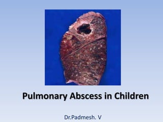
- 2. Lung infection Destroys lung parenchyma Cavitations and central necrosis Localized areas of thick-walled purulent material Lung abscess
- 3. • Primary lung abscess: • Occur in previously healthy patients with no underlying medical disorders. • Usually solitary. • Secondary lung abscess: • Occur in patients with underlying or predisposing conditions • May be multiple.
- 4. • PATHOLOGY AND PATHOGENESIS • Predisposing conditions: • Aspiration (of infected material or FB) • Pneumonia / Hematogenous seeding from other sites • Cystic fibrosis • Gastroesophageal reflux • Tracheoesophageal fistula • Immunodeficiencies • Postoperative complications of tonsillectomy and adenoidectomy • Seizures • Neurologic and other conditions associated with impaired mucociliary defense.
- 5. • PATHOLOGY AND PATHOGENESIS: Aspiration of infected material or foreign body Pneumonitis impairs drainage of fluid or aspirated material Inflammatory vascular obstruction Tissue necrosis, liquefaction Abscess formation
- 6. • PATHOLOGY AND PATHOGENESIS Aspiration in recumbent position Right & Left upper lobes and apical segment of the right lower lobes most likely. Aspiration in upright position Posterior segments of upper lobes most likely
- 7. • PATHOLOGY AND PATHOGENESIS • Primary abscesses: Most often on Right side. • Secondary lung abscesses, esp in immunocompromised : Predilection for left side.
- 8. • Organisms: Both anaerobic and aerobic organisms. • Anaerobic bacteria: • Bacteroides spp., • Fusobacterium spp., • Peptostreptococcus spp., • Aerobic bacteria: • Streptococcus spp., • Staphylococcus aureus • Escherichia coli • Klebsiella pneumoniae • Pseudomonas aeruginosa • Mycoplasma pneumoniae (Very rare) • Fungi can cause lung abscess,esp in immunocompromised.
- 9. • CLINICAL MANIFESTATIONS: Symptoms • Cough, • Fever, • Dyspnea, • Chest pain, • Vomiting, • Sputum production, • Weight loss, • Hemoptysis.
- 10. • CLINICAL MANIFESTATIONS: Signs • Tachypnea • Dyspnea • Retractions with accessory muscle use • Decreased breath sounds • Dullness to percussion in affected area • Crackles • Occasionally a prolonged expiratory phase on auscultation
- 11. • Diagnosis: • Chest X Ray: Parenchymal inflammation with a cavity containing an air–fluid level .
- 12. • Diagnosis: • Chest CT scan: Abscess is usually a thick-walled lesion with a low-density center progressing to an air–fluid level.
- 13. • Diagnosis: • Gram stain of sputum: Early clue. • Sputum cultures: Yield mixed bacteria, therefore not always reliable. • Attempts to avoid contamination from oral flora include direct lung puncture, percutaneous (aided by CT guidance) or transtracheal aspiration, and bronchoalveolar lavage specimens obtained bronchoscopically.
- 14. • Diagnosis: • Bronchoscopic aspiration should be avoided as it can be complicated by massive intrabronchial aspiration. • To avoid invasive procedures in previously normal hosts, empiric therapy can be initiated in the absence of culturable material.
- 15. • Differential Diagnosis: • Abscesses should be distinguished from pneumatoceles. • Pneumatoceles often complicate severe bacterial pneumonias. • Pneumatoceles: Thin- and Smooth-walled, localized air collections with or without air–fluid level. • Pneumatoceles often resolve spontaneously with treatment of specific cause of the pneumonia.
- 16. • TREATMENT: • Conservative management. • 2-3 wk course of parenteral antibiotics for uncomplicated cases, followed by oral antibiotics to complete a Total of 4-6 wk. • Aerobic and anaerobic coverage. • Include penicillinase-resistant agent active against S.aureus and anaerobic coverage, typically clindamycin or ticarcillin/clavulanic acid. • If Gram-negative bacteria are suspected or isolated, an aminoglycoside should be added.
- 17. • TREATMENT • Early CT-guided percutaneous aspiration or drainage. • Severely ill or those who fail to improve after 7-10 days of antibiotics Surgical intervention. • Minimally invasive CT guided percutaneous aspiration. • Thorascopic drainage. • In rare complicated cases Thoracotomy with surgical drainage or lobectomy and/or decortication.
- 18. • PROGNOSIS • Excellent. • Presence of aerobic organisms may be a negative prognostic indicator, particularly in those with secondary lung abscesses. • Most become asymptomatic within 7-10 days, although fever can persist for as long as 3 wk. • Radiologic abnormalities usually resolve in 1-3 mo but can persist for years.