Objective: To test the hypotheses that (1) the distal angulation of unerupted mandibular premolar
(MnP2) is significantly greater in children with palatally displaced canines (PDC) than in those in
a control sample; and (2) delayed tooth formation is significantly more frequent in children with
both malposed MnP2 and PDC than in children with PDC only.
Materials and Methods: We examined retrospectively panoramic radiographs from 43 patients
with PDC who had no previous orthodontics. A control sample consisted of age- and sex-matched
patients. The distal angle formed between the long axis of MnP2 and the tangent to the inferior
border was measured. Dental age was evaluated using the Koch classification.
Results: A significant difference was observed between the mean inclination of the right side
MnP2 in the PDC group (75.4 degrees) and that of the control group (85.8 degrees). This difference
was highly statistically significant (P < .0001). The same evaluation was carried out for the
left side, with similar results. The average dental age was found to be delayed in patients who
showed both abnormalities (malposed MnP2 and PDC) compared with patients who showed the
PDC anomaly only.
Conclusion: Both hypotheses are retained. Statistically, PDC and MnP2 malposition are significantly
associated suggesting a common genetic etiology, despite taking place on opposite jaws.
While the presence of PDC or MnP2 anomaly has been associated with a delay in tooth formation,
we find the presence of both anomalies to show a more profound delay. Our findings suggest a
delay in tooth formation as a possible common genetic mechanism for these 2 malposition anomalies.
Chandigarh Call Girls Service ❤️🍑 9809698092 👄🫦Independent Escort Service Cha...
Malposition of unerupted mandibular second premolar in children with palatally displaced canines
1. 796Angle Orthodontist, Vol 79, No 4, 2009 DOI: 10.2319/081808-435.1
Original Article
Malposition of Unerupted Mandibular Second Premolar in Children with
Palatally Displaced Canines
Miri Shalisha
; Stella Chaushua
; Atalia Wassersteinb
ABSTRACT
Objective: To test the hypotheses that (1) the distal angulation of unerupted mandibular premolar
(MnP2) is significantly greater in children with palatally displaced canines (PDC) than in those in
a control sample; and (2) delayed tooth formation is significantly more frequent in children with
both malposed MnP2 and PDC than in children with PDC only.
Materials and Methods: We examined retrospectively panoramic radiographs from 43 patients
with PDC who had no previous orthodontics. A control sample consisted of age- and sex-matched
patients. The distal angle formed between the long axis of MnP2 and the tangent to the inferior
border was measured. Dental age was evaluated using the Koch classification.
Results: A significant difference was observed between the mean inclination of the right side
MnP2 in the PDC group (75.4 degrees) and that of the control group (85.8 degrees). This differ-
ence was highly statistically significant (P Ͻ .0001). The same evaluation was carried out for the
left side, with similar results. The average dental age was found to be delayed in patients who
showed both abnormalities (malposed MnP2 and PDC) compared with patients who showed the
PDC anomaly only.
Conclusion: Both hypotheses are retained. Statistically, PDC and MnP2 malposition are signifi-
cantly associated suggesting a common genetic etiology, despite taking place on opposite jaws.
While the presence of PDC or MnP2 anomaly has been associated with a delay in tooth formation,
we find the presence of both anomalies to show a more profound delay. Our findings suggest a
delay in tooth formation as a possible common genetic mechanism for these 2 malposition anom-
alies. (Angle Orthod. 2009;79:796–799.)
KEY WORDS: Malposition; Dental anomalies; Mandibular second premolar; PDC, Genetic etiol-
ogy; Late development
INTRODUCTION
Orthodontists treat malposed teeth. As orthodon-
tists, we are interested in knowing what causes teeth
to assume abnormal positions during their develop-
ment. To gain this knowledge, we study malposition
anomalies. In a recent such study, it was discovered
that exaggerated distoangular malposition of the un-
erupted mandibular second premolar (MnP2) was as-
Department of Orthodontics, Hebrew University–Hadassah
School of Dental Medicine, Jerusalem, Israel.
Department of Orthodontics, School of Dental Medicine, Tel
Aviv University, Israel Defence Forces, Tel-Aviv, Israel.
Corresponding author: Dr Miri Shalish, Hebrew University–
Hadassah School of Dental Medicine, PO B 12272 Jerusalem,
Israel
(e-mail: mshalish@hadassah.org.il)
Accepted: October 2008. Submitted: August 2008.
ᮊ 2009 by The EH Angle Education and Research Foundation,
Inc.
sociated with agenesis of its antimere.1
This finding
relates the MnP2 malposition to a group of tooth de-
velopment abnormalities of possible common genetic
origin, including agenesis (hypodontia), peg-shaped
maxillary lateral incisors, small maxillary lateral inci-
sors, palatally displaced canines (PDC), infraocclusion
of primary molars, and transpositions of various
teeth.2–8
Accumulated evidence on associations
among this group go well beyond coincidence,8
sug-
gesting that they are part of a broader genetically re-
lated pattern of dental anomalies.
A genetic basis of PDC is widely acknowledged.4,9,10
Interestingly, delayed tooth formation was reported
both in children with PDC11,12
and in children with mal-
position of MnP2.13
This study was undertaken to test
the hypotheses that (1) the distal angulation of the
MnP2 is significantly greater in children with PDC than
in age- and sex-matched controls, and (2) delayed
tooth formation is a significantly more frequent finding
2. 797MALPOSITION OF MANDIBULAR SECOND PREMOLAR IN PDC
Angle Orthodontist, Vol 79, No 4, 2009
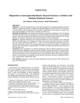
Figure 1. The distal angle between the long axis of the mandibular
second premolar and the tangent to the lower border of the mandible
defined on a typical drawing of the relevant part of an orthopanto-
mogram.
in children with both malposed MnP2 and PDC than
in children with PDC but with a normal inclination of
MnP2.
MATERIALS AND METHODS
Two samples, an experimental group and a control
group, were selected from the pretreatment records of
patients. The experimental sample consisted of 43 pa-
tients (15 males and 28 females). Age ranged from 8
to 14 years (mean, 11.5; standard deviation [SD], 1.4).
Criteria for inclusion in this sample included (1) unilat-
eral or bilateral PDC, (2) no previous orthodontic treat-
ment, (3) mandibular deciduous second molars pres-
ent, and (4) development of the MnP2 tooth bud in
stages D to G of tooth formation, according to the clas-
sification of Koch et al.14
Unerupted stage D is defined
with crown formation completed down to the cemento-
enamel junction, unerupted stage E is defined with
root length smaller than crown length, unerupted stage
F is defined with root length equal or larger than crown
length, and unerupted stage G is defined with walls of
the root canal parallel and the root apex still partially
open. Of 43 patients with PDC, 25 patients had bilat-
eral PDC, 10 had PDC on the right side only, and 8
had PDC on the left side only.
The control sample consisted of 43 patients with
normally erupting canines and was collected from the
same orthodontic patient pool to match age (rounded
to half year) and sex in the study group.
We developed a unique method by which to mea-
sure the inclination of the MnP2.1
In both samples,
panoramic radiographs were used to trace each
MnP2, along with the neighboring mandibular first mo-
lar, the deciduous second molar, and a tangent to the
inferior border of the mandibular body on that side.
The long axis of the MnP2 was determined as the line
connecting the uppermost point of the pulp with the
point bisecting the distance between the mesial and
distal points of the apex. A protractor was used to
measure the distal angle formed between the long axis
of the MnP2 and the line drawn tangent to the inferior
border of the mandible. Figure 1 shows a typical draw-
ing with assigned lines and the resulting angle. All
tracings were made independently by a single exam-
iner using 0.003 inch frosted acetate paper and a 0.5
mm pencil.
Descriptive statistics, including mean, standard de-
viation, and range, were calculated for the unerupted
MnP2 angles measured in the experimental and the
control groups. The significance of the differences be-
tween compared means was evaluated using the Stu-
dent t-test for paired samples. The significance level
was set at P Ͻ .05. Some patients had unerupted
MnP2 on both sides. However, because one may not
include more than 1 data point per patient in the same
statistics, the question of which side to choose arises,
as well as whether this arbitrary choice influences the
result and how. To avoid any possible inconsistency
while showing all the data, we collected 2 independent
sets of data: one for all the right sides of the sample
group, and another for the left sides of the same
group, each matched with the same side in the control
sample.
To quantify the error of the method, a second set of
data was traced and measured 1 month later by the
same examiner. Standard deviations calculated for 2
repeated measurements of 2 tracings of 6 different
panoramic roentgenograms were used as intraexami-
ner error. This procedural error was found to be 1.0
degree, within reasonable limits in the context of this
study.
To test the second hypothesis, we had to define the
developmental stage of MnP2 and single out patients
with malposed MnP2. The developmental stage of
MnP2 was evaluated using the Koch classification.13
MnP2 malposition was defined as the distal angle (be-
tween the long axis of MnP2 and the tangent to the
3. 798 SHALISH, CHAUSHU, WASSERSTEIN
Angle Orthodontist, Vol 79, No 4, 2009
Table 1. Interpatient Test: Sample vs. Control—Right Sidea,b
Group N Range Mean SD t-Test Value P Value
Right side MnP2 in the experimental sample (PDC) 40 51.0–108.0 75.4 10.2 Ϫ5.53 Ͻ.0001
Right side MnP2 in the control sample (reference) 40 71.0–98.0 85.8 6.2
a
Comparison of distoangular malposition of the MnP2 (ie, distal angle formed at intersection of long axis of unerupted MnP2 and tangent
to inferior border of the mandible) in degrees, between the right side MnP2 in the PDC sample and that in the control sample.
b
MnP2 indicates unerupted mandibular second premolar tooth; PDC, palatally displaced canine.
Table 2. Interpatient Test: Sample vs. Control—Left Sidea,b
Group N Range Mean SD t-Test Value P Value
Left side MnP2 in the experimental sample (PDC) 37 61.0–97.0 77.9 8.5 Ϫ4.54 Ͻ.0001
Left side MnP2 in the control sample (reference) 37 76.0–97.0 85.1 4.5
a
Comparison of distoangular malposition of the MnP2 (ie, the distal angle formed at the intersection of the long axis of unerupted MnP2
and tangent to the inferior border of the mandible) in degrees, between the left-side MnP2 in the PDC sample and the control sample.
b
MnP2 indicates unerupted mandibular second premolar tooth; PDC, palatally displaced canine.
lower border of the mandible) when it was smaller than
75 degrees. This value is about the mean angle of
malposed MnP2, as observed in previous studies.1,15
RESULTS
Table 1 shows descriptive statistics of the right-side
MnP2 in the experimental group and the same side in
the age- and sex-matched paired control group. Of 43
patients with PDC, 40 had an unerupted MnP2 on the
right side. The mean distal inclination of the MnP2 in
the right side of the experimental sample was 75.4 de-
grees, compared with a mean of 85.8 degrees ob-
tained for the same side in the matched control group.
The mean increase of 10.4 degrees in the distoangular
malposition of the developing MnP2 in PDC patients
was highly statistically significant (P Ͻ .0001).
Table 2 shows the descriptive statistics of the left-
side MnP2 in the experimental group and the same
side in the age- and sex-matched paired control group.
Of 43 patients with PDC, 37 had an unerupted MnP2
on the left side. The mean distal inclination of the
MnP2 in the left side of the experimental sample was
77.9 degrees, compared with a mean of 85.1 degrees
obtained for the same side in the matched control
group. The mean increase of 7.2 degrees in the dis-
toangular malposition of the developing MnP2 in PDC
patients was highly statistically significant (P Ͻ .0001).
Thus, the first hypothesis, that the distal angulation
of the MnP2 is significantly greater in children with
PDC than in age- and sex-matched controls, is retained.
The difference between sides within the PDC sample
calls for an intrapatient comparison to test whether this
difference is of significance. Using a paired t-test, we
found no significant difference between the right side
and the left side. Pearson correlation was found to be
0.57, significant at the 0.01 level (P ϭ .0004).
The distribution of the MnP2 dental developmental
stage in the PDC sample according to Koch classifi-
cation was as follows: Of 23 patients showing MnP2
malposition, 61% were at stage E and 39% at stage
F, while out of 20 patients showing normal inclination
of the MnP2, 25% were at stage E, and 75% were at
stage F. These results show that the average dental
age is delayed in patients who show both abnormali-
ties (malposed MnP2 and PDC) compared with pa-
tients who show PDC anomaly but with normal incli-
nation of MnP2. Our second hypothesis that delayed
tooth formation is a significantly more frequent finding
in children with both malposed MnP2 and PDC is thus
retained.
DISCUSSION
This study was designed to test the null hypothesis
that angular malposition of unerupted MnP2 is not di-
rectly associated with PDC. The results suggest a sta-
tistically significant association between these 2 con-
ditions.
The palatally displaced canine is a maxillary dental
anomaly, whereas MnP2 is a mandibular anomaly.
Hence common mechanical cause is unlikely. The ab-
sence of a shared mechanical cause suggests asso-
ciation through a common genetic disorder. Peck et
al16
have already suggested that the homeobox gene
MSX1 may be involved in the genetic control of PDC.
The association of MSX1 with agenesis17
and with
clefting18
has been established in genetic linkage anal-
yses. Both agenesis and clefting have been shown to
be associated with the MnP2 malposition anomaly.1,15
Results of this work, along with results from Shalish et
al,1,15
associate the MnP2 angulation anomaly with
PDC, agenesis, and clefting, suggesting the MnP2
anomaly is a variable in a genetically related group of
dental anomalies likely to be associated with MSX1
mutations. It is likely that the MnP2 anomaly may ap-
4. 799MALPOSITION OF MANDIBULAR SECOND PREMOLAR IN PDC
Angle Orthodontist, Vol 79, No 4, 2009
pear in combination with any other of these inter-as-
sociated anomalies (eg, infraocclusion, mesially ectop-
ic maxillary first molar, tooth transposition, tooth rota-
tion, tooth size reduction, peg-shaped maxillary lateral
incisor), perhaps because all of these anomalies are
caused by the same mechanism. What could be this
mechanism?
Delayed tooth formation was reported in children
with clefting,19
with PDC,11,12
and with malposition of
MnP2.13
It therefore seems possible that the common
mechanism is a delay in tooth formation. If this is cor-
rect, one should expect a longer delay to increase the
likelihood of anomalies and thereby the likelihood that
more than 1 anomaly will be observed in the same
patient. This means that children who show more than
1 anomaly should also show a greater delay in tooth
formation. Indeed, an average greater delay was con-
firmed in this study in children showing both PDC and
MnP2 anomalies, compared with children showing
PDC but with a normal inclination of MnP2.
CONCLUSION
• PDC and MnP2 malposition anomalies are signifi-
cantly statistically associated, despite their taking
place on opposite jaws, suggesting a common ge-
netic etiology.
• Although the presence of PDC or MnP2 anomaly
has been associated with a delay in tooth formation,
we find the presence of both anomalies to show a
more profound delay.
• These findings suggest a delay in tooth formation as
a possible common genetic mechanism for these 2
malposition anomalies.
REFERENCES
1. Shalish M, Peck S, Wasserstein A, Peck L. Malposition of
unerupted mandibular second premolar associated with
agenesis of its antimere. Am J Orthod Dentofacial Orthop.
2001;121:53–55.
2. Alvesalo L, Portin P. The inheritance pattern of missing,
peg-shaped and strongly mesio-distally reduced upper lat-
eral incisors. Acta Odontol Scand. 1969;27:563–573.
3. Garn SM, Lewis AB. The gradient and the pattern of crown-
size reduction in simple hypodontia. Angle Orthod. 1970;40:
51–58.
4. Peck S, Peck L, Kataja M. The palatally displaced canine
as a dental anomaly of genetic origin. Angle Orthod. 1994;
64:249–256.
5. Peck S, Peck L, Kataja M. Prevalence of tooth agenesis
and peg-shaped maxillary lateral incisor associated with
palatally displaced canine (PDC) anomaly. Am J Orthod
Dentofacial Orthop. 1996;110:441–443.
6. Peck L, Peck S, Attia Y. Maxillary canine–first premolar
transposition, associated dental anomalies and genetic ba-
sis. Angle Orthod. 1993;63:99–109.
7. Symons AL, Stritzel F, Stamation J. Anomalies associated
with hypodontia of the permanent lateral incisor and second
premolar. J Clin Ped Dent. 1993;17:109–111.
8. Baccetti T. A controlled study of associated dental anoma-
lies. Angle Orthod. 1998;68:267–274.
9. Zilberman Y, Cohen B, Becker A. Familial trends in palatal
canines, anomalous lateral incisors, and related phenome-
na. Eur J Orthod. 1990;12:135–139.
10. Pirinen S, Arte S, Apajalahti S. Palatal displacement of ca-
nine is genetic and related to congenital absence of teeth.
J Dent Res. 1996;75:1742–1746.
11. Becker A, Chaushu S. Dental age in maxillary canine ec-
topia. Am J Orthod Dentofacial Orthop. 2000;117:657–662.
12. Chaushu S, Sharabi S, Becker A. Dental morphologic char-
acteristics of normal versus delayed dentitions with palatally
displaced canines. Am J Orthod Dentofacial Orthop. 2002;
121:339–346.
13. Wasserstein A, Brezniak N, Shalish M, Heller M, Rakocz M.
Angular changes and their rates in concurrence to devel-
opmental stages of the mandibular second premolar. Angle
Orthod. 2004;74:332–336.
14. Koch G, Modeer T, Poulsen S, Rasmussen P. Pedodontics:
A Clinical Approach. Copenhagen: Munksgaard; 1991:60.
15. Shalish M, Will LA, Shusterman S. Malposition of unerupted
mandibular second premolar in children with cleft lip and
palate. Angle Orthod. 2007;77:1062–1066.
16. Peck S, Peck L, Kataja M. Concomitant occurrence of ca-
nine malposition and tooth agenesis: evidence of orofacial
genetic fields. Am J Orthod Dentofacial Orthop. 2002;122:
657–660.
17. Vastardis H, Karimbux N, Guthua SW, Seidman JG, Seid-
man CE. A human MSX1 homeodomain missense mutation
causes selective tooth agenesis. Nat Genet. 1996;13:417–
421.
18. Van den Boogaard MJ, Dorland M, Beemer FA, van Amstel
HK. MSX1 mutation is associated with orofacial clefting and
tooth agenesis in humans. Nat Genet. 2000;24:342–343.
19. Ranta R. A review of tooth formation in children with cleft
lip/plate. Am J Orthod Dentofacial Orthop. 1986;90:11–18.
![797MALPOSITION OF MANDIBULAR SECOND PREMOLAR IN PDC
Angle Orthodontist, Vol 79, No 4, 2009
Figure 1. The distal angle between the long axis of the mandibular
second premolar and the tangent to the lower border of the mandible
defined on a typical drawing of the relevant part of an orthopanto-
mogram.
in children with both malposed MnP2 and PDC than
in children with PDC but with a normal inclination of
MnP2.
MATERIALS AND METHODS
Two samples, an experimental group and a control
group, were selected from the pretreatment records of
patients. The experimental sample consisted of 43 pa-
tients (15 males and 28 females). Age ranged from 8
to 14 years (mean, 11.5; standard deviation [SD], 1.4).
Criteria for inclusion in this sample included (1) unilat-
eral or bilateral PDC, (2) no previous orthodontic treat-
ment, (3) mandibular deciduous second molars pres-
ent, and (4) development of the MnP2 tooth bud in
stages D to G of tooth formation, according to the clas-
sification of Koch et al.14
Unerupted stage D is defined
with crown formation completed down to the cemento-
enamel junction, unerupted stage E is defined with
root length smaller than crown length, unerupted stage
F is defined with root length equal or larger than crown
length, and unerupted stage G is defined with walls of
the root canal parallel and the root apex still partially
open. Of 43 patients with PDC, 25 patients had bilat-
eral PDC, 10 had PDC on the right side only, and 8
had PDC on the left side only.
The control sample consisted of 43 patients with
normally erupting canines and was collected from the
same orthodontic patient pool to match age (rounded
to half year) and sex in the study group.
We developed a unique method by which to mea-
sure the inclination of the MnP2.1
In both samples,
panoramic radiographs were used to trace each
MnP2, along with the neighboring mandibular first mo-
lar, the deciduous second molar, and a tangent to the
inferior border of the mandibular body on that side.
The long axis of the MnP2 was determined as the line
connecting the uppermost point of the pulp with the
point bisecting the distance between the mesial and
distal points of the apex. A protractor was used to
measure the distal angle formed between the long axis
of the MnP2 and the line drawn tangent to the inferior
border of the mandible. Figure 1 shows a typical draw-
ing with assigned lines and the resulting angle. All
tracings were made independently by a single exam-
iner using 0.003 inch frosted acetate paper and a 0.5
mm pencil.
Descriptive statistics, including mean, standard de-
viation, and range, were calculated for the unerupted
MnP2 angles measured in the experimental and the
control groups. The significance of the differences be-
tween compared means was evaluated using the Stu-
dent t-test for paired samples. The significance level
was set at P Ͻ .05. Some patients had unerupted
MnP2 on both sides. However, because one may not
include more than 1 data point per patient in the same
statistics, the question of which side to choose arises,
as well as whether this arbitrary choice influences the
result and how. To avoid any possible inconsistency
while showing all the data, we collected 2 independent
sets of data: one for all the right sides of the sample
group, and another for the left sides of the same
group, each matched with the same side in the control
sample.
To quantify the error of the method, a second set of
data was traced and measured 1 month later by the
same examiner. Standard deviations calculated for 2
repeated measurements of 2 tracings of 6 different
panoramic roentgenograms were used as intraexami-
ner error. This procedural error was found to be 1.0
degree, within reasonable limits in the context of this
study.
To test the second hypothesis, we had to define the
developmental stage of MnP2 and single out patients
with malposed MnP2. The developmental stage of
MnP2 was evaluated using the Koch classification.13
MnP2 malposition was defined as the distal angle (be-
tween the long axis of MnP2 and the tangent to the](data:image/gif;base64,R0lGODlhAQABAIAAAAAAAP///yH5BAEAAAAALAAAAAABAAEAAAIBRAA7)