2. WHAT IS A TISSUE?
A tissue is a group of similar cells which work
together to perform a specific function in the body.
All cells in the body can be categorized into Four (4)
major types of tissues (Basic/Primary Tissues):
1. Epithelial Tissue
2. Connective Tissue
3. Muscular Tissue
4. Nervous Tissue
3. TISSUE
The study of tissues and how they are arranged into
organs – Histology or Microscopic Anatomy
Aside cells, tissues are also made up of extracellular
material which surrounds the cells – Matrix
The 4 basic tissue types differ in the types and functions
of their cells and characteristics of their matrix.
The matrix is composed of fibrous proteins and usually a
clear gel (ground substance/ tissue fluid/ extracellular
fluid/ interstitial fluid/ tissue gel)
4. Embryonic Tissues
The first tissues (embryonic tissues) arise in the early
embryo when similar cells organise together into
layers (3) called Primary Germ Layers.
1. Ectoderm
2. Mesoderm
3. Endoderm
Germ layers give rise to all the body’s mature tissues
5. Embryonic Tissues
Endoderm – Inner layer which gives rise to various
membranes of the digestive and respiratory tracts.
Ectoderm – Outer layer which forms the outer
covering of the body (epidermis).
Mesoderm – Middle layer that forms the skeleton
and muscles of the body.
6. TISSUE
Body organs usually contain all four tissue types.
Example the stomach
Lined with epithelial tissue
Walls contain smooth muscle and connective tissue
Nerve tissue supply which control motility and
gastric secretions.
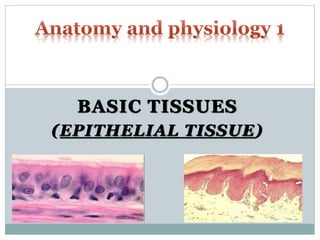
7. EPITHELIAL TISSUE
(EPITHELIUM)
This group of tissues is found covering the body and
lining cavities and tubes.
It is also found in glands.
Functions of epithelial tissues include:
1. Protection e.g. skin
2. Absorption e.g. small intestines
3. Filtration e.g. kidneys
4. Secretion e.g. glands such as salivary
8. CHARACTERISTICS OF EPITHELIAL TISSUE
1. Limited intercellular spaces
Flat sheet of closely packed cells with very little extracellular
material (matrix) between the cells.
2. Free apical surface
Tissue always has one free surface (apical surface) which is
exposed to the body’s exterior or to the cavity of an internal
organ.
3. Basement Membrane
Lower surface of tissue rests on a basement membrane which
anchors the tissue to the underlying connective tissue
Basement membrane usually very thin
Contains an aggregate of carbohydrates and proteins
9. CHARACTERISTICS OF EPITHELIAL TISSUE
4. Avascular
Contain no blood vessels.
Depend on blood vessels in underlying connective tissue for
nourishment and waste removal
5. Mitotic Capabilities
Epithelial cells readily divide to produce new cells that replace
lost or damaged ones
10. CLASSIFICATION OF EPITHELIUM
Epithelial tissues are classified according to 3 key
criteria which describe their unique traits:
1. Number of Cell Layers
2. Shape of Cells
3. Surface modifications
11. CLASSIFICATION OF EPITHELIUM
Number of Cell Layers:
Two types:
1. Simple Epithelium:
Composed of a single layer with each cell touching the basement
membrane
2. Stratified Epithelium:
Composed of two or more layers.
Here some of the cells rest on top of other cells and do not touch
the basement membrane.
12. CLASSIFICATION OF EPITHELIUM
Shape of Cell:
Three categories:
1. Squamous Epithelium:
Flattened cells.
2. Cuboidal Epithelium:
Cube shaped cells.
3. Columnar Epithelium:
Elongated cells.
Cells longer than wide.
13. CLASSIFICATION OF EPITHELIUM
Surface Modification:
Usually 2 types:
1. Cilia and Stereocilia:
Cilia and stereocilia are similar filament-like structures
projecting from the apical cell surfaces.
Cilia beat in rhythmic motions to propel substances across the
apical surfaces of cells. E.g. Fallopian tube
Stereocilia are longer and not uniform in length and do not beat
like cilia. E.g. Epididymis and Inner Ear
2. Microvilli
Uniform folds of the membrane on the apical cell surfaces.
Much shorter than cilia
14. CLASSIFICATION OF EPITHELIUM
Epithelial tissues are given at least two names.
The first name indicates the number of cell layers
The second name describes the shape of the cell
Where applicable, a third name is given which
denotes the type surface modification.
Type of surface modification is a prefix to the first two names
Naming:
(Surface modification) – No. of layers – Shape of Cell
15. EPITHELIAL TISSUES
Simple Epithelial:
Usually concerned with absorption, secretion and filtration.
Very thin and protection is not one of their specialties
16. EPITHELIAL TISSUES
Stratified Epithelial:
Consists of two or more cell layers.
More durable than the simple epithelial and as such function
primarily to protect.
Locations which have to withstand mechanical or chemical wear
Named according to cells on the outer layer (superficial layer)
2 Specialisations occur in stratified epithelial tissues
1. Keratinized/ (Non- Keratinized):
Cells in apical layers are dead and lose their nucleus and
cytoplasm and instead contain keratin
Keratin is a tough resistant protein which is waterproof and
provides strength
2. Transitional:
17. SQUAMOUS EPITHELIUM
Simple Squamous:
Composed of a single layer of flattened cells.
Usually found in areas where filtration or exchange of
substances by rapid diffusion occurs
Forms tiny air sacs of lungs, where O2 and CO2 exchange occurs
Forms walls of capillaries, where nutrients and waste exchange
occurs
Forms serous membranes that line the ventral body cavity and
organs in that cavity
18. SQUAMOUS EPITHELIUM
Stratified Squamous:
Most common type of stratified epithelium in the body
Has many layers and plays a protective role
Found in sites that receive a good deal of abuse or
friction e.g. oesophagus, mouth and outer part of skin.
In the skin, stratified squamous epithelium is keratinised
Non-keratinised found in oesophagus, rectum, vagina,
cervix
19. CUBOIDAL EPITHELIUM
Simple Cuboidal Epithelium:
Consists of a single layer of cube-shaped cells
attached to a basement membrane
Commonly found in glands and their ducts
Salivary gland, thyroid gland, pancreas
Also forms the walls of kidney tubules and surface of
ovaries
20. CUBOIDAL EPITHELIUM
Stratified Cuboidal Epithelium:
Mostly found lining larger ducts of certain glands
Mammary gland, salivary gland
Usually has just two layers
21. COLUMNAR EPITHELIUM
Simple Columnar Epithelium:
Columnar cells are longer than they are wide.
Simple columnar epithelium made up of a single
layer of tall cells that fit closely together
Ultimate cells for absorption and secretion
Largest cytoplasmic volumes of all epithelial cells
Line digestive organs (stomach – Rectum)
Have microvilli (small intestines) to increase surface area
Line small bronchioles and uterine tubes
Have cilia which aid in movement of mucous and reproductive
cells respectively
22. COLUMNAR EPITHELIUM
Goblet cells
Specialised simple columnar cells which secrete
mucous onto the free surface of epithelium
23. COLUMNAR EPITHELIUM
Stratified Columnar Epithelium:
Very rare
Lines ducts of large glands (e.g. salivary)
Parts of pharynx
Male urethra
24. COLUMNAR EPITHELIUM
Pseudostratified Columnar Epithelium:
Pseudo – false
Appear to be layered but all cells touch the basement
membrane
Nuclei appear at different heights above the
basement membrane giving false impression that it
is stratified.
25. TRANSITIONAL EPITHELIUM
Special type of epithelium
Change shape in response to tension
Main locations are urinary bladder, ureters and part
of urethra
All above sites need to stretch
Appear to be stratified cuboidal when not stretched and as
stratified squamous when the organ is distended and tissue is
stretched
Also known as urothelium since almost exclusively
found in the urinary system
27. GLANDULAR EPITHELIUM
A gland consists of one or more cells that produce
and secrete a particular product
Most glands consist primarily of epithelial tissue
Two major types of glands develop from epithelial
tissue:
1. Endocrine Glands
2. Exocrine Glands
28. ENDOCRINE GLANDS
Secrete products (hormones) directly into the
bloodstream
Have no ducts (ductless)
Examples: thyroid and adrenal gland
29. EXOCRINE GLANDS
Have ducts
Secretions empty through the ducts to the epithelial
surface
Example sweat glands, mammary gland
Glands are multicellular except goblet cells which are
unicellular
Various ways of classifying but mainly done according to
1. Structure
2. Mode or method of secretion
3. Product or nature of secretion
30. EXOCRINE GLANDS
STRUCTURE:
Exocrine glands contain a glandular (secretory)
portion and a duct portion
Based on duct portion
Simple – Unbranched
Compound – Branched
Based on glandular portion
Tubular – forms a tube
Acinar – form a bulblike sac
If the glandular portion is branched, the gland is
called a branched gland.
32. EXOCRINE GLANDS
MODE OF SECRETION:
Secretory cells release products into ducts in 3
different ways
1. Merocrine
2. Apocrine
3. Holocrine
33. EXOCRINE GLAND
MEROCRINE GLAND:
Secretions pass through the cell membrane of the
secretory cells – through exocytosis mainly
Most glands of this type
Salivary, Pancreas
34. EXOCRINE GLAND
APOCRINE GLAND:
Apical portions of secretory cells pinched off and lost
during secretory process
Secretory product contains a variety of molecular
components including those of membrane
Mammary gland
35. EXOCRINE GLAND
HOLOCRINE GLAND:
Entire secretory cell disintegrates and released along
with the content.
Results in most complex secretory product
Sebaceous gland, some sweat glands located in axillae, pubic
area and around areola of breasts
37. PRODUCT/ NATURE OF SECRETION
Nature or product of secretion is also used to classify
exocrine glands:
1. Mucous glands
Secrete thick mucus
Brunner’s glands (in duodenum)
2. Serous glands
Secrete a thinner, watery substance and usually contain enzymes
E.g. Chief cells in stomach
3. Mixed glands
Produce both mucous and serous secretions (have both mucous
and serous cells)
E.g. Salivary glands (submandibular gland)