Acute ppendicitis case
•Télécharger en tant que DOCX, PDF•
1 j'aime•778 vues

Appendicitis Differential Diagnosis

Recommandé
Contenu connexe
Tendances
Tendances (20)
Similaire à Acute ppendicitis case
Similaire à Acute ppendicitis case (20)
Plus de Timothy Zagada
Plus de Timothy Zagada (17)
Dernier
Dernier (20)
Acute ppendicitis case
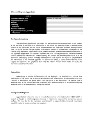
- 1. Differential Diagnosis: Appendicitis RULE IN RULE OUT Initial Periumbilical pain Cannot be ruled out Progression to RLQ pain Anorexia Vomitting Loose stool Rebound tenderness Progression to muscle guarding Leukocytosis The Appendix: Anatomy The appendix is derived from the midgut just like the ileum and ascending colon. It first appears at the 8th week of gestation as an outpouching of the cecum and gradually rotates to a more medial location as the gut rotates and the cecum becomes fixed in the right lower quadrant. Its length varies from 2 to 20 cm, and the average length is 9 cm in adults. Its base is located at the convergence of the taeniae along the inferior aspect of the cecum, and this anatomic relationship facilitates identification of the appendix at operation. The tip of the appendix may lie in a variety of locations. The most common location is retrocecal but within the peritoneal cavity. It is pelvic in 30% and retroperitoneal in 7% of the population. The varying location of the tip of the appendix likely explains the myriad of symptoms that are attributable to the inflamed appendix. The appendiceal artery, a branch of the ileocolic artery, supplies the appendix. The lymphatics drain into the anterior ileocolic lymph nodes. In adults, the appendix has no known function. Appendicitis Appendicitis is swelling (inflammation) of the appendix. The appendix is a normal true diverticulum of the cecum that is prone to acute and chronic inflammation. Acute appendicitis is most common in adolescents and young adults, but may occur in any age group. The lifetime rate of appendectomy is 12% for men and 25% for women, with approximately 7% of all people undergoing appendectomy for acute appendicitis during their lifetime Etiology and Pathogenesis Appendicitis is believed to occur as a result of appendiceal luminal obstruction in 50% to 80% of cases.It is thought to initiateprogressive increase in intraluminal pressure that compromise venous outflow. This may be due to inspissated stool (fecalith or appendicolith), lymphoid hyperplasia, vegetable matter or seeds, parasites, or a neoplasm.
- 2. The lumen of the appendix is small in relation to its length, and this configuration may predispose to closed-loop obstruction. Obstruction of the appendiceal lumen contributes to bacterial overgrowth, and continued secretion of mucus leads to intraluminal distention and increased wall pressure. Subsequent impairment of lymphatic and venous drainage leads to mucosal ischemia. Ischemic injury and stasis of luminal contents, which favor bacterial proliferation, trigger inflammatory responses including tissue edema and neutrophilic infiltration of the lumen, muscular wall, and periappendiceal soft tissues. If the process evolves slowly, adjacent organs such as the terminal ileum, cecum, and omentum may wall off the appendiceal area so that a localized abscess will develop, whereas rapid progression of vascular impairment may cause perforation with free access to the peritoneal cavity. Subsequent rupture of primary appendiceal abscesses may produce fistulas between the appendix and bladder, small intestine, sigmoid, or cecum. Clinical Manifestations Abdominal pain is the prime symptom of acute appendicitis. The pain is described as being located in the periumbilical region initially after a period varying from 1 to 12 hours, but usually within 4 to 6 hours, the pain localizes to the right lower quadrant.. This classic sequence of symptoms occurs in only 66% of patients.Luminal distention of the appendix produces the visceral pain sensation experienced by the patient as periumbilical pain. Inflammation of the adjacent peritoneum gives rise to localized pain in the right lower quadrant. This classic pattern of migratory pain is the most reliable symptom of acute appendicitis. Fever ensues, followed by the development of leukocytosis.Although moderate leukocytosis of 10,000–18,000 cells/uL is frequent (with a concomitant left shift), the absence of leukocytosis does not rule out acute appendicitis. Leukocytosis of >20,000 cells/uL suggests probable
- 3. perforation.In more severe cases a prominent neutrophilic exudate generates a serosalfibrinopurulent reaction. As the process continues, focal abscesses may form within the wall (acute suppurative appendicitis). Further appendiceal compromise leads to large areas of hemorrhagic ulceration and gangrenous necrosis that extends to the serosa creating acute gangrenous appendicitis, which is often followed by rupture and suppurative peritonitis. The temperature is usually normal or slightly elevated [37.2°–38°C (99°–100.5°F)], but a temperature >38.3°C (101°F) should suggest perforation. Anorexia is very common; a hungry patient does not have acute appendicitis. It is so constant that the diagnosis should be questioned if the patient is not anorectic. Nausea and vomiting occur in 50– 60% of cases, but vomiting is usually self-limited in contrast to the repeated bouts of vomiting that typically accompany viral gastroenteritis or small bowel obstruction. Vomiting is caused by both neural stimulation and the presence of ileus. Diarrhea occurs in some patients, however, particularly children, so that the pattern of bowel function is of little differential diagnostic value. Examination of the abdomen usually reveals diminished bowel sounds and focal tenderness with voluntary guarding. The exact location of the tenderness is directly over the appendix, which is most commonly at McBurney's point (located one third of the distance along a line drawn from the anterior superior iliac spine to the umbilicus).Direct rebound tenderness usually is present. In addition, referred or indirect rebound tenderness is present. This referred tenderness is felt maximally in the right lower quadrant, which indicates localized peritoneal irritation. Muscular resistance to palpation of the abdominal wall roughly parallels the severity of the inflammatory process. Early in the disease, resistance, if present, consists mainly of voluntary guarding. As peritoneal irritation progresses, appendix perforates, abdominal pain becomes intense and more diffuse, and abdominal muscular spasm increases, producing rigidity and guarding becomes largely involuntary, that is, true reflex rigidity due to contraction of muscles directly beneath the inflamed parietal peritoneum. References: Townsend: Sabiston Textbook of Surgery, 18th ed. Copyright © 2007 Saunders. Copyright © 2008 Elsevier Inc. Schwartz's Principles of Surgery, Ninth Edition Copyright © 2010, 2005, 1999, 1994, 1989, 1984, 1979, 1974, 1969 by The McGraw-Hill Companies, Inc. Robbins and Cotran pathologic basis of disease. – 8th ed. / Vinay Kumar, Copyright © 2010 by Saunders, an imprint of Elsevier Inc. Harrison’s Internal Medicine 18th Edition