SlideShare une entreprise Scribd logo
1  sur  62
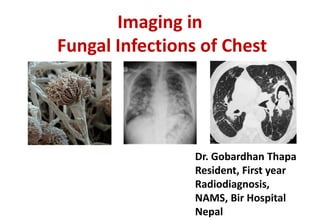
Imaging in
Fungal Infections of Chest
Dr. Gobardhan Thapa
Resident, First year
Radiodiagnosis,
NAMS, Bir Hospital
Nepal
Outline of Presentation
• Introduction
• Classification of Fungal Infections
• Overview of specific Fungal organism and the
current Imaging modalities
• Summary
• references
A case report
• A 71-year-old Chinese male was admitted with complaint of
chronic cough and malaise for two months. He had been in
Tucson, Arizona, USA, visiting for four months right before
the symptoms occurred. He had transient low-grade fever,
but denied hemoptysis, night sweats, skin rashes, or
headache. He did not smoke or abuse drugs. Physical
examination revealed no abnormalities. Lab tests showed
an elevated erythrocyte sedimentation rate (ESR) of 46
mm/h, while complete blood count (CBC), eosinophil count,
serum chemistry, and tumor biomarkers were all in normal
range. Human immunodeficiency virus (HIV) antibody was
found to be negative. Sputum culture showed normal flora
growth.
• Chest computed tomographic (CT) scan revealed
an irregular-margined opacity measuring 3.0
cm×3.8 cm in diameter in the sub-pleura region of
right middle lobe. Right hilar and mediastinal
lymphadenopathy was noted. Lung cancer was
considered as the most likely diagnosis.
Subsequent bronchoscopy and brush cytology
were negative. The patient received a procedure
of right middle lobe and lower lobectomy on the
eighth day after admission.
Chest computed tomographic (CT) scan
-Journal of Zhejiang university science, April, 2014
• Histopathological examination of lung specimen
showed focal necrotic granulomatous
inflammation with multinucleated giant cells
containing fungal spherules and infiltrations of
massive neutrophils, eosinophils, and
lymphocytes.. The final diagnosis thus was
confirmed pathologically as pulmonary
Coccidioides infection. The patient was totally free
from discomfort in the course of two years follow-
up after the lobectomy
Introduction
• Fungal Pneumonia
– Are now seen with increased frequency
• Increase in the incidence of disease caused by pathogenic
fungi in healthy hosts
• Emergence of opportunistic species in immuno-
compromsied hosts
Fungal Infections:
Saprophytic Fungi
Candidiasis
Pneumocystis
Cryptococcosis
Mucormycosis
Aspergillus
Pathogenic Fungi
Histoplasmosis
Coccidioidomycosis
Blastomycosis
Paracoccidioidomycosis
In all cases, fungi elicit a “Necrotizing granulomatous reaction”
1. Histoplasmosis
• Histoplasma capsulatum
• In moist soil and bird/bat excreta
• Mostly subclinical infection:
– Heals spontaneously
– CXR: may be normal
• Or sometimes well-defined, calcific nodules <1 mm in size
• Calcified hilar of mediastinal nodes
• Multiple miliary calcified nodules
• Progression of the infective foci:
– Leading to a larger nodule
– Hilar nodes enlargement is common
– Locally progressive: may have consolidative changes,
later associated with Fibrosis and cavitation.
• Massive inhalation of organisms:
– May show fairly discrete, nodular opacities 3-4 mm in
diameter with hilar adenopathy
Fig. acute histoplasmosis
widespread bilateral well-defined 3-5 mm nodules
Fig. disseminated histoplasmosis in a HIV patient
axial CT shows multiple small pulmonary nodules distributed
uniformly throughout the both lungs
Fig. Histoplasmosis
incidental finding of multiple calcified
pulmonary nodules
• Histoplasmoma:
– A solitary, sharply defined nodule <3 cm
– Most common in lower lobes-frequently calcify
• Fibrosing Mediastinitis (chronic pulmonary disease):
– Uncommon late manifestation
– Stenosis of venacava, oesophagus, trachea, bronchi or
central pulmonary vessels
– CXR: widened mediastinum
Fig. Histoplasmoma
Axial CT shows right lower lobe Histoplasmoma with central
calcification
Fig. fibrosing mediastinitis following Histoplasmosis
radiographs shows widening of the mediastinum
• blood-borne dissemination
– Asymptomatic blood-borne dissemination is common
– Eg calcified granulomas in patients of endemic area
– Clinically apparent disseminated histoplasmosis
• Extremely rare
2. Coccidioidomycosis
• Coccidioides immitis
• Found in soil in arid/semi-arid areas
• 4 types of clinical and radiographic pulmonary
infections:
a) Acute Coccidioidomycosis
b) Persistent Coccidioidomycosis
c) Chronic progressive disease
d) Disseminated (Miliary) Coccidioidomycosis
a) Acute coccidioidomycosis
• Develops in 40% of infected adults
• Self-limiting viral type illness: Valley fever
• Associated with erythema nodosum and Arthralgia
• CXR: may be normal or
 Focal or multifocal segmental air-space opacities
 Associated with Hilar and mediastinal adenopathy and pleural
effusion
b) Persistent coccidioidomycosis (infection beyond 6-8
weeks)
• Coccidioidal masses or nodules (coccidioidomas)
• Areas of round pneumonia- subpleural regions of upper lobes
• Cavitate rapidly-produce characteristic thin-walled cavities
c) Chronic progressive disease
• Upper lobe fibro-cavitatory disease
– Thin-walled cyst : Grape-skin sign
• Similar to Post-primary TB and Histoplasmosis
d) Disseminated (Miliary) coccidioidomycosis
• Relatively rare
• Affects the immuno-compromised patients
Fig. coccidioidomycosis
a non-specific patch of consolidation present in left lower
lobe. One year later a thin-walled cavity is evident
Fig. primary coccidioides infection
frontal radiograph in a female with a clinical diagnosis of
valley fever reveals a mass like opacity in the right lower
lung with enlarged right hilar nodes
Fig. primary coccidioides infection
coronal reformatted CT of same patient
confirms a right middle lobe nodule
Fig. Chest x-ray showing Grape-skin sign
thin-walled grape-skin cyst
over time cavity may deflate and acquire slightly thicker wall
3. Blastomycois
• Caused by Blastomyces dermatidis
• Chronic systemic disease
• Primarily affects the lungs and the skin
• Pulmonary infections often asymptomatic
• Symptomatic infection:
– Resembles that of an Acute bacterial pneumonia
• Radiographically:
– Usually non-specific
• Most common presentation:
– Homogeneous non-segmental air space opacification with
propensity for upper lobes
• Less common presentation:
– Single or multiple masses
– Cavitate in 15% of cases
– Tend to occur in patients with prolonged symptoms (1
months)-may mimic Bronchogenic Ca
• Less common presentation:
– Diffuse reticulo-nodular opacities
• Pleural effusion and lymph node enlargement –
uncommon
• Disseminated miliary form
– In immunocompromised hosts
Fig. Blastomyces dermatidis infection
chest radiograph shows an ill-defined mass in the left upper
lobe. CT scan through the upper lobes shows an irregular mass
in the left upper lobe with surrounding ground glass opacity.
Biopsy revealed Blastomyces dermatidis infection
4. Paracoccidioidomycosis(PCM)
• Also known as South American Blastomycosis
• Endemic disease caused by dimorphic fungi
– Paracoccidioides brasiliensis
• Most frequent systemic mycosis in Latin America
esp. in Brazil
• HRCT:
– Areas of ground-glass opacities, nodules, interlobular
septal thickening, air-space consolidation, cavitation
and fibrosis
5. Candidiasis
• Candida albicans
• Important pathogen esp. in Immunocompromised
patients:
– Particularly in patients with underlying malignancy, IV
drugs abuser, AIDS, following Bone marrow transplant
• Lung infection is usually due to hematogeneous
spread
• Radiographically, may present as
– Chronic pneumonia
– Abscess formation
– Mycetoma formation
• CT
– Multiple bilateral nodular opacities often associated with
areas of consolidation and ground glass opacities-CT halo
sign
• Less common presentations:
– Pleural effusion, thickening of bronchial walls, cavitation
Fig. disseminated candidiasis with CT halo
sign
6. Pneumocystis jiroveci
• Opportunistic fungal pathogen
• Cause pneumonia in patients with
– AIDs
– Organ transplant
– Undergoing chemotherapy
– Immunosuppressive treatment
– Long term corticosteroids
• Radiographically,
– May have normal findings
– Classic features: diffuse, bilateral interstitial infiltrates in
peri-hilar distribution
• CT:
– Done in a highly suspicious case for confirming the
diagnosis
– Peri-hilar ground glass opacities, in a patchy or geographic
distribution with areas of superimposed interlobular septal
thickening: Crazy Paving pattern
– May rapidly progress to involve entire lung
Fig. Pneumocystis Pneumonia in an HIV patient:
Crazy paving sign
• Complications:
– Thin-walled cavities
– Pneumothorax
• Less common presentations
– pleural effusion, nodules, miliary disease, calcified
lymph nodes
7. Cryptococcosis (Torulosis)
• Cryptococcus neoformans (yeast form fungi)
• Found in soil or bird droppings
• Mostly asymptomatic
• Cryptococcal pneumonia
– Common in AIDS (when CD4 <100/cu. mm)
• Chest radiography:
– Homogeneous, segmental or lobar opacifications
– Miliary, reticular or reticulo-nodular interstitial
patterns
– Pulmonary masses-5 mm to large (usually pleura-
based) with ill-defined edge known as Torulosis
• May show Halo sign
• May cavitate
– Lymph node enlargement and calcification is unusual
Fig. cryptococcus
a pleurally based mass like area of consolidation in the left
upper lobe is present in a patient who also had cryptococcal
meningitis
8. Mucormycosis
• Opportunistic fungal infection of order Mucorales
• Broad, non-septated hyphae that randomly branch
at right angles
• Spreading destructive infections in Diabetics and
immuno-compromised
• Radiographically,
– Lobar or multi-lobar areas of consolidation and solitary or
pulmonary nodules and masses with Cavitation in 26-40 %
cases-air crescent sign suggestive of invasive fungal
infection in 5-12.5 % cases
• Dense cavitating bronchopneumonia
• CT:
– Non-specific
– Solitary or multiple areas of consolidation or
– solitary of multiple nodules surrounded by a Halo of
ground-glass attenuation and cavitation
Fig. pulmonary mucormycosis in a patient
reverse halo or bird nest or Atoll sign
axial (left) and coronal (right) images show peripheral rim of consolidation
surrounding central ground glass opacity, reticulation and nodularity
9. Aspergillus infection
• Caused by Aspergillus species, usually A.
fumigatus
• Can take different forms, depending on an
individual’s immune response to the organism,
classically:
– Aspergilloma or Mycetoma formation or Saprophytic
forms
– Invasive forms
– Allergic forms
Aspergilloma
• Also known as fungus ball
• a ball of hyphae, mucus and cellular debris that
colonizes a pre-existing bulla or a parenchymal cavity
created by some other pathogen or destructive
process
• Invasion into lung parenchyma does not occur unless
the host defense mechanisms are compromised
• Usually asymptomatic
• May cause Hemoptysis-which can be massive
• Radiographs or CT findings:
– Solid round mass within an upper
lobe cavity, with an area of Air-
crescent separating the mycetoma
from the cavity wall- roll
dependently on decubitus
radiographs
– Progressive apical pleural
thickening adjacent to a cavity is
common
• should prompt a search for a
complicating mycetoma
Fig. Air-crescent or Monad sign of Aspergillus
gravity dependence of fungus ball
Semi-invasive or Chronic necrotizing Aspergillosis
• In patients with mildly impaired immunity e.g.
Chronic illness, Diabetes, malnutrition, alcoholism,
advanced age, steroid administration, chronic
obstructive disease
• Radiographically- variable appearance
– Most common:
• One or more rounded, poorly marginated areas of homogeneous
opacification with or without Air-bronchograms and or cavitation
• With time, the lesions margins may become discrete
– May resemble a mass
Fig. aspergillosis
a necrotizing pneumonia in both lower zones has
cavitated mimicking the formation of fungus balls
Invasive Aspergillosis
• Angio-invasive:
– Occlusion of small-to-medium pulmonary arteries
– Developing necrotic hemorrhagic nodules or infarcts
– CT:
• Multiple nodules surrounded by a Halo of ground glass
attenuation CT HALO sign or
• Pleural-based wedge-shaped areas of consolidation
Halo sign: Angio-Invasive aspergillosis
PA radiograph and axial CT image show right upper lobe mass with
peripheral ground glass opacity constituting Halo sign
• Broncho-invasive:
– In patients with severe
neutropenia and in patients
with AIDS
– Chest X-ray: large nodular
opacities to diffuse
parenchymal consolidation
Allergic Bronchopulmonary Aspergillosis
(ABPA)
• A hypersensitivity reaction-occurring in major airways
• Associated with asthma, elevated serum IgE levels,
positive precipitins and skin reactivity to aspergillus
• Chest X-ray:
– Non-segmental areas of opacities most common in upper
lobes
– Lobar collapse
– thick tubular opacities due to bronchi distended with
mucus and fungus- Finger-in-gloves sign
– Occasional cavitation
Fig. chest x-ray PA view:
branching tubular opacities emanating from the hila -
FINGER-IN-GLOVE appearance
• CT:
– Usually, hypodense mucus plugs
– 20% cases- hyperdense mucus plugs
– Branching tubular opacities with high attenuation
– Nearly 30% cases: calcified
– Recurrent attacks:
• Pulmonary fibrosis and bronchiectatic changes
Fig. allergic broncho-pulmonary aspergillosis
NECT axial (left) and oblique sagittal (right) chest
Fig. allergic bronchopulmonary aspergillosis
HRCT scan demonstrating finger-like opacities due
to dilated mucus-filled bronchi
Summary
Fungal pneumonia Specific imaging findings
1 Histoplasmosis • Central, lamellated or diffuse calcification of a nodule
< 3 cm virtually diagnostic
• Acute histoplasma pneumonia: Airspace opacities
any lobe, solitary or multiple; usually lower lungs
• Ipsilateral hilar mediastinal lymphadenopathy
• Fibrosing mediastinitis
2 Coccodioidomycosis Cavitating segmental or lobar consolidation in an
endemic area
Solitary or multifocal segmental or lobar consolidation
Solitary or multiple lung nodules
Mediastinal and hilar nodes
3 Blastomycosis • Airspace disease or mass in an outdoorsman from an
endemic area
• Cavitation
Fungal pneumonia Specific imaging findings
4 Paracoccidioidomycosis • Areas of ground glass opacities, interlobular septal
thickening, consolidation, cavitation and fibrosis
5 Candidiasis Chronic pneumonia, abscess, mycetoma formation
Multiple bilateral nodular opacities with areas of
consolidation and ground glass opacities
6 Pneumocystis Peri-hilar ground-glass opacited in a patchy or
geographic distribution with thickened septa
Fungal pneumonia Specific imaging findings
7 Cryptococcosis Homogeneous segmental or lobar opacifications
Miliary, reticular or reticulo-nodular interstitial patterns
8 Mucormycosis Lobar or multilobar areas of consolidation
cavitation
9 Aspergillus Aspergilloma of mycetoma formation
Chronic necrotizing aspergillosis
Broncho-invasive with diffuse parenchymal
consolidation
Necrotic nodules surrounded by ground glass
attenuaiton- halo sign
ABPA with non-segmental areas of opacities mainly in
upper lobes, branching thick tubular opacities due to
bronchi distended with mucus- finger-in-glove
appearance
Role of the radiologists
• Integrating the clinical data and the radiological
data enables in substantial narrowing of the
differential diagnosis
• Need of guided biopsy in selected cases for
providing a presumptive final diagnosis, especially
when dealing with immuno-compromised patients
References
• Text book of Imaging and radiology, David sutton
• Fundamentals of diagnostic radiology, Brant and
Helms
• Grainger and Allison’s Diagnostic radiology
• Christopher M. et al, Imaging Pulmonary Infection:
classic signs and patterns (2014) American journal
of radiology
• Images from Various websites
Imaging in fungal infection of chest

Contenu connexe

Tendances

Diagnostic Imaging of Pulmonary infections
Diagnostic Imaging of Pulmonary infectionsDiagnostic Imaging of Pulmonary infections
Diagnostic Imaging of Pulmonary infectionsMohamed M.A. Zaitoun
 
Ascites and Pleural Effusion
 Ascites and Pleural Effusion Ascites and Pleural Effusion
Ascites and Pleural EffusionMedia Genie
 
Interstitial lung disease Radiology
Interstitial lung disease RadiologyInterstitial lung disease Radiology
Interstitial lung disease RadiologyHenock Negasi
 
IMAGING IN ABDOMINAL TUBERCULOSIS
IMAGING IN ABDOMINAL TUBERCULOSISIMAGING IN ABDOMINAL TUBERCULOSIS
IMAGING IN ABDOMINAL TUBERCULOSISNavni Garg
 
Medastinal lymphadenopathy
Medastinal lymphadenopathyMedastinal lymphadenopathy
Medastinal lymphadenopathyGamal Agmy
 
Role of hrct in interstitial lung diseases pk upload
Role of hrct in interstitial lung diseases pk uploadRole of hrct in interstitial lung diseases pk upload
Role of hrct in interstitial lung diseases pk uploadDr pradeep Kumar
 
Interstitial lung diseases radiology
Interstitial lung diseases radiologyInterstitial lung diseases radiology
Interstitial lung diseases radiologyShrikant Nagare
 
Patterns in HRCT chest
Patterns in HRCT chest Patterns in HRCT chest
Patterns in HRCT chest Ranjith Kumar
 
Differential diagnosis of cavitary lung lesions
Differential diagnosis of cavitary lung lesionsDifferential diagnosis of cavitary lung lesions
Differential diagnosis of cavitary lung lesionsDr.Bijay Yadav
 
Presentation1.pptx, radiological imaging of bronchiectasis.
Presentation1.pptx, radiological imaging of bronchiectasis.Presentation1.pptx, radiological imaging of bronchiectasis.
Presentation1.pptx, radiological imaging of bronchiectasis.Abdellah Nazeer
 
Mediastinum-RADIOLOGY
Mediastinum-RADIOLOGYMediastinum-RADIOLOGY
Mediastinum-RADIOLOGYNavdeep Shah
 
Approach to ct chest 578
Approach to ct chest  578Approach to ct chest  578
Approach to ct chest 578divitto1
 
Presentation1.pptx, radiological imaging of diffuse lung disease.
Presentation1.pptx, radiological imaging of diffuse lung disease.Presentation1.pptx, radiological imaging of diffuse lung disease.
Presentation1.pptx, radiological imaging of diffuse lung disease.Abdellah Nazeer
 
Signs in Chest Xray
Signs in Chest Xray Signs in Chest Xray
Signs in Chest Xray Archana Koshy
 
The basic interpretation of hrct
The basic interpretation of hrctThe basic interpretation of hrct
The basic interpretation of hrctUmar Tauqir
 
Solitary pulmonary nodule
Solitary pulmonary noduleSolitary pulmonary nodule
Solitary pulmonary nodulePRIYANKA SINGH
 
Radiographic Presentation of Congenital Heart Disease
Radiographic Presentation of Congenital Heart DiseaseRadiographic Presentation of Congenital Heart Disease
Radiographic Presentation of Congenital Heart DiseaseTarique Ajij
 

Tendances (20)

Diagnostic Imaging of Pulmonary infections
Diagnostic Imaging of Pulmonary infectionsDiagnostic Imaging of Pulmonary infections
Diagnostic Imaging of Pulmonary infections
 
Ascites and Pleural Effusion
 Ascites and Pleural Effusion Ascites and Pleural Effusion
Ascites and Pleural Effusion
 
Interstitial lung disease Radiology
Interstitial lung disease RadiologyInterstitial lung disease Radiology
Interstitial lung disease Radiology
 
IMAGING IN ABDOMINAL TUBERCULOSIS
IMAGING IN ABDOMINAL TUBERCULOSISIMAGING IN ABDOMINAL TUBERCULOSIS
IMAGING IN ABDOMINAL TUBERCULOSIS
 
Medastinal lymphadenopathy
Medastinal lymphadenopathyMedastinal lymphadenopathy
Medastinal lymphadenopathy
 
Role of hrct in interstitial lung diseases pk upload
Role of hrct in interstitial lung diseases pk uploadRole of hrct in interstitial lung diseases pk upload
Role of hrct in interstitial lung diseases pk upload
 
Interstitial lung diseases radiology
Interstitial lung diseases radiologyInterstitial lung diseases radiology
Interstitial lung diseases radiology
 
Patterns in HRCT chest
Patterns in HRCT chest Patterns in HRCT chest
Patterns in HRCT chest
 
11 cavitary lesions of the lungs
11 cavitary lesions of the lungs11 cavitary lesions of the lungs
11 cavitary lesions of the lungs
 
Differential diagnosis of cavitary lung lesions
Differential diagnosis of cavitary lung lesionsDifferential diagnosis of cavitary lung lesions
Differential diagnosis of cavitary lung lesions
 
Presentation1.pptx, radiological imaging of bronchiectasis.
Presentation1.pptx, radiological imaging of bronchiectasis.Presentation1.pptx, radiological imaging of bronchiectasis.
Presentation1.pptx, radiological imaging of bronchiectasis.
 
Mediastinum-RADIOLOGY
Mediastinum-RADIOLOGYMediastinum-RADIOLOGY
Mediastinum-RADIOLOGY
 
Approach to ct chest 578
Approach to ct chest  578Approach to ct chest  578
Approach to ct chest 578
 
Presentation1.pptx, radiological imaging of diffuse lung disease.
Presentation1.pptx, radiological imaging of diffuse lung disease.Presentation1.pptx, radiological imaging of diffuse lung disease.
Presentation1.pptx, radiological imaging of diffuse lung disease.
 
Signs in Chest Xray
Signs in Chest Xray Signs in Chest Xray
Signs in Chest Xray
 
Ring enhancing lesions
Ring enhancing lesionsRing enhancing lesions
Ring enhancing lesions
 
The basic interpretation of hrct
The basic interpretation of hrctThe basic interpretation of hrct
The basic interpretation of hrct
 
Solitary pulmonary nodule
Solitary pulmonary noduleSolitary pulmonary nodule
Solitary pulmonary nodule
 
Radiographic Presentation of Congenital Heart Disease
Radiographic Presentation of Congenital Heart DiseaseRadiographic Presentation of Congenital Heart Disease
Radiographic Presentation of Congenital Heart Disease
 
Pavm
PavmPavm
Pavm
 

Similaire à Imaging in fungal infection of chest

Imaging in pulmonary infections (non bacterial)
Imaging in pulmonary infections (non bacterial)Imaging in pulmonary infections (non bacterial)
Imaging in pulmonary infections (non bacterial)devrajkandel1
 
Chapter 5 systemic mycoses.pptx
Chapter 5 systemic mycoses.pptxChapter 5 systemic mycoses.pptx
Chapter 5 systemic mycoses.pptxRodasGetachew1
 
Chapter 5 systemic mycoses.pptx.ppt
Chapter 5 systemic mycoses.pptx.pptChapter 5 systemic mycoses.pptx.ppt
Chapter 5 systemic mycoses.pptx.pptalhabobaarajhi
 
Hiv manifestations in ent
Hiv manifestations in entHiv manifestations in ent
Hiv manifestations in entNayeem Baig
 
Infeksi jamur pada paru 6.
Infeksi jamur pada paru 6.Infeksi jamur pada paru 6.
Infeksi jamur pada paru 6.su darto
 
Dimorphic Systemic Mycoses.pptx
Dimorphic Systemic Mycoses.pptxDimorphic Systemic Mycoses.pptx
Dimorphic Systemic Mycoses.pptxhabtamu biazin
 
Pulmonary tuberculosis
Pulmonary tuberculosisPulmonary tuberculosis
Pulmonary tuberculosisMilan Silwal
 
DISEASES CAUSED BY FUNGI
DISEASES CAUSED BY FUNGIDISEASES CAUSED BY FUNGI
DISEASES CAUSED BY FUNGIDr. Roopam Jain
 
Neuroradiology of cns funfal infections
Neuroradiology of cns funfal infectionsNeuroradiology of cns funfal infections
Neuroradiology of cns funfal infectionsNeurologyKota
 
Subcutaneous,Systemic, Oppurtunistic mycosis
Subcutaneous,Systemic, Oppurtunistic mycosisSubcutaneous,Systemic, Oppurtunistic mycosis
Subcutaneous,Systemic, Oppurtunistic mycosisAman Ullah
 
Cryptococcal Meningitis
Cryptococcal MeningitisCryptococcal Meningitis
Cryptococcal MeningitisMbi Mbi
 

Similaire à Imaging in fungal infection of chest (20)

Imaging in pulmonary infections (non bacterial)
Imaging in pulmonary infections (non bacterial)Imaging in pulmonary infections (non bacterial)
Imaging in pulmonary infections (non bacterial)
 
Chapter 5 systemic mycoses.pptx
Chapter 5 systemic mycoses.pptxChapter 5 systemic mycoses.pptx
Chapter 5 systemic mycoses.pptx
 
Chapter 5 systemic mycoses.pptx.ppt
Chapter 5 systemic mycoses.pptx.pptChapter 5 systemic mycoses.pptx.ppt
Chapter 5 systemic mycoses.pptx.ppt
 
Hiv manifestations in ent
Hiv manifestations in entHiv manifestations in ent
Hiv manifestations in ent
 
1_TUBERCULOSIS_2.pdf
1_TUBERCULOSIS_2.pdf1_TUBERCULOSIS_2.pdf
1_TUBERCULOSIS_2.pdf
 
Tuberculosis.pptx
Tuberculosis.pptxTuberculosis.pptx
Tuberculosis.pptx
 
RS Lecture 3.ppt
RS Lecture 3.pptRS Lecture 3.ppt
RS Lecture 3.ppt
 
Infeksi jamur pada paru 6.
Infeksi jamur pada paru 6.Infeksi jamur pada paru 6.
Infeksi jamur pada paru 6.
 
Dimorphic Systemic Mycoses.pptx
Dimorphic Systemic Mycoses.pptxDimorphic Systemic Mycoses.pptx
Dimorphic Systemic Mycoses.pptx
 
Pulmonary tuberculosis
Pulmonary tuberculosisPulmonary tuberculosis
Pulmonary tuberculosis
 
Inflammation(3)
Inflammation(3)Inflammation(3)
Inflammation(3)
 
DISEASES CAUSED BY FUNGI
DISEASES CAUSED BY FUNGIDISEASES CAUSED BY FUNGI
DISEASES CAUSED BY FUNGI
 
Airborrne and vectorborne
Airborrne and vectorborneAirborrne and vectorborne
Airborrne and vectorborne
 
Systemic mycoses
Systemic mycosesSystemic mycoses
Systemic mycoses
 
lymphangitis.ppt
lymphangitis.pptlymphangitis.ppt
lymphangitis.ppt
 
Meningococci
MeningococciMeningococci
Meningococci
 
Neuroradiology of cns funfal infections
Neuroradiology of cns funfal infectionsNeuroradiology of cns funfal infections
Neuroradiology of cns funfal infections
 
Subcutaneous,Systemic, Oppurtunistic mycosis
Subcutaneous,Systemic, Oppurtunistic mycosisSubcutaneous,Systemic, Oppurtunistic mycosis
Subcutaneous,Systemic, Oppurtunistic mycosis
 
Cryptococcal Meningitis
Cryptococcal MeningitisCryptococcal Meningitis
Cryptococcal Meningitis
 
Lung abscess
Lung abscessLung abscess
Lung abscess
 

Plus de Gobardhan Thapa

Imaging in white matter disorders gt
Imaging in white matter disorders gtImaging in white matter disorders gt
Imaging in white matter disorders gtGobardhan Thapa
 
Congenital skeletal anomalies
Congenital skeletal anomaliesCongenital skeletal anomalies
Congenital skeletal anomaliesGobardhan Thapa
 
Polyps and malignancy of large bowel
Polyps and malignancy of large bowelPolyps and malignancy of large bowel
Polyps and malignancy of large bowelGobardhan Thapa
 
Normal anatomy and congenital anomalies of vena cavae
Normal anatomy and congenital anomalies of vena cavaeNormal anatomy and congenital anomalies of vena cavae
Normal anatomy and congenital anomalies of vena cavaeGobardhan Thapa
 
Mri shoulder joint with common pathologies
Mri shoulder joint with common pathologiesMri shoulder joint with common pathologies
Mri shoulder joint with common pathologiesGobardhan Thapa
 
Mediastinal lesions final
Mediastinal lesions   finalMediastinal lesions   final
Mediastinal lesions finalGobardhan Thapa
 
Malignant lesions of uterus
Malignant lesions of uterusMalignant lesions of uterus
Malignant lesions of uterusGobardhan Thapa
 
Imaging of congenital cns lesions
Imaging of congenital cns lesionsImaging of congenital cns lesions
Imaging of congenital cns lesionsGobardhan Thapa
 

Plus de Gobardhan Thapa (11)

Imaging in white matter disorders gt
Imaging in white matter disorders gtImaging in white matter disorders gt
Imaging in white matter disorders gt
 
Echocardiography
EchocardiographyEchocardiography
Echocardiography
 
Congenital skeletal anomalies
Congenital skeletal anomaliesCongenital skeletal anomalies
Congenital skeletal anomalies
 
Polyps and malignancy of large bowel
Polyps and malignancy of large bowelPolyps and malignancy of large bowel
Polyps and malignancy of large bowel
 
Ovarian tumors
Ovarian tumorsOvarian tumors
Ovarian tumors
 
Normal anatomy and congenital anomalies of vena cavae
Normal anatomy and congenital anomalies of vena cavaeNormal anatomy and congenital anomalies of vena cavae
Normal anatomy and congenital anomalies of vena cavae
 
Mri shoulder joint with common pathologies
Mri shoulder joint with common pathologiesMri shoulder joint with common pathologies
Mri shoulder joint with common pathologies
 
Mediastinal lesions final
Mediastinal lesions   finalMediastinal lesions   final
Mediastinal lesions final
 
Mdct in cad
Mdct in cadMdct in cad
Mdct in cad
 
Malignant lesions of uterus
Malignant lesions of uterusMalignant lesions of uterus
Malignant lesions of uterus
 
Imaging of congenital cns lesions
Imaging of congenital cns lesionsImaging of congenital cns lesions
Imaging of congenital cns lesions
 

Dernier

Locating and isolating a gene, FISH, GISH, Chromosome walking and jumping, te...
Locating and isolating a gene, FISH, GISH, Chromosome walking and jumping, te...Locating and isolating a gene, FISH, GISH, Chromosome walking and jumping, te...
Locating and isolating a gene, FISH, GISH, Chromosome walking and jumping, te...Silpa
 
Pulmonary drug delivery system M.pharm -2nd sem P'ceutics
Pulmonary drug delivery system M.pharm -2nd sem P'ceuticsPulmonary drug delivery system M.pharm -2nd sem P'ceutics
Pulmonary drug delivery system M.pharm -2nd sem P'ceuticssakshisoni2385
 
Pests of mustard_Identification_Management_Dr.UPR.pdf
Pests of mustard_Identification_Management_Dr.UPR.pdfPests of mustard_Identification_Management_Dr.UPR.pdf
Pests of mustard_Identification_Management_Dr.UPR.pdfPirithiRaju
 
Bacterial Identification and Classifications
Bacterial Identification and ClassificationsBacterial Identification and Classifications
Bacterial Identification and ClassificationsAreesha Ahmad
 
Module for Grade 9 for Asynchronous/Distance learning
Module for Grade 9 for Asynchronous/Distance learningModule for Grade 9 for Asynchronous/Distance learning
Module for Grade 9 for Asynchronous/Distance learninglevieagacer
 
GBSN - Microbiology (Unit 2)
GBSN - Microbiology (Unit 2)GBSN - Microbiology (Unit 2)
GBSN - Microbiology (Unit 2)Areesha Ahmad
 
Conjugation, transduction and transformation
Conjugation, transduction and transformationConjugation, transduction and transformation
Conjugation, transduction and transformationAreesha Ahmad
 
Introduction to Viruses
Introduction to VirusesIntroduction to Viruses
Introduction to VirusesAreesha Ahmad
 
Call Girls Ahmedabad +917728919243 call me Independent Escort Service
Call Girls Ahmedabad +917728919243 call me Independent Escort ServiceCall Girls Ahmedabad +917728919243 call me Independent Escort Service
Call Girls Ahmedabad +917728919243 call me Independent Escort Serviceshivanisharma5244
 
FAIRSpectra - Enabling the FAIRification of Analytical Science
FAIRSpectra - Enabling the FAIRification of Analytical ScienceFAIRSpectra - Enabling the FAIRification of Analytical Science
FAIRSpectra - Enabling the FAIRification of Analytical ScienceAlex Henderson
 
Pests of cotton_Borer_Pests_Binomics_Dr.UPR.pdf
Pests of cotton_Borer_Pests_Binomics_Dr.UPR.pdfPests of cotton_Borer_Pests_Binomics_Dr.UPR.pdf
Pests of cotton_Borer_Pests_Binomics_Dr.UPR.pdfPirithiRaju
 
development of diagnostic enzyme assay to detect leuser virus
development of diagnostic enzyme assay to detect leuser virusdevelopment of diagnostic enzyme assay to detect leuser virus
development of diagnostic enzyme assay to detect leuser virusNazaninKarimi6
 
High Profile 🔝 8250077686 📞 Call Girls Service in GTB Nagar🍑
High Profile 🔝 8250077686 📞 Call Girls Service in GTB Nagar🍑High Profile 🔝 8250077686 📞 Call Girls Service in GTB Nagar🍑
High Profile 🔝 8250077686 📞 Call Girls Service in GTB Nagar🍑Damini Dixit
 
PSYCHOSOCIAL NEEDS. in nursing II sem pptx
PSYCHOSOCIAL NEEDS. in nursing II sem pptxPSYCHOSOCIAL NEEDS. in nursing II sem pptx
PSYCHOSOCIAL NEEDS. in nursing II sem pptxSuji236384
 
Factory Acceptance Test( FAT).pptx .
Factory Acceptance Test( FAT).pptx       .Factory Acceptance Test( FAT).pptx       .
Factory Acceptance Test( FAT).pptx .Poonam Aher Patil
 
FAIRSpectra - Enabling the FAIRification of Spectroscopy and Spectrometry
FAIRSpectra - Enabling the FAIRification of Spectroscopy and SpectrometryFAIRSpectra - Enabling the FAIRification of Spectroscopy and Spectrometry
FAIRSpectra - Enabling the FAIRification of Spectroscopy and SpectrometryAlex Henderson
 
pumpkin fruit fly, water melon fruit fly, cucumber fruit fly
pumpkin fruit fly, water melon fruit fly, cucumber fruit flypumpkin fruit fly, water melon fruit fly, cucumber fruit fly
pumpkin fruit fly, water melon fruit fly, cucumber fruit flyPRADYUMMAURYA1
 

Dernier (20)

Locating and isolating a gene, FISH, GISH, Chromosome walking and jumping, te...
Locating and isolating a gene, FISH, GISH, Chromosome walking and jumping, te...Locating and isolating a gene, FISH, GISH, Chromosome walking and jumping, te...
Locating and isolating a gene, FISH, GISH, Chromosome walking and jumping, te...
 
Pulmonary drug delivery system M.pharm -2nd sem P'ceutics
Pulmonary drug delivery system M.pharm -2nd sem P'ceuticsPulmonary drug delivery system M.pharm -2nd sem P'ceutics
Pulmonary drug delivery system M.pharm -2nd sem P'ceutics
 
Pests of mustard_Identification_Management_Dr.UPR.pdf
Pests of mustard_Identification_Management_Dr.UPR.pdfPests of mustard_Identification_Management_Dr.UPR.pdf
Pests of mustard_Identification_Management_Dr.UPR.pdf
 
Bacterial Identification and Classifications
Bacterial Identification and ClassificationsBacterial Identification and Classifications
Bacterial Identification and Classifications
 
Module for Grade 9 for Asynchronous/Distance learning
Module for Grade 9 for Asynchronous/Distance learningModule for Grade 9 for Asynchronous/Distance learning
Module for Grade 9 for Asynchronous/Distance learning
 
GBSN - Microbiology (Unit 2)
GBSN - Microbiology (Unit 2)GBSN - Microbiology (Unit 2)
GBSN - Microbiology (Unit 2)
 
Conjugation, transduction and transformation
Conjugation, transduction and transformationConjugation, transduction and transformation
Conjugation, transduction and transformation
 
Clean In Place(CIP).pptx .
Clean In Place(CIP).pptx                 .Clean In Place(CIP).pptx                 .
Clean In Place(CIP).pptx .
 
Site Acceptance Test .
Site Acceptance Test                    .Site Acceptance Test                    .
Site Acceptance Test .
 
Introduction to Viruses
Introduction to VirusesIntroduction to Viruses
Introduction to Viruses
 
Call Girls Ahmedabad +917728919243 call me Independent Escort Service
Call Girls Ahmedabad +917728919243 call me Independent Escort ServiceCall Girls Ahmedabad +917728919243 call me Independent Escort Service
Call Girls Ahmedabad +917728919243 call me Independent Escort Service
 
FAIRSpectra - Enabling the FAIRification of Analytical Science
FAIRSpectra - Enabling the FAIRification of Analytical ScienceFAIRSpectra - Enabling the FAIRification of Analytical Science
FAIRSpectra - Enabling the FAIRification of Analytical Science
 
Pests of cotton_Borer_Pests_Binomics_Dr.UPR.pdf
Pests of cotton_Borer_Pests_Binomics_Dr.UPR.pdfPests of cotton_Borer_Pests_Binomics_Dr.UPR.pdf
Pests of cotton_Borer_Pests_Binomics_Dr.UPR.pdf
 
development of diagnostic enzyme assay to detect leuser virus
development of diagnostic enzyme assay to detect leuser virusdevelopment of diagnostic enzyme assay to detect leuser virus
development of diagnostic enzyme assay to detect leuser virus
 
High Profile 🔝 8250077686 📞 Call Girls Service in GTB Nagar🍑
High Profile 🔝 8250077686 📞 Call Girls Service in GTB Nagar🍑High Profile 🔝 8250077686 📞 Call Girls Service in GTB Nagar🍑
High Profile 🔝 8250077686 📞 Call Girls Service in GTB Nagar🍑
 
+971581248768>> SAFE AND ORIGINAL ABORTION PILLS FOR SALE IN DUBAI AND ABUDHA...
+971581248768>> SAFE AND ORIGINAL ABORTION PILLS FOR SALE IN DUBAI AND ABUDHA...+971581248768>> SAFE AND ORIGINAL ABORTION PILLS FOR SALE IN DUBAI AND ABUDHA...
+971581248768>> SAFE AND ORIGINAL ABORTION PILLS FOR SALE IN DUBAI AND ABUDHA...
 
PSYCHOSOCIAL NEEDS. in nursing II sem pptx
PSYCHOSOCIAL NEEDS. in nursing II sem pptxPSYCHOSOCIAL NEEDS. in nursing II sem pptx
PSYCHOSOCIAL NEEDS. in nursing II sem pptx
 
Factory Acceptance Test( FAT).pptx .
Factory Acceptance Test( FAT).pptx       .Factory Acceptance Test( FAT).pptx       .
Factory Acceptance Test( FAT).pptx .
 
FAIRSpectra - Enabling the FAIRification of Spectroscopy and Spectrometry
FAIRSpectra - Enabling the FAIRification of Spectroscopy and SpectrometryFAIRSpectra - Enabling the FAIRification of Spectroscopy and Spectrometry
FAIRSpectra - Enabling the FAIRification of Spectroscopy and Spectrometry
 
pumpkin fruit fly, water melon fruit fly, cucumber fruit fly
pumpkin fruit fly, water melon fruit fly, cucumber fruit flypumpkin fruit fly, water melon fruit fly, cucumber fruit fly
pumpkin fruit fly, water melon fruit fly, cucumber fruit fly
 

Imaging in fungal infection of chest

  • 1. Imaging in Fungal Infections of Chest Dr. Gobardhan Thapa Resident, First year Radiodiagnosis, NAMS, Bir Hospital Nepal
  • 2. Outline of Presentation • Introduction • Classification of Fungal Infections • Overview of specific Fungal organism and the current Imaging modalities • Summary • references
  • 3. A case report • A 71-year-old Chinese male was admitted with complaint of chronic cough and malaise for two months. He had been in Tucson, Arizona, USA, visiting for four months right before the symptoms occurred. He had transient low-grade fever, but denied hemoptysis, night sweats, skin rashes, or headache. He did not smoke or abuse drugs. Physical examination revealed no abnormalities. Lab tests showed an elevated erythrocyte sedimentation rate (ESR) of 46 mm/h, while complete blood count (CBC), eosinophil count, serum chemistry, and tumor biomarkers were all in normal range. Human immunodeficiency virus (HIV) antibody was found to be negative. Sputum culture showed normal flora growth.
  • 4. • Chest computed tomographic (CT) scan revealed an irregular-margined opacity measuring 3.0 cm×3.8 cm in diameter in the sub-pleura region of right middle lobe. Right hilar and mediastinal lymphadenopathy was noted. Lung cancer was considered as the most likely diagnosis. Subsequent bronchoscopy and brush cytology were negative. The patient received a procedure of right middle lobe and lower lobectomy on the eighth day after admission.
  • 6. -Journal of Zhejiang university science, April, 2014 • Histopathological examination of lung specimen showed focal necrotic granulomatous inflammation with multinucleated giant cells containing fungal spherules and infiltrations of massive neutrophils, eosinophils, and lymphocytes.. The final diagnosis thus was confirmed pathologically as pulmonary Coccidioides infection. The patient was totally free from discomfort in the course of two years follow- up after the lobectomy
  • 7. Introduction • Fungal Pneumonia – Are now seen with increased frequency • Increase in the incidence of disease caused by pathogenic fungi in healthy hosts • Emergence of opportunistic species in immuno- compromsied hosts
  • 8. Fungal Infections: Saprophytic Fungi Candidiasis Pneumocystis Cryptococcosis Mucormycosis Aspergillus Pathogenic Fungi Histoplasmosis Coccidioidomycosis Blastomycosis Paracoccidioidomycosis In all cases, fungi elicit a “Necrotizing granulomatous reaction”
  • 9. 1. Histoplasmosis • Histoplasma capsulatum • In moist soil and bird/bat excreta • Mostly subclinical infection: – Heals spontaneously – CXR: may be normal • Or sometimes well-defined, calcific nodules <1 mm in size • Calcified hilar of mediastinal nodes • Multiple miliary calcified nodules
  • 10. • Progression of the infective foci: – Leading to a larger nodule – Hilar nodes enlargement is common – Locally progressive: may have consolidative changes, later associated with Fibrosis and cavitation. • Massive inhalation of organisms: – May show fairly discrete, nodular opacities 3-4 mm in diameter with hilar adenopathy
  • 11. Fig. acute histoplasmosis widespread bilateral well-defined 3-5 mm nodules
  • 12. Fig. disseminated histoplasmosis in a HIV patient axial CT shows multiple small pulmonary nodules distributed uniformly throughout the both lungs
  • 13. Fig. Histoplasmosis incidental finding of multiple calcified pulmonary nodules
  • 14. • Histoplasmoma: – A solitary, sharply defined nodule <3 cm – Most common in lower lobes-frequently calcify • Fibrosing Mediastinitis (chronic pulmonary disease): – Uncommon late manifestation – Stenosis of venacava, oesophagus, trachea, bronchi or central pulmonary vessels – CXR: widened mediastinum
  • 15. Fig. Histoplasmoma Axial CT shows right lower lobe Histoplasmoma with central calcification
  • 16. Fig. fibrosing mediastinitis following Histoplasmosis radiographs shows widening of the mediastinum
  • 17. • blood-borne dissemination – Asymptomatic blood-borne dissemination is common – Eg calcified granulomas in patients of endemic area – Clinically apparent disseminated histoplasmosis • Extremely rare
  • 18. 2. Coccidioidomycosis • Coccidioides immitis • Found in soil in arid/semi-arid areas • 4 types of clinical and radiographic pulmonary infections: a) Acute Coccidioidomycosis b) Persistent Coccidioidomycosis c) Chronic progressive disease d) Disseminated (Miliary) Coccidioidomycosis
  • 19. a) Acute coccidioidomycosis • Develops in 40% of infected adults • Self-limiting viral type illness: Valley fever • Associated with erythema nodosum and Arthralgia • CXR: may be normal or  Focal or multifocal segmental air-space opacities  Associated with Hilar and mediastinal adenopathy and pleural effusion b) Persistent coccidioidomycosis (infection beyond 6-8 weeks) • Coccidioidal masses or nodules (coccidioidomas) • Areas of round pneumonia- subpleural regions of upper lobes • Cavitate rapidly-produce characteristic thin-walled cavities
  • 20. c) Chronic progressive disease • Upper lobe fibro-cavitatory disease – Thin-walled cyst : Grape-skin sign • Similar to Post-primary TB and Histoplasmosis d) Disseminated (Miliary) coccidioidomycosis • Relatively rare • Affects the immuno-compromised patients
  • 21. Fig. coccidioidomycosis a non-specific patch of consolidation present in left lower lobe. One year later a thin-walled cavity is evident
  • 22. Fig. primary coccidioides infection frontal radiograph in a female with a clinical diagnosis of valley fever reveals a mass like opacity in the right lower lung with enlarged right hilar nodes
  • 23. Fig. primary coccidioides infection coronal reformatted CT of same patient confirms a right middle lobe nodule
  • 24. Fig. Chest x-ray showing Grape-skin sign thin-walled grape-skin cyst over time cavity may deflate and acquire slightly thicker wall
  • 25. 3. Blastomycois • Caused by Blastomyces dermatidis • Chronic systemic disease • Primarily affects the lungs and the skin • Pulmonary infections often asymptomatic • Symptomatic infection: – Resembles that of an Acute bacterial pneumonia
  • 26. • Radiographically: – Usually non-specific • Most common presentation: – Homogeneous non-segmental air space opacification with propensity for upper lobes • Less common presentation: – Single or multiple masses – Cavitate in 15% of cases – Tend to occur in patients with prolonged symptoms (1 months)-may mimic Bronchogenic Ca
  • 27. • Less common presentation: – Diffuse reticulo-nodular opacities • Pleural effusion and lymph node enlargement – uncommon • Disseminated miliary form – In immunocompromised hosts
  • 28. Fig. Blastomyces dermatidis infection chest radiograph shows an ill-defined mass in the left upper lobe. CT scan through the upper lobes shows an irregular mass in the left upper lobe with surrounding ground glass opacity. Biopsy revealed Blastomyces dermatidis infection
  • 29. 4. Paracoccidioidomycosis(PCM) • Also known as South American Blastomycosis • Endemic disease caused by dimorphic fungi – Paracoccidioides brasiliensis • Most frequent systemic mycosis in Latin America esp. in Brazil • HRCT: – Areas of ground-glass opacities, nodules, interlobular septal thickening, air-space consolidation, cavitation and fibrosis
  • 30. 5. Candidiasis • Candida albicans • Important pathogen esp. in Immunocompromised patients: – Particularly in patients with underlying malignancy, IV drugs abuser, AIDS, following Bone marrow transplant • Lung infection is usually due to hematogeneous spread
  • 31. • Radiographically, may present as – Chronic pneumonia – Abscess formation – Mycetoma formation • CT – Multiple bilateral nodular opacities often associated with areas of consolidation and ground glass opacities-CT halo sign • Less common presentations: – Pleural effusion, thickening of bronchial walls, cavitation
  • 32. Fig. disseminated candidiasis with CT halo sign
  • 33. 6. Pneumocystis jiroveci • Opportunistic fungal pathogen • Cause pneumonia in patients with – AIDs – Organ transplant – Undergoing chemotherapy – Immunosuppressive treatment – Long term corticosteroids
  • 34. • Radiographically, – May have normal findings – Classic features: diffuse, bilateral interstitial infiltrates in peri-hilar distribution • CT: – Done in a highly suspicious case for confirming the diagnosis – Peri-hilar ground glass opacities, in a patchy or geographic distribution with areas of superimposed interlobular septal thickening: Crazy Paving pattern – May rapidly progress to involve entire lung
  • 35. Fig. Pneumocystis Pneumonia in an HIV patient: Crazy paving sign
  • 36. • Complications: – Thin-walled cavities – Pneumothorax • Less common presentations – pleural effusion, nodules, miliary disease, calcified lymph nodes
  • 37. 7. Cryptococcosis (Torulosis) • Cryptococcus neoformans (yeast form fungi) • Found in soil or bird droppings • Mostly asymptomatic • Cryptococcal pneumonia – Common in AIDS (when CD4 <100/cu. mm)
  • 38. • Chest radiography: – Homogeneous, segmental or lobar opacifications – Miliary, reticular or reticulo-nodular interstitial patterns – Pulmonary masses-5 mm to large (usually pleura- based) with ill-defined edge known as Torulosis • May show Halo sign • May cavitate – Lymph node enlargement and calcification is unusual
  • 39. Fig. cryptococcus a pleurally based mass like area of consolidation in the left upper lobe is present in a patient who also had cryptococcal meningitis
  • 40. 8. Mucormycosis • Opportunistic fungal infection of order Mucorales • Broad, non-septated hyphae that randomly branch at right angles • Spreading destructive infections in Diabetics and immuno-compromised
  • 41. • Radiographically, – Lobar or multi-lobar areas of consolidation and solitary or pulmonary nodules and masses with Cavitation in 26-40 % cases-air crescent sign suggestive of invasive fungal infection in 5-12.5 % cases • Dense cavitating bronchopneumonia • CT: – Non-specific – Solitary or multiple areas of consolidation or – solitary of multiple nodules surrounded by a Halo of ground-glass attenuation and cavitation
  • 42. Fig. pulmonary mucormycosis in a patient reverse halo or bird nest or Atoll sign axial (left) and coronal (right) images show peripheral rim of consolidation surrounding central ground glass opacity, reticulation and nodularity
  • 43. 9. Aspergillus infection • Caused by Aspergillus species, usually A. fumigatus • Can take different forms, depending on an individual’s immune response to the organism, classically: – Aspergilloma or Mycetoma formation or Saprophytic forms – Invasive forms – Allergic forms
  • 44. Aspergilloma • Also known as fungus ball • a ball of hyphae, mucus and cellular debris that colonizes a pre-existing bulla or a parenchymal cavity created by some other pathogen or destructive process • Invasion into lung parenchyma does not occur unless the host defense mechanisms are compromised • Usually asymptomatic • May cause Hemoptysis-which can be massive
  • 45. • Radiographs or CT findings: – Solid round mass within an upper lobe cavity, with an area of Air- crescent separating the mycetoma from the cavity wall- roll dependently on decubitus radiographs – Progressive apical pleural thickening adjacent to a cavity is common • should prompt a search for a complicating mycetoma
  • 46. Fig. Air-crescent or Monad sign of Aspergillus gravity dependence of fungus ball
  • 47. Semi-invasive or Chronic necrotizing Aspergillosis • In patients with mildly impaired immunity e.g. Chronic illness, Diabetes, malnutrition, alcoholism, advanced age, steroid administration, chronic obstructive disease • Radiographically- variable appearance – Most common: • One or more rounded, poorly marginated areas of homogeneous opacification with or without Air-bronchograms and or cavitation • With time, the lesions margins may become discrete – May resemble a mass
  • 48. Fig. aspergillosis a necrotizing pneumonia in both lower zones has cavitated mimicking the formation of fungus balls
  • 49. Invasive Aspergillosis • Angio-invasive: – Occlusion of small-to-medium pulmonary arteries – Developing necrotic hemorrhagic nodules or infarcts – CT: • Multiple nodules surrounded by a Halo of ground glass attenuation CT HALO sign or • Pleural-based wedge-shaped areas of consolidation
  • 50. Halo sign: Angio-Invasive aspergillosis PA radiograph and axial CT image show right upper lobe mass with peripheral ground glass opacity constituting Halo sign
  • 51. • Broncho-invasive: – In patients with severe neutropenia and in patients with AIDS – Chest X-ray: large nodular opacities to diffuse parenchymal consolidation
  • 52. Allergic Bronchopulmonary Aspergillosis (ABPA) • A hypersensitivity reaction-occurring in major airways • Associated with asthma, elevated serum IgE levels, positive precipitins and skin reactivity to aspergillus • Chest X-ray: – Non-segmental areas of opacities most common in upper lobes – Lobar collapse – thick tubular opacities due to bronchi distended with mucus and fungus- Finger-in-gloves sign – Occasional cavitation
  • 53. Fig. chest x-ray PA view: branching tubular opacities emanating from the hila - FINGER-IN-GLOVE appearance
  • 54. • CT: – Usually, hypodense mucus plugs – 20% cases- hyperdense mucus plugs – Branching tubular opacities with high attenuation – Nearly 30% cases: calcified – Recurrent attacks: • Pulmonary fibrosis and bronchiectatic changes
  • 55. Fig. allergic broncho-pulmonary aspergillosis NECT axial (left) and oblique sagittal (right) chest
  • 56. Fig. allergic bronchopulmonary aspergillosis HRCT scan demonstrating finger-like opacities due to dilated mucus-filled bronchi
  • 57. Summary Fungal pneumonia Specific imaging findings 1 Histoplasmosis • Central, lamellated or diffuse calcification of a nodule < 3 cm virtually diagnostic • Acute histoplasma pneumonia: Airspace opacities any lobe, solitary or multiple; usually lower lungs • Ipsilateral hilar mediastinal lymphadenopathy • Fibrosing mediastinitis 2 Coccodioidomycosis Cavitating segmental or lobar consolidation in an endemic area Solitary or multifocal segmental or lobar consolidation Solitary or multiple lung nodules Mediastinal and hilar nodes 3 Blastomycosis • Airspace disease or mass in an outdoorsman from an endemic area • Cavitation
  • 58. Fungal pneumonia Specific imaging findings 4 Paracoccidioidomycosis • Areas of ground glass opacities, interlobular septal thickening, consolidation, cavitation and fibrosis 5 Candidiasis Chronic pneumonia, abscess, mycetoma formation Multiple bilateral nodular opacities with areas of consolidation and ground glass opacities 6 Pneumocystis Peri-hilar ground-glass opacited in a patchy or geographic distribution with thickened septa
  • 59. Fungal pneumonia Specific imaging findings 7 Cryptococcosis Homogeneous segmental or lobar opacifications Miliary, reticular or reticulo-nodular interstitial patterns 8 Mucormycosis Lobar or multilobar areas of consolidation cavitation 9 Aspergillus Aspergilloma of mycetoma formation Chronic necrotizing aspergillosis Broncho-invasive with diffuse parenchymal consolidation Necrotic nodules surrounded by ground glass attenuaiton- halo sign ABPA with non-segmental areas of opacities mainly in upper lobes, branching thick tubular opacities due to bronchi distended with mucus- finger-in-glove appearance
  • 60. Role of the radiologists • Integrating the clinical data and the radiological data enables in substantial narrowing of the differential diagnosis • Need of guided biopsy in selected cases for providing a presumptive final diagnosis, especially when dealing with immuno-compromised patients
  • 61. References • Text book of Imaging and radiology, David sutton • Fundamentals of diagnostic radiology, Brant and Helms • Grainger and Allison’s Diagnostic radiology • Christopher M. et al, Imaging Pulmonary Infection: classic signs and patterns (2014) American journal of radiology • Images from Various websites