Glycolysis
•Télécharger en tant que PPT, PDF•
6 j'aime•3,943 vues

Reading: Harper's Biochemistry pp. 190-198 Lehninger Principles of Biochemistry 3rd Ed. pp. 527-566

Recommandé
Contenu connexe
Tendances
Tendances (20)
Similaire à Glycolysis
Similaire à Glycolysis (20)
Plus de Hamid Ur-Rahman
Plus de Hamid Ur-Rahman (20)
Dernier
Dernier (20)
Glycolysis
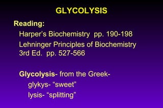
- 1. GLYCOLYSISGLYCOLYSIS Reading: Harper’s Biochemistry pp. 190-198 Lehninger Principles of Biochemistry 3rd Ed. pp. 527-566 Glycolysis- from the Greek- glykys- “sweet” lysis- “splitting”
- 2. OBJECTIVESOBJECTIVES To understand how the glycolytic pathway is used to convert glucose to pyruvate (and lactate) with conservation of chemical potential energy in the form of ATP and NADH. To learn the intermediates, enzymes, and cofactors of the glycolytic pathway.
- 3. Major pathways of glucose utilization inMajor pathways of glucose utilization in cells of higher plants and animalscells of higher plants and animals Although not the only possible fates for glucose, these three pathways are the most significant in terms of the amount of glucose that flows through them in most cells.
- 4. Glucose is the major fuel of most organisms. It is relatively rich in potential energy- complete oxidation to CO2 and H2O proceeds with a free- energy change of -2,840 kJ/mol. By storing glucose as high molecular weight polymers (starch/glycogen) a cell can stockpile large quantities of hexose units. When energy demands increase, glucose can be released quickly from storage and used to produce ATP either aerobically or anaerobically.
- 5. Glycolysis occurs in the cytosol of cells, is common to most organisms, and in humans occurs in virtually all tissues. Most tissues have at least a minimal requirement for glucose. In the brain, the requirement for glucose is substantial, in erythrocytes, it is nearly total. In glycolysis, a molecule of glucose is degraded in a series of steps catalyzed by ten cytosolic enzymes, to yield two molecules of the 3 carbon compound, pyruvate. During those sequential reactions, some of the free energy released is conserved in the form of ATP and NADH
- 6. Biomedical ImportanceBiomedical Importance Of crucial biomedical significance is the ability of glycolysis to provide ATP in the absence of oxygen. This allows skeletal muscle to perform at high levels when aerobic oxidation becomes insufficient, and allows cells to survive anoxic episodes. Diseases associated with impaired glycolysis: ♦Hemolytic anemia: - of the defects in glycolysis that cause hemolytic anemia, pyruvate kinase deficiency (genetic mutations) is the most common. - mature erythrocytes contain no mitochondria, totally dependent upon glycolysis for ATP. - ATP is required for Na/K-ATPase-ion transport system which maintain the proper shape of the erythrocyte membrane. ♦Lactic Acidosis: - can be due to several causes of improper utilization of lactate.
- 7. Glycolysis is only the first step in theGlycolysis is only the first step in the degradation of glucosedegradation of glucose Three possible catabolic fates of the pyruvate formed in glycolysis. Pyruvate also serves as a precursor in many anabolic reactions, not shown here.
- 8. Where glycolysis fits inWhere glycolysis fits in the big picture ofthe big picture of catabolismcatabolism
- 12. Energy Transformations during GlycolysisEnergy Transformations during Glycolysis Glucose + 2 NAD+ + 2 ADP + 2 Pi → 2 Pyruvate + 2 NADH + 2 H+ + 2 ATP + H2O (Note: Much, 95%, of the energy remains in pyruvate) Resolve into two processes: Glucose + 2 NAD+ → 2 pyruvate + 2 NADH + 2 H+ ∆Go´ 1 = -146 kJ/mol 2 ADP + 2 Pi → 2 ATP + 2 H2O ∆Go´ = 2(30.5 kJ/mol) = 61 kJ/mol Overall free energy change = -146 + 61 = -85 kJ/mol
- 13. Why phosphorylated intermediates?Why phosphorylated intermediates? Each of the nine glycolytic intermediates between glucose and pyruvate is phosphorylated 1. Phosphate groups are ionized at pH 7, giving each glycolytic intermediate a net negative charge. Because the plasma membrane is impermeable to charged molecules, the phosphorylated intermediates cannot disperse out of the cell. 2. Energy used in the formation of the phosphate ester is partially conserved. High energy phosphate compounds formed in glycolysis (1,3-bisphosphoglycerate and phosphoenolpyruvate) donate phosphoryl groups to ADP to form ATP. 3. Binding energy resulting from the binding of phosphate groups to the active sites of enzymes lowers the activation energy and increases the specificity of the enzymatic reactions.
- 14. Step 1: Phosphorylation of glucoseStep 1: Phosphorylation of glucose Glucose is activated by phosphorylation at C6 Reaction is catalyzed by hexokinase, present in virtually all extrahepatic cells. Has high affinity (low Km) for glucose, so phosphorylates essentially all the glucose that enters cell, maintaining a large glucose gradient. Will also phosphorylate other hexose sugars. Under physiological conditions, reaction is essentially irreversible. In liver, glucose is phosphorylated by glucokinase. This enzyme has a low affinity (high Km) for glucose, is specific for glucose, and its
- 15. Step 2: Conversion of glucose 6-phosphateStep 2: Conversion of glucose 6-phosphate to fructose 6-phosphateto fructose 6-phosphate The enzyme phosphohexose isomerase catalyzes the reversible isomerization of glucose 6-phosphate (an aldose) to fructose 6-phosphate (a ketose) As predicted for the relatively small change in standard free energy, the reaction proceeds readily in either direction, and requires Mg2+
- 16. Step 3: Phosphorylation of fructose 6-Step 3: Phosphorylation of fructose 6- phosphate to fructose 1, 6-bisphosphatephosphate to fructose 1, 6-bisphosphate Phosphofructokinase-1 catalyzes the transfer of a phosphoryl group from ATP to fructose 6-phosphate to yield fructose 1, 6- bisphosphate. This reaction is essentially irreversible under cellular conditions. Phosphofructokinase-1 is a regulated enzyme at a major point in the regulation of glycolysis. PFK-1 activity is increased whenever the cell’s ATP supply is depleted or when ADP/Pi are in excess. Activity is inhibited whenever the cell has ample ATP and is well supplied by other fuels such as fatty acids.
- 17. Step 4: Cleavage of fructose 1, 6-Step 4: Cleavage of fructose 1, 6- bisphosphatebisphosphate The enzyme fructose 1, 6-bisphosphate aldolase, called just aldolase, catalyzes the cleavage of fructose 1, 6-bisphosphate into two different triose phosphate, glyceraldehyde 3-phosphate and dihydroxyacetone phosphate In cells, this reaction can proceed in either direction, and proceeds to the right during glycolysis because products are quickly removed.
- 18. Step 5: Interconverstion of the trioseStep 5: Interconverstion of the triose phosphatesphosphates Dihydroxyacetone phosphate is rapidly and reversibly converted to glyceraldehyde 3-phosphate by triose phosphate isomerase The C-1, C-2, C-3 of the starting glucose now become chemically indistinguishable for the C-6, C-5, and C-4, respectively. This reaction completes the prepatory phase of glycolysis Other hexoses (fructose, mannose, galactose) can also be
- 19. The payoff phase of glycolysis producingThe payoff phase of glycolysis producing ATP + NADHATP + NADH 2 molecules of glyceraldehyde 3-phosphate → 2 molecules of pyruvate Step 6: Glyceraldehyde 3-phosphate dehydrogenase catalyzes the oxidation of glyceraldehyde 3-phosphate to 1, 3- bisphosphoglycerate Note that the aldehyde group is dehydrogenated to an acyl phosphate, which has a very high standard free energy of hydrolysis (-49.3 kJ/mol). Glyceraldehyde 3-phosphate dehydrogenase is inhibited by iodoacetetate
- 20. Step 7: Phosphoryl transfer from 1, 3-Step 7: Phosphoryl transfer from 1, 3- bisphospho-glycerate to ADPbisphospho-glycerate to ADP The enzyme phosphoglycerate kinase transfers the high-energy phosphoryl group from the carboxyl group to ADP, forming ATP and 3-phosphoglycerate. Steps 6 and 7 represent an energy-coupling process in which 1, 3- phosphoglycerate is the common intermediate. Glyceraldehyde 3-phosphate + ADP + Pi +NAD+ 3-phosphoglycerate + ATP + NADH + H+ ∆Go´ 1= -12.5 kJ/mol
- 21. Step 8: Conversion of 3-phosphoglycerateStep 8: Conversion of 3-phosphoglycerate to 2-phosphoglycerateto 2-phosphoglycerate The enzyme phosphoglycerate mutase catalyzes a reversible shift of the phosphoryl group between C- 2 and C-3 of glycerate. Mg2+ is essential
- 22. Step 9: Dehydration of 2-phosphoglycerateStep 9: Dehydration of 2-phosphoglycerate to phosphoenolpyruvateto phosphoenolpyruvate Enolase promotes reversible removal of a molecule of water from 2-phosphoglycerate to yield phosphoenolpyruvate Standard free energy of hydrolysis of the phosphate groups of the reactant and product are -17.6 kJ/mol and -61.9 kJ/mol, respectively. ie. The loss of the water molecule causes a redistribution of energy within the molecule, generating a super high-energy phosphate compound.
- 23. Step 10: Transfer of the phosphoryl groupStep 10: Transfer of the phosphoryl group from phosphoenolpyruvate to ADPfrom phosphoenolpyruvate to ADP This last step in glycolysis is catalyzed by pyruvate kinase, which requires K+ and Mg 2+ or Mn 2+ This step is also an important site of regulation The product pyruvate undergoes tautomerization from its enol to keto form which is more stable at pH 7
- 24. Overall balance sheet - net gain of ATPOverall balance sheet - net gain of ATP Glucose + 2 ATP+ 2 NAD+ + 4 ADP + 2 Pi → 2 Pyruvate + 2 ADP + 2 NADH + 2 H+ + 4 ATP + 2 H2O or Glucose + 2 NAD+ + 2 ADP + 2 Pi → 2 Pyruvate + 2 NADH + 2 H+ + 2 ATP + 2 H2O Under aerobic conditions, the two molecules of NADH are reoxidized to NAD+ by transfer of their electrons to the respiratory chain in the mitochondrion 2 NADH + 2 H+ + O2 → 2 NAD+ + 2 H2O During glycolysis: *Carbon pathway - Glucose → 2x pyruvate *Phosphate pathway - 2 ADP + 2 Pi → 2 ATP *Electron pathway - Four electrons (two hydride ions) are transferred from 2 molecules of glyceraldehyde 3-phosphate to two of NAD+
- 25. Conversion of pyruvate to lactateConversion of pyruvate to lactate Under hypoxic or anaerobic conditions, NADH generated by glycolysis cannot be reoxidized by O2 - NAD+ is required during glycolysis as electron acceptor in step 6. In these cases, NAD+ is regenerated from NADH by the reduction of pyruvate to lactate, catalyzed by lactate dehydrogenase. This allows glycolysis to occur in the absence of oxygen Lactate produced in muscle during a short burst of physical activity is converted back to glucose in the liver.
- 26. Glycolysis is regulated at 3 steps involvingGlycolysis is regulated at 3 steps involving non equilibrium reactionsnon equilibrium reactions Step 1: hexokinase glucose → glucose 6-phosphate Step 3: phosphofructokinase fructose 6-phosphate → fructose 1, 6-bisphosphate Step 10: pyruvate kinase phosphoenolpyruvate → pyruvate These are all exergonic and physiologically irreversible These enzymes function as “valves”, regulating the flow of carbon through glycolysis. The rates of these steps are limited not by the substrate but by the activity of the enzymes. Enzymes that catalyze these exergonic, rate-limiting steps are commonly the targets of metabolic regulation. Examples of regulation: Phosphofructokinase-1 - inhibited by high levels of ATP. ATP binds to an allosteric site and lowers affinity for fructose 6-phosphate Hexokinase - allosterically inhibited by its product. Pyruvate kinase - inhibited by ATP
- 27. Regulation occurs at steps that are enzyme-limited. At each of these steps (orange arrows), which are generally exergonic, the substrate is not in equilibrium with the product because the reaction is relatively slow; the substrate tends to accumulate, just as river water accumulates behind a dam. In the substrate- limited reactions (blue arrows), the substrate and product are essentially at their equilibrium concentrations. In the steady state, all reactions in the sequence occur at the same rate, which is determined by the rate- limiting step. Regulation of the flux through a multistepRegulation of the flux through a multistep pathwaypathway
- 28. Feeder pathways for glycolysisFeeder pathways for glycolysis
- 29. SUMMARYSUMMARY Glycolysis is a universal metabolic pathway for the catabolism of glucose to pyruvate accompanied by the formation of ATP. The process is catalyzed by 10 cytosolic enzymes and there is a net gain of two ATPs per molecule of glucose. The NADH formed must be recycled to regenerate NAD+ . Under aerobic conditions this occurs during mitochondrial respiration; under anaerobic conditions, NAD+ is regenerated by the conversion of pyruvate to lactate. Other organisms such as yeast regenerate NAD+ by reducing pyruvate to ethanol + CO2 (fermentation) A variety of D-hexoses, including fructose, mannose, and galactose, can be funneled into glycolysis. Enzyme limited, regulated steps are catalyzed by hexokinase, phosphofructokinase-1, and pyruvate kinase.