Variant Flexor Carpi Ulnaris Muscle and Variant Course of Ulnar Artery in Fore Arm
•
1 j'aime•604 vues

(IJMER) www.ijmer.com International Journal of Modern Engineering Research

Recommandé
Contenu connexe
Tendances
Tendances (20)
En vedette
En vedette (20)
Similaire à Variant Flexor Carpi Ulnaris Muscle and Variant Course of Ulnar Artery in Fore Arm
Similaire à Variant Flexor Carpi Ulnaris Muscle and Variant Course of Ulnar Artery in Fore Arm (20)
Plus de IJMER
Plus de IJMER (20)
Variant Flexor Carpi Ulnaris Muscle and Variant Course of Ulnar Artery in Fore Arm
- 1. International Journal of Modern Engineering Research (IJMER) www.ijmer.com Vol.2, Issue.6, Nov-Dec. 2012 pp-4108-4110 ISSN: 2249-6645 Variant Flexor Carpi Ulnaris Muscle and Variant Course of Ulnar Artery in Fore Arm 1 Dr.Sharadkumar Pralhad Sawant, 2Dr.Shaguphta T. Shaikh, 3Dr.Rakhi Milind More 1,2,3 Department of Anatomy, K.J.Somaiya Medical College, Somaiya Ayurvihar, Eastern Express Highway, Sion, Mumbai- 400 022. ABSTRACT: During routine dissection, of the right upper limb of a 70 years old donated embalmed male cadaver in the Department of Anatomy, K.J. Somaiya Medical College, Sion, Mumbai, India, we observed a separate humeral and ulnar heads of flexor carpi ulnaris muscle. To recognise Anatomical variations it is necessary to know the normal Anatomy. Normally the flexor carpi ulnaris muscle arises by two heads, humeral and ulnar, connected by a tendinous arch. The humeral head arises from the medial epicondyle via the common flexor tendon. The ulnar head arises from the medial margin of the olecranon process and an aponeurosis attached to the posterior sub cutaneous border of the ulna. The tendon of flexor carpi ulnaris inserted into the hamate and the fifth metacarpal bone through pisohamate and pisometacarpal ligaments. In the present case the ulnar head of flexor carpi ulnaris muscle was more bulky. It separated ulnar nerve and artery. The humeral and ulnar heads were separated from each other by ulnar nerve. These two heads fused with each other just before their insertion, where the ulnar artery came in contact wih ulnar nerve. The further course and distribution of ulnar artery and ulnar nerve were normal. The knowledge of such unusual ulnar head separating ulnar artery and ulnar nerve may be clinically important for plastic surgeons doing flap surgeries and for the surgeon dealing with cubital tunnel syndrome. KEYWORDS: Flexor carpi ulnaris, Ulnar artery, Ulnar Head, Ulnar nerve, Humeral head, plastic surgeons, cubital tunnel syndrome. I. INTRODUCTION: Flexor carpi ulnaris muscle is the medial most muscle of the superficial flexor group. It arises by two heads, humeral and ulnar, connected by a tendinous arch. The small humeral head arises from the medial epicondyle via the common flexor tendon. The ulnar head has an extensive origin from the medial margin of the olecranon process and proximal two-thirds of the posterior border of the ulna, an aponeurosis (along with flexor digitorum profundus and extensor carpi ulnaris) and from the intermuscular septum between it and flexor digitorum superficialis. A thick tendon forms along its anterolateral border in its distal half. The tendon is attached to the pisiform, and thence prolonged to the hamate and the fifth metacarpal bone by pisohamate and pisometacarpal ligaments (pisiform is the sesamoid bone developing in the tendon of flexor carpi ulnaris). Acting with the flexor carpi radialis, it flexes the wrist and acting with the extensor carpi ulnaris it adducts the wrist (1). Flexor carpi ulnaris muscle is innervated by the ulnar nerve (C7, C8 and T1). The line between the medial humeral epicondyle and the pisiform, along the anterior palmar margin of the muscle, is used as a reference point for locating the ulnar neurovascular bundle. The ulnar artery reaches the muscle in its middle third, whereas the ulnar nerve is covered by the muscle throughout its entire course running under the tendon in the wrist region. The ulnar artery, the larger of the two terminal branches of the brachial, begins a little below the bend of the elbow, and, passing obliquely downward, reaches the ulnar side of the forearm at a point about midway between the elbow and the wrist. It then runs along the ulnar border to the wrist, crosses the transverse carpal ligament on the radial side of the pisiform bone, and immediately beyond this bone divides into two branches, which enter into the formation of the superficial and deep palmar arches. The ulnar nerve, after descending in the forearm between the flexor digitorum profundus and flexor carpi ulnaris muscles, pierces the deep fascia and enters the wrist through the Guyon’s canal. In the distal part of the canal, the ulnar nerve divides into a superficial sensory branch and a deep motor branch, which supplies the hypothenar muscles and then passes across the palm, distributing to other intrinsic hand muscles. II. MATERIALS AND METHODS: The right upper limb of a donated embalmed 70 years old male cadaver was dissected during routine dissection in the department of Anatomy at K.J. Somaiya Medical College, Sion, Mumbai. All the superficial flexor muscles were exposed. The humeral and ulnar heads of flexor carpi ulnaris muscle was dissected carefully to observe the arrangement of ulnar artery, ulnar head, ulnar nerve and humeral head. The course of ulnar artery and ulnar nerve were also dissected. The photographs of the variations were taken for proper documentation. III. RESULTS: The variation was observed in the forearm of right upper limb. However, the left upper limb was normal. The right forearm showed separate ulnar and humeral heads of flexor carpi ulnaris muscle. The ulnar head of flexor carpi ulnaris muscle separated ulnar artery and nerve. The humeral and ulnar heads were separated from each other by ulnar nerve. The tendons of both the heads of flexor carpi ulnaris fused with each other just before their insertion. The ulnar artery comes in contact wih ulnar nerve in the lower part of the forearm near the wrist where the two heads of flexor carpi ulnaris fused with each other . The ulnar artery in the hand takes part in the formation of superficial and deep palmar arches. The ulnar nerve in the hand bifurcates into superficial sensory branch and a deep motor branch. The course and distribution of the terminal branches of both ulnar nerve and ulnar artery were normal. www.ijmer.com 4108 | Page
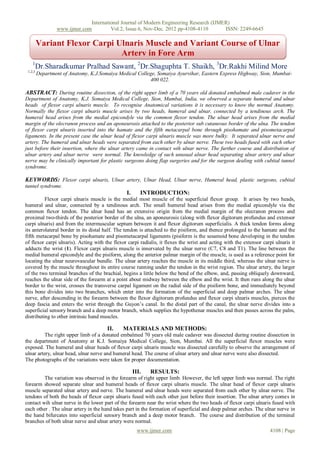
- 2. International Journal of Modern Engineering Research (IJMER) www.ijmer.com Vol.2, Issue.6, Nov-Dec. 2012 pp-4108-4110 ISSN: 2249-6645 . Figure : Photographic presentation of the right forearm showing separate ulnar and humeral heads of flexor carpi ulnaris muscle. The ulnar artery running in between ulnar and humeral heads of flexor carpi ulnaris muscle. IV. DISCUSSION: The variations of flexor carpi ulnaris muscle have been reported previously by many authors. These variations include: (i) an additional slip of flexor carpi ulnaris (2). (ii) variations in musculotendinous junction of the flexor carpi ulnaris muscle (3), (iii) variant flexor carpi ulnaris causing ulnar nerve compression (4), In the present case the two heads of flexor carpi ulnaris muscle remain separate. The ulnar head separated ulnar artery and nerve. The ulnar nerve runs in between the two heads of the flexor carpi ulnaris muscle in the forearm. Such variation is not yet reported in literature. The flexor carpi ulnaris muscle acts as an anatomical guideline for finding the neurovascular bundle (ulnar nerve, ulnar artery and accompanying venae comitantes), it can be easily palpated in its distal course if the wrist is flexed and adducted. The present variation need to be taken into account when interpreting ultrasound and MR images, as well as during dissection of the ulnar neurovascular bundle when using flexor carpi ulnaris as a guideline. The flexor carpi ulnaris is a useful local muscle flap in the forearm and elbow. It is, however, an important palmar flexor and ulnar deviator of the wrist, and functional loss may arise from the use of this muscle in its entirety. The flexor carpi ulnaris is made up of two distinct neuromuscular compartments. This arrangement allows for splitting of the muscle and the potential use of the larger ulnar compartment as a local muscle flap while maintaining the humeral compartment as an ulnar deviator and palmar flexor of the wrist (5). After multiple efforts to heal an infected nonunion of the proximal ulna, a flexor carpi ulnaris muscle pedicle flap was used to improve blood supply and softtissue coverage at the nonunion site. It was observed that it promoted bone healing and restoration of useful elbow function (6). The course and distribution of ulnar nerve and ulnar artery can assist the surgeon in the diagnosis and effective management of the more common pain syndromes conditions associated with the ulnar aspect of the hand (7). As the two heads of flexor carpi ulnaris muscles were separate the tendinous arch between them was absent. The possibility of entrapment of ulnar nerve in cubital tunnel in such cases is rare. V. CONCLUSION: The knowledge of such unusual variations of flexor carpi ulnaris is a must before any operative procedures of the forearm and hand. The ulnar head of flexor carpi ulnaris separating ulnar artery and ulnar nerve seen in present case is important for Anatomists. It may be clinically important for plastic surgeons doing flap surgeries and for the surgeons dealing with cubital tunnel syndrome. Orthopaedicians have used this muscle flap for treating non union of proximal ulna and hence knowledge of this variable head is important. Competing Interests: The authors declare that they have no competing interest. Authors' contributions: SPS wrote the case report, performed the literature review & obtained the photograph for the study. RMM performed the literature search and assisted with writing the paper. STS conceived the study and helped to draft the manuscript. All authors have read and approved the final version manuscript. VI. Acknowledgement: All the authors wish to convey our sincere thanks to Dr. Arif A. Faruqui for his valuable help, support and inspiration. Authors also acknowledge the immense help received from the scholars whose articles are cited and included in references of this manuscript. The authors are also grateful to authors / editors / publishers of all those articles, journals and books from where the literature for this article has been reviewed and discussed. www.ijmer.com 4109 | Page
- 3. International Journal of Modern Engineering Research (IJMER) www.ijmer.com Vol.2, Issue.6, Nov-Dec. 2012 pp-4108-4110 ISSN: 2249-6645 REFERENCES: 1. Susan, Standring, Gray’s Anatomy, 39th Edition Elsevier Churchill Livingstone, 2005. pg: 877. 2. Bergman, R. A.; Thomson, S. A.; Afifi, A. K. & Saadesh, F. A. Compendium of human anatomic variations. Urban & Schwarzenberg , Baltimore – Munich, 13, 1988. 3. Grechenig, W.; Clement, H.; Egner, S.; Tesch, N. P.; Weiglein, A. & Peicha, G. Musculo-tendinous junction of the flexor carpi ulnaris muscle. An anatomical study. Surg. Radiol. Anat., 22:255-60, 2000. 4. Al-Qattan, M. M. & Duerksen, F. A variant of flexor carpi ulnaris causing ulnar nerve compression. J. Anat., 180:189- 190, 1992. 5. Lingaraj, K.; Lim, A. Y.; Puhaindran, M. E. & Kumar, P. V. The split flexor carpi ulnaris as a local muscle flap. Clin. Orthop. Relat. Res., 455:262-6, 2007. 6. Meals, R. A. The use of a flexor carpi ulnaris muscle flap in the treatment of an infected nonunion of the proximal ulna. A case report. Clin. Orthop. Relat. Res., 240:168- 72, 1989. 7. Kleinert, H. and Hayes, J. The ulnar tunnel syndrome. Plastic Reconstructive Surgery, 1971, Vol.47, pg. 21-24. Corresponding author full mailing address: - Dr. Sharadkumar Pralhad Sawant, 25/2, Samrat Ashok Nagar Society, Shell Colony Road, Chembur, Mumbai – 400 071, Maharashtra, India. www.ijmer.com 4110 | Page