Sds page Electrophoresis Troubleshooting Tips
•
1 j'aime•3,471 vues

Electrophoresis is a simple, rapid, and highly sensitive analytical technique to study the properties of proteins and nucleic acids, and has become a principle tool in analytical chemistry, biochemistry, and molecular biology. Polyacrylamide gel electrophoresis (PAGE) can be used to analyze the size, amount, purity, and isoelectric point of polypeptides and proteins. Sodium dodecyl sulfate polyacrylamide discontinuous gel electrophoresis (SDS PAGE) is the most commonly used system whereby proteins become separated strictly by their size, but there are different variations of this technique.

Recommandé
Contenu connexe
Tendances
Tendances (20)
Similaire à Sds page Electrophoresis Troubleshooting Tips
Similaire à Sds page Electrophoresis Troubleshooting Tips (20)
Plus de Expedeon
Plus de Expedeon (20)
Dernier
Dernier (20)
Sds page Electrophoresis Troubleshooting Tips
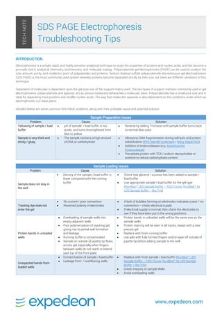
- 1. www.expedeon.com INTRODUCTION Electrophoresis is a simple, rapid, and highly sensitive analytical technique to study the properties of proteins and nucleic acids, and has become a principle tool in analytical chemistry, biochemistry, and molecular biology. Polyacrylamide gel electrophoresis (PAGE) can be used to analyze the size, amount, purity, and isoelectric point of polypeptides and proteins. Sodium dodecyl sulfate polyacrylamide discontinuous gel electrophoresis (SDS PAGE) is the most commonly used system whereby proteins become separated strictly by their size, but there are different variations of this technique. Separation of molecules is dependent upon the gel pore size of the support matrix used. The two types of support matrices commonly used in gel electrophoresis, polyacrylamide and agarose, act as porous media and behave like a molecular sieve. Polyacrylamide has a small pore size and is ideal for separating most proteins and smaller nucleic acids. The way that molecules separate is also dependent on the conditions under which an electrophoretic run takes place. Detailed below are some common SDS PAGE problems, along with their probable cause and potential solution: Sample Preparation Issues Problem Cause Solution Yellowing of sample / load buffer pH of sample / load buffer is too acidic, and turns bromophenol from blue to yellow Reverse by adding Tris base until sample buffer turns back to normal blue color Sample is very thick and sticky / gluey The sample contains a high amount of DNA or carbohydrate Ultrasonic DNA fragmentation during cell lysis and protein solubilization (PPS Silent® Surfactant / NVoy Stabil-PAC) Addition of endonucleases (e.g. BaseMuncher Endonuclease) Precipitate protein with TCA / sodium deoxycholate or acetone to reduce carbohydrate content Sample Loading Issues Problem Cause Solution Sample does not stay in the well Density of the sample / load buffer is lower compared with the running buffer Check that glycerol / sucrose has been added to sample / load buffer Use appropriate sample / load buffer for the gel type (RunBlue™ LDS Sample Buffer – TEO-Tricine, RunBlue™ 4x LDS Sample Buffer – Bis-Tris) Tracking dye does not enter the gel No current / poor connection Reversed polarity of electrodes A lack of bubbles forming on electrodes indicates a poor / no connection – check electrical supply If electrical supply is normal, then check the electrodes to see if they have been put in the wrong positions Protein bands in unloaded wells Overloading of sample wells into empty adjacent wells Poor polymerization of stacking gel, giving rise to partial well formation and leakage Running buffer is contaminated Sample on outside of pipette tip flows across gel, especially when fingers between wells do not reach or extend past top of the front plate Protein bands in unloaded wells will be the same size as the sample wells Protein staining will be seen in all tracks, repeat with a new precast gel Replace with fresh running buffer Use gels with fully formed fingers and/or wipe off outside of pipette tip before adding sample to the well Unexpected bands from loaded wells Contamination of sample / load buffer Leakage from / overflowing wells Replace with fresh sample / load buffer (RunBlue™ LDS Sample Buffer – TEO-Tricine, RunBlue™ 4x LDS Sample Buffer – Bis-Tris) Check integrity of sample Wells Avoid overloading wells SDS PAGE Electrophoresis Troubleshooting Tips TECHNOTE
- 2. www.expedeon.com Sample Preparation Issues Problem Cause Solution High / uneven background staining Samples with a high degree of proteolysis Overloaded wells Sample / load buffer impurities / contamination Dirty equipment Impure reagents Too long in stain Use protease inhibitor in sample buffer (Proteoloc™) Do not overload wells, but use the suggested volume / concentrate the sample Use new sample / buffer Use appropriate load buffer for the gel type (RunBlue™ LDS Sample Buffer – TEO- Tricine, RunBlue™ 4x LDS Sample Buffer – Bis-Tris) Ensure clean equipment is used Ensure use of high purity water and reagents Restrict length of time in stain, wash gel in water or destain for a minimum of 30 mins Staining is uneven Gel not shaken / agitated sufficiently during staining Increase agitation and / or time during staining Protein Separation Issues Problem Cause Solution No bands observed Gel does not contain protein Imager not working / incorrect parameters used to visualize gel Use alternative stain to confirm presence of protein Refer to instrument manual to troubleshoot / contact imager manufacturer Poor resolution Impure reagents Overloading of sample Incorrect buffer pH Incorrect running buffer Incorrect sample / load buffer High temperature Ensure use of high purity water and reagents Do not overload sample Adjust buffer pH Ensure that the running buffer is correct one for gel buffer system (RunBlue™ SDS Running Buffer, RunBlue™ MES Run Buffer, RunBlue™ MOPS Run Buffer) Use appropriate sample / load buffer for the gel type (RunBlue™ LDS Sample Buffer – TEO-Tricine, RunBlue™ 4x LDS Sample Buffer – Bis-Tris) Reduce the current to slow down the gel run, reduce temperature and obtain an even gel temperature Distorted / fuzzy bands Temperature gradient from center of gel to gel surface Sample has a high salt content Poor polymerization, bubbles or insoluble materials in the gel Insufficient reducing agent / sample not reduced Ensure proper cooling of gel to reduce temperature gradient Desalt sample Repeat with a new gel Revise concentration of reducing agent / use β- mercaptoethanol (β-ME) or dithiothreitol (DTT) (RunBlue™ DTT Reducer) and heat sample (70–80°C for 10 minutes) Protein does not enter the resolving gel / streaking of protein in the gel Protein aggregation / precipitation Dissolution of the precipitate during the electrophoretic run When running both reduced and non- reduced samples on the same gel, lateral diffusion of reducing agent during the run causing reduction during the run Centrifuge samples following denaturation to remove insoluble materials Decrease the amount of sample loaded Dilute sample preparation and use a continuous buffer system Keep one or two empty lanes between reduced and non- reduced samples Protein dimer or double bands in gel Due to disulf ide bonds Sample not sufficiently reduced or not reduced Use fresh sample / load buffer (RunBlue™ LDS Sample Buffer – TEO-Tricine, RunBlue™ 4x LDS Sample Buffer – Bis-Tris) Ensure that the sample has been heated with a reducing agent such as β-mercaptoethanol (β-ME) or dithiothreitol (DTT) (RunBlue™ DTT Reducer) Electrophoretic Run Issues Problem Cause Solution Run time is too short Gel buffer may be too dilute Running buffer may be too concentrated Use gel buffer that is more concentrated Dilute the running buffer (RunBlue™ SDS Running Buffer, RunBlue™ MES Run Buffer, RunBlue™ MOPS Run Buffer) Gel shrinkage Gel has become dehydrated Rehydrate gel in water Run time is too long Running buffer may be too dilute Gel buffer may be too concentrated Use running buffer that is more concentrated Dilute the gel buffer Leaking from electrophoresis tank Improper assembly of tank Damage to tank Assemble correctly according to manufacturer’s instructions Check for damage to the tank
- 3. www.expedeon.com RELATED PRODUCTS FROM EXPEDEON: Expedeon produce a range of products to assist you with your SDS PAGE electrophoresis. Please click here to view our extensive range. References: Gel Electrophoresis of Proteins – A Practical Approach. 3rd edition. BD. Hames. Series ed. Oxford, United Kingdom: Oxford University Press. http://www.oup.co.uk/PAS